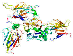
CHEK2

Last updated
CHEK2
Protein CHEK2 PDB 1gxc.png
Available structures
PDB Ortholog search: PDBe RCSB
Identifiers
Aliases CHEK2 , CDS1, CHK2, HuCds1, LFS2, PP1425, RAD53, hCds1, checkpoint kinase 2
External IDs OMIM: 604373 MGI: 1355321 HomoloGene: 38289 GeneCards: CHEK2
Orthologs
SpeciesHumanMouse
Entrez
Ensembl
UniProt
RefSeq (mRNA)

NM_001005735
NM_001257387
NM_007194
NM_145862
NM_001349956

NM_016681
NM_001363308

RefSeq (protein)

NP_001005735
NP_001244316
NP_009125
NP_665861
NP_001336885

NP_057890
NP_001350237

Location (UCSC) Chr 22: 28.69 – 28.74 Mb Chr 5: 110.99 – 111.02 Mb
PubMed search [3] [4]
Wikidata
View/Edit Human View/Edit Mouse

CHEK2 (Checkpoint kinase 2) is a tumor suppressor gene that encodes the protein CHK2, a serine-threonine kinase. CHK2 is involved in DNA repair, cell cycle arrest or apoptosis in response to DNA damage. Mutations to the CHEK2 gene have been linked to a wide range of cancers. [5]

Gene location

The CHEK2 gene is located on the long (q) arm of chromosome 22 at position 12.1. Its location on chromosome 22 stretches from base pair 28,687,742 to base pair 28,741,904. [5]

Protein structure

The CHEK2 protein encoded by the CHEK2 gene is a serine threonine kinase. The protein consists of 543 amino acids and the following domains:

The SCD domain contains multiple SQ/TQ motifs that serve as sites for phosphorylation in response to DNA damage. The most notable and frequently phosphorylated site being Thr68. [6]

CHK2 appears as a monomer in its inactive state. However, in the event of DNA damage SCD phosphorylation causes CHK2 dimerization. The phosphorylated Thr68 (located on the SCD) interacts with the FHA domain to form the dimer. After the protein dimerizes the KD is activated via autophosphorylation. Once the KD is activated the CHK2 dimer dissociates. [6]

Function and mechanism

The CHEK2 gene encodes for checkpoint kinase 2 (CHK2), a protein that acts as a tumor suppressor. CHK2 regulates cell division, and has the ability to prevent cells from dividing too rapidly or in an uncontrolled manner. [5]

When DNA undergoes a double-strand break, CHK2 is activated. Specifically, DNA damage-activated phosphatidylinositol kinase family protein (PIKK) ATM phosphorylates site Thr68 and activates CHK2. [6] Once activated, CHK2 phosphorylates downstream targets including CDC25 phosphatases, responsible for dephosphorylating and activating the cyclin-dependent kinases (CDKs). Thus, CHK2's inhibition of the CDC25 phosphatases prevents entry of the cell into mitosis. Furthermore, the CHK2 protein interacts with several other proteins including p53 (p53). Stabilization of p53 by CHK2 leads to cell cycle arrest in phase G1. Furthermore, CHK2 is known to phosphorylate the cell-cycle transcription factor E2F1 and the promyelocytic leukemia protein (PML) involved in apoptosis (programmed cell death). [6]

Association with cancer

The CHK2 protein plays a critical role in the DNA damage checkpoint. Thus, mutations to the CHEK2 gene have been labeled as causes to a wide range of cancers.

In 1999, genetic variations of CHEK2 were found to correspond to inherited cancer susceptibility. [7]

Bell et al. (1999) discovered three CHEK2 germline mutations among four Li–Fraumeni syndrome (LFS) and 18 Li–Fraumeni-like (LFL) families. Since the time of this discovery, two of the three variants (a deletion in the kinase domain in exon 10 and a missense mutation in the FHA domain in exon 3) have been linked to inherited susceptibility to breast as well as other cancers. [8]

Beyond initial speculations, screening of LFS and LFL patients has revealed no or very rare individual missense variants in the CHEK2 gene. Additionally, the deletion in the kinase domain on exon 10 has been found rare among LFS/LFL patients. The evidence from these studies has suggests that CHEK2 is not a predisposition gene to Li–Fraumeni syndrome. [8]

Breast cancer

Inherited mutations in the CHEK2 gene have been linked to certain cases of breast cancer. Most notably, the deletion of a single DNA nucleotide at position 1100 in exon 10 (1100delC) produces a nonfunctional version of the CHK2 protein, truncated at the kinase domain. The loss of normal CHK2 protein function leads to unregulated cell division, accumulated damage to DNA and in many cases, tumor development. [5] The CHEK2*1100del mutation is most commonly seen in individuals of Eastern and Northern European descent. Within these populations the CHEK2*1100delC mutation is seen in 1 out of 100 to 1 out of 200 individuals. However, in North America the frequency drops to 1 out of 333 to 1 out of 500. The mutation is almost absent in Spain and India. [9] Studies show that a CHEK2 1100delC corresponds to a two-fold increased risk of breast cancer and a 10-fold increased risk of breast cancer in males. [10]

A CHEK2 mutation known as the I157T variant to the FHA domain in exon 3 has also been linked to breast cancer but at a lower risk than the CHEK2*1100delC mutation. The estimated fraction of breast cancer attributed to this variant is reported to be around 1.2% in the US. [8]

Two more CHEK2 gene mutations, CHEK2*S428F, an amino-acid substitution to the kinase domain in exon 11 and CHEK2*P85L, an amino-acid substitution in the N-terminal region (exon 1) have been found in the Ashkenazi Jewish population. [9] Suggestion of a Hispanic founder mutation has also been described. [11]

Other cancers

Mutations to CHEK2 have been found in hereditary and nonhereditary cases of cancer. Studies link the mutation to cases of prostate, lung, colon, kidney, and thyroid cancers. Links have also been drawn to certain brain tumors and osteosarcoma. [5]

Unlike BRCA1 and BRCA2 mutations, CHEK2 mutations do not appear to cause an elevated risk for ovarian cancer. [10] However, a large-effect genome-wide association for squamous lung cancer has been described for a rare variant in CHEK2 (p.Ile157Thr, rs17879961, OR = 0.38). [12]

Meiosis

CHEK2 regulates cell cycle progression and spindle assembly during mouse oocyte maturation and early embryo development. [13] [14] Although CHEK2 is a down stream effector of the ATM kinase that responds primarily to double-strand breaks it can also be activated by ATR (ataxia-telangiectasia and Rad3 related) kinase that responds primarily to single-strand breaks. In mice, CHEK2 is essential for DNA damage surveillance in female meiosis. The response of oocytes to DNA double-strand break damage involves a pathway hierarchy in which ATR kinase signals to CHEK2 which then activates p53 and p63 proteins. [15]

In the fruit fly Drosophila, irradiation of germ line cells generates double-strand breaks that result in cell cycle arrest and apoptosis. The Drosophila CHEK2 ortholog mnk and the p53 ortholog dp53 are required for much of the cell death observed in early oogenesis when oocyte selection and meiotic recombination occur. [16]

Interactions

CHEK2 has been shown to interact with:

Related Research Articles

p53 Mammalian protein found in Homo sapiens

p53, also known as Tumor protein P53, cellular tumor antigen p53, or transformation-related protein 53 (TRP53) is a regulatory protein that is often mutated in human cancers. The p53 proteins are crucial in vertebrates, where they prevent cancer formation. As such, p53 has been described as "the guardian of the genome" because of its role in conserving stability by preventing genome mutation. Hence TP53 is classified as a tumor suppressor gene.

<span class="mw-page-title-main">BRCA1</span> Gene known for its role in breast cancer

Breast cancer type 1 susceptibility protein is a protein that in humans is encoded by the BRCA1 gene. Orthologs are common in other vertebrate species, whereas invertebrate genomes may encode a more distantly related gene. BRCA1 is a human tumor suppressor gene and is responsible for repairing DNA.

<span class="mw-page-title-main">DNA repair</span> Cellular mechanism

DNA repair is a collection of processes by which a cell identifies and corrects damage to the DNA molecules that encodes its genome. In human cells, both normal metabolic activities and environmental factors such as radiation can cause DNA damage, resulting in tens of thousands of individual molecular lesions per cell per day. Many of these lesions cause structural damage to the DNA molecule and can alter or eliminate the cell's ability to transcribe the gene that the affected DNA encodes. Other lesions induce potentially harmful mutations in the cell's genome, which affect the survival of its daughter cells after it undergoes mitosis. As a consequence, the DNA repair process is constantly active as it responds to damage in the DNA structure. When normal repair processes fail, and when cellular apoptosis does not occur, irreparable DNA damage may occur, including double-strand breaks and DNA crosslinkages. This can eventually lead to malignant tumors, or cancer as per the two-hit hypothesis.

<span class="mw-page-title-main">Li–Fraumeni syndrome</span> Autosomal dominant cancer syndrome

Li–Fraumeni syndrome is a rare, autosomal dominant, hereditary disorder that predisposes carriers to cancer development. It was named after two American physicians, Frederick Pei Li and Joseph F. Fraumeni Jr., who first recognized the syndrome after reviewing the medical records and death certificates of 648 childhood rhabdomyosarcoma patients. This syndrome is also known as the sarcoma, breast, leukaemia and adrenal gland (SBLA) syndrome.

<span class="mw-page-title-main">ATM serine/threonine kinase</span>

ATM serine/threonine kinase or Ataxia-telangiectasia mutated, symbol ATM, is a serine/threonine protein kinase that is recruited and activated by DNA double-strand breaks, oxidative stress, topoisomerase cleavage complexes, splicing intermediates, R-loops and in some cases by single-strand DNA breaks. It phosphorylates several key proteins that initiate activation of the DNA damage checkpoint, leading to cell cycle arrest, DNA repair or apoptosis. Several of these targets, including p53, CHK2, BRCA1, NBS1 and H2AX are tumor suppressors.

<span class="mw-page-title-main">Cell cycle checkpoint</span> Control mechanism in the eukaryotic cell cycle

Cell cycle checkpoints are control mechanisms in the eukaryotic cell cycle which ensure its proper progression. Each checkpoint serves as a potential termination point along the cell cycle, during which the conditions of the cell are assessed, with progression through the various phases of the cell cycle occurring only when favorable conditions are met. There are many checkpoints in the cell cycle, but the three major ones are: the G1 checkpoint, also known as the Start or restriction checkpoint or Major Checkpoint; the G2/M checkpoint; and the metaphase-to-anaphase transition, also known as the spindle checkpoint. Progression through these checkpoints is largely determined by the activation of cyclin-dependent kinases by regulatory protein subunits called cyclins, different forms of which are produced at each stage of the cell cycle to control the specific events that occur therein.

<span class="mw-page-title-main">Ataxia telangiectasia and Rad3 related</span> Protein kinase that detects DNA damage and halts cell division

Serine/threonine-protein kinase ATR, also known as ataxia telangiectasia and Rad3-related protein (ATR) or FRAP-related protein 1 (FRP1), is an enzyme that, in humans, is encoded by the ATR gene. It is a large kinase of about 301.66 kDa. ATR belongs to the phosphatidylinositol 3-kinase-related kinase protein family. ATR is activated in response to single strand breaks, and works with ATM to ensure genome integrity.

<span class="mw-page-title-main">CHEK1</span> Protein-coding gene in humans

Checkpoint kinase 1, commonly referred to as Chk1, is a serine/threonine-specific protein kinase that, in humans, is encoded by the CHEK1 gene. Chk1 coordinates the DNA damage response (DDR) and cell cycle checkpoint response. Activation of Chk1 results in the initiation of cell cycle checkpoints, cell cycle arrest, DNA repair and cell death to prevent damaged cells from progressing through the cell cycle.

<span class="mw-page-title-main">BARD1</span> Protein-coding gene in the species Homo sapiens

BRCA1-associated RING domain protein 1 is a protein that in humans is encoded by the BARD1 gene. The human BARD1 protein is 777 amino acids long and contains a RING finger domain, four ankyrin repeats, and two tandem BRCT domains.

<span class="mw-page-title-main">RAD9A</span> Protein-coding gene in the species Homo sapiens

Cell cycle checkpoint control protein RAD9A is a protein that in humans is encoded by the RAD9A gene.Rad9 has been shown to induce G2 arrest in the cell cycle in response to DNA damage in yeast cells. Rad9 was originally found in budding yeast cells but a human homolog has also been found and studies have suggested that the molecular mechanisms of the S and G2 checkpoints are conserved in eukaryotes. Thus, what is found in yeast cells are likely to be similar in human cells.

<span class="mw-page-title-main">TP53BP1</span> Protein-coding gene in the species Homo sapiens

Tumor suppressor p53-binding protein 1 also known as p53-binding protein 1 or 53BP1 is a protein that in humans is encoded by the TP53BP1 gene.

<span class="mw-page-title-main">HIPK2</span> Protein-coding gene in the species Homo sapiens

Homeodomain-interacting protein kinase 2 is an enzyme that in humans is encoded by the HIPK2 gene. HIPK2 can be categorized as a Serine/Threonine Protein kinase, specifically one that interacts with homeodomain transcription factors. It belongs to a family of protein kinases known as the DYRK kinases. Within this family HIPK2 belongs to a group of homeodomain-interacting protein kinases (HIPKs), including HIPK1 and HIPK3. HIPK2 can be found in a wide variety of species and its functions in gene expression and apoptosis are regulated by several different mechanisms.

<span class="mw-page-title-main">TOPBP1</span> Protein-coding gene in the species Homo sapiens

DNA topoisomerase 2-binding protein 1 (TOPBP1) is a scaffold protein that in humans is encoded by the TOPBP1 gene.

<span class="mw-page-title-main">PLK3</span> Protein-coding gene in the species Homo sapiens

Polo-like kinase 3 (Drosophila), also known as PLK3, is an enzyme which in humans is encoded by the PLK3 gene.

<span class="mw-page-title-main">MDC1</span> Protein-coding gene in the species Homo sapiens

Mediator of DNA damage checkpoint protein 1 is a 2080 amino acid long protein that in humans is encoded by the MDC1 gene located on the short arm (p) of chromosome 6. MDC1 protein is a regulator of the Intra-S phase and the G2/M cell cycle checkpoints and recruits repair proteins to the site of DNA damage. It is involved in determining cell survival fate in association with tumor suppressor protein p53. This protein also goes by the name Nuclear Factor with BRCT Domain 1 (NFBD1).

<span class="mw-page-title-main">BRIP1</span> Mammalian protein found in Homo sapiens

Fanconi anemia group J protein is a protein that in humans is encoded by the BRCA1-interacting protein 1 (BRIP1) gene.

<span class="mw-page-title-main">FANCL</span> Protein-coding gene in the species Homo sapiens

E3 ubiquitin-protein ligase FANCL is an enzyme that in humans is encoded by the FANCL gene.

<span class="mw-page-title-main">Meiotic recombination checkpoint</span>

The meiotic recombination checkpoint monitors meiotic recombination during meiosis, and blocks the entry into metaphase I if recombination is not efficiently processed.

Anticancer genes exhibit a preferential ability to kill cancer cells while leaving healthy cells unharmed. This phenomenon is achieved through various processes such as apoptosis following a mitotic catastrophe, necrosis, and autophagy. In the late 1990s, extensive research in the field of cancer cells led to the discovery of anticancer genes. Currently, 291 anticancer genes have been identified. The deregulation of these genes due to base substitutions leading to insertions, deletions, or alterations in missense amino acids can cause frameshifts, thereby altering the protein. A change in gene copy number or rearrangements is also essential for deregulating these genes. The loss or alteration of these anticancer genes due to mutations or rearrangements may lead to the development of cancer.

Antineoplastic resistance, often used interchangeably with chemotherapy resistance, is the resistance of neoplastic (cancerous) cells, or the ability of cancer cells to survive and grow despite anti-cancer therapies. In some cases, cancers can evolve resistance to multiple drugs, called multiple drug resistance.

References

  1. 1 2 3 GRCh38: Ensembl release 89: ENSG00000183765 - Ensembl, May 2017
  2. 1 2 3 GRCm38: Ensembl release 89: ENSMUSG00000029521 - Ensembl, May 2017
  3. "Human PubMed Reference:". National Center for Biotechnology Information, U.S. National Library of Medicine.
  4. "Mouse PubMed Reference:". National Center for Biotechnology Information, U.S. National Library of Medicine.
  5. 1 2 3 4 5 "CHEK2". Genetics Home Reference. August 2007. Archived from the original on 2015-04-25. Retrieved 2015-04-22.
  6. 1 2 3 4 Cai Z, Chehab NH, Pavletich NP (September 2009). "Structure and activation mechanism of the CHK2 DNA damage checkpoint kinase". Molecular Cell. 35 (6): 818–29. doi: 10.1016/j.molcel.2009.09.007 . PMID   19782031.
  7. Bell DW, Varley JM, Szydlo TE, Kang DH, Wahrer DC, Shannon KE, et al. (December 1999). "Heterozygous germ line hCHK2 mutations in Li-Fraumeni syndrome". Science. 286 (5449): 2528–31. doi:10.1126/science.286.5449.2528. PMID   10617473.
  8. 1 2 3 Nevanlinna H, Bartek J (September 2006). "The CHEK2 gene and inherited breast cancer susceptibility". Oncogene. 25 (43): 5912–9. doi:10.1038/sj.onc.1209877. PMID   16998506. S2CID   7343321.
  9. 1 2 Offit K, Garber JE (February 2008). "Time to check CHEK2 in families with breast cancer?". Journal of Clinical Oncology. 26 (4): 519–20. doi: 10.1200/JCO.2007.13.8503 . PMID   18172189.
  10. 1 2 Meijers-Heijboer H, van den Ouweland A, Klijn J, Wasielewski M, de Snoo A, Oldenburg R, et al. (May 2002). "Low-penetrance susceptibility to breast cancer due to CHEK2(*)1100delC in noncarriers of BRCA1 or BRCA2 mutations". Nature Genetics. 31 (1): 55–9. doi: 10.1038/ng879 . PMID   11967536. S2CID   195216803.
  11. Weitzel JN, Neuhausen SL, Adamson A, Tao S, Ricker C, Maoz A, et al. (August 2019). "Pathogenic and likely pathogenic variants in PALB2, CHEK2, and other known breast cancer susceptibility genes among 1054 BRCA-negative Hispanics with breast cancer". Cancer. 125 (16): 2829–2836. doi:10.1002/cncr.32083. PMC   7376605 . PMID   31206626.
  12. Wang Y, McKay JD, Rafnar T, Wang Z, Timofeeva MN, Broderick P, et al. (July 2014). "Rare variants of large effect in BRCA2 and CHEK2 affect risk of lung cancer". Nature Genetics. 46 (7): 736–41. doi: 10.1038/ng.3002 . PMC   4074058 . PMID   24880342.
  13. Dai XX, Duan X, Liu HL, Cui XS, Kim NH, Sun SC (February 2014). "Chk2 regulates cell cycle progression during mouse oocyte maturation and early embryo development". Molecules and Cells. 37 (2): 126–32. doi:10.14348/molcells.2014.2259. PMC   3935625 . PMID   24598997.
  14. Ruth KS, Day FR, Hussain J, Martínez-Marchal A, Aiken CE, Azad A, et al. (August 2021). "Genetic insights into biological mechanisms governing human ovarian ageing". pp. 393–397. medRxiv   10.1101/2021.01.11.20248322v1 .
  15. Bolcun-Filas E, Rinaldi VD, White ME, Schimenti JC (January 2014). "Reversal of female infertility by Chk2 ablation reveals the oocyte DNA damage checkpoint pathway". Science. 343 (6170): 533–6. Bibcode:2014Sci...343..533B. doi:10.1126/science.1247671. PMC   4048839 . PMID   24482479.
  16. Shim HJ, Lee EM, Nguyen LD, Shim J, Song YH (2014). "High-dose irradiation induces cell cycle arrest, apoptosis, and developmental defects during Drosophila oogenesis". PLOS ONE. 9 (2): e89009. Bibcode:2014PLoSO...989009S. doi: 10.1371/journal.pone.0089009 . PMC   3923870 . PMID   24551207.
  17. Lee JS, Collins KM, Brown AL, Lee CH, Chung JH (March 2000). "hCds1-mediated phosphorylation of BRCA1 regulates the DNA damage response". Nature. 404 (6774): 201–4. Bibcode:2000Natur.404..201L. doi:10.1038/35004614. PMID   10724175. S2CID   4345911.
  18. Chabalier-Taste C, Racca C, Dozier C, Larminat F (December 2008). "BRCA1 is regulated by Chk2 in response to spindle damage". Biochimica et Biophysica Acta (BBA) - Molecular Cell Research. 1783 (12): 2223–33. doi:10.1016/j.bbamcr.2008.08.006. PMID   18804494.
  19. Matsuoka S, Ballif BA, Smogorzewska A, McDonald ER, Hurov KE, Luo J, et al. (May 2007). "ATM and ATR substrate analysis reveals extensive protein networks responsive to DNA damage". Science. 316 (5828): 1160–6. Bibcode:2007Sci...316.1160M. doi:10.1126/science.1140321. PMID   17525332. S2CID   16648052.
  20. Lou Z, Minter-Dykhouse K, Wu X, Chen J (February 2003). "MDC1 is coupled to activated CHK2 in mammalian DNA damage response pathways". Nature. 421 (6926): 957–61. Bibcode:2003Natur.421..957L. doi:10.1038/nature01447. PMID   12607004. S2CID   4411622.
  21. Adamson AW, Beardsley DI, Kim WJ, Gao Y, Baskaran R, Brown KD (March 2005). "Methylator-induced, mismatch repair-dependent G2 arrest is activated through Chk1 and Chk2". Molecular Biology of the Cell. 16 (3): 1513–26. doi:10.1091/mbc.E04-02-0089. PMC   551512 . PMID   15647386.
  22. Brown KD, Rathi A, Kamath R, Beardsley DI, Zhan Q, Mannino JL, Baskaran R (January 2003). "The mismatch repair system is required for S-phase checkpoint activation". Nature Genetics. 33 (1): 80–4. doi:10.1038/ng1052. PMID   12447371. S2CID   20616220.
  23. Chen XB, Melchionna R, Denis CM, Gaillard PH, Blasina A, Van de Weyer I, et al. (November 2001). "Human Mus81-associated endonuclease cleaves Holliday junctions in vitro". Molecular Cell. 8 (5): 1117–27. doi: 10.1016/s1097-2765(01)00375-6 . PMID   11741546.
  24. Tsvetkov L, Xu X, Li J, Stern DF (March 2003). "Polo-like kinase 1 and Chk2 interact and co-localize to centrosomes and the midbody". The Journal of Biological Chemistry. 278 (10): 8468–75. doi: 10.1074/jbc.M211202200 . PMID   12493754.
  25. Bahassi EM, Conn CW, Myer DL, Hennigan RF, McGowan CH, Sanchez Y, Stambrook PJ (September 2002). "Mammalian Polo-like kinase 3 (Plk3) is a multifunctional protein involved in stress response pathways". Oncogene. 21 (43): 6633–40. doi:10.1038/sj.onc.1205850. PMID   12242661. S2CID   24106070.

Further reading

This article incorporates text from the United States National Library of Medicine, which is in the public domain.