SDHB

Last updated
SDHB
PDB 2h89 EBI.png
Identifiers
Aliases SDHB , CWS2, IP, PGL4, SDH, SDH1, SDH2, SDHIP, succinate dehydrogenase complex iron sulfur subunit B, MC2DN4
External IDs OMIM: 185470 MGI: 1914930 HomoloGene: 2255 GeneCards: SDHB
Orthologs
SpeciesHumanMouse
Entrez
Ensembl
UniProt
RefSeq (mRNA)

NM_003000

NM_023374
NM_001355515

RefSeq (protein)

NP_002991

NP_075863
NP_001342444

Location (UCSC) Chr 1: 17.02 – 17.05 Mb Chr 4: 140.69 – 140.71 Mb
PubMed search [3] [4]
Wikidata
View/Edit Human View/Edit Mouse

Succinate dehydrogenase [ubiquinone] iron-sulfur subunit, mitochondrial (SDHB) also known as iron-sulfur subunit of complex II (Ip) is a protein that in humans is encoded by the SDHB gene. [5] [6] [7]

Contents

The succinate dehydrogenase (also called SDH or Complex II) protein complex catalyzes the oxidation of succinate (succinate + ubiquinone => fumarate + ubiquinol). SDHB is one of four protein subunits forming succinate dehydrogenase, the other three being SDHA, SDHC and SDHD. The SDHB subunit is connected to the SDHA subunit on the hydrophilic, catalytic end of the SDH complex. It is also connected to the SDHC/SDHD subunits on the hydrophobic end of the complex anchored in the mitochondrial membrane. The subunit is an iron-sulfur protein with three iron-sulfur clusters. It weighs 30 kDa.
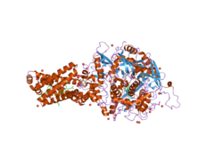
Structure

The gene that codes for the SDHB protein is nuclear, not mitochondrial DNA. However, the expressed protein is located in the inner membrane of the mitochondria. The location of the gene in humans is on the first chromosome at locus p36.1-p35. The gene is coded in 1,162 base pairs, partitioned in 8 exons. [5] The expressed protein weighs 31.6 kDa and is composed of 280 amino acids. [8] [9] SDHB contains the iron-sulphur clusters necessary for tunneling electrons through the complex. It is located between SDHA and the two transmembrane subunits SDHC and SDHD. [10]

Function

Figure 1: Function of the SDHB protein. Electrons are transferred from the Citric Acid Cycle to the Respiratory Chain. Electron path is shown by red arrows. SDHBfunction.svg
Figure 1: Function of the SDHB protein. Electrons are transferred from the Citric Acid Cycle to the Respiratory Chain. Electron path is shown by red arrows.

The SDH complex is located on the inner membrane of the mitochondria and participates in both the Citric Acid Cycle and Respiratory chain. SDHB acts as an intermediate in the basic SDH enzyme action shown in Figure 1:

  1. SDHA converts succinate to fumarate as part of the Citric Acid Cycle. This reaction also converts FAD to FADH2.
  2. Electrons from the FADH2 are transferred to the SDHB subunit iron clusters [2Fe-2S],[4Fe-4S],[3Fe-4S].
  3. Finally the electrons are transferred to the Ubiquinone (Q) pool via the SDHC/SDHD subunits. This function is part of the Respiratory chain.

Initially, SDHA oxidizes succinate via deprotonation at the FAD binding site, forming FADH2 and leaving fumarate, loosely bound to the active site, free to exit the protein. Electrons from FADH2 are transferred to the SDHB subunit iron clusters [2Fe-2S],[4Fe-4S],[3Fe-4S] and tunnel along the [Fe-S] relay until they reach the [3Fe-4S] iron sulfur cluster. The electrons are then transferred to an awaiting ubiquinone molecule at the Q pool active site in the SDHC/SDHD dimer. The O1 carbonyl oxygen of ubiquinone is oriented at the active site (image 4) by hydrogen bond interactions with Tyr83 of SDHD. The presence of electrons in the [3Fe-4S] iron sulphur cluster induces the movement of ubiquinone into a second orientation. This facilitates a second hydrogen bond interaction between the O4 carbonyl group of ubiquinone and Ser27 of SDHC. Following the first single electron reduction step, a semiquinone radical species is formed. The second electron arrives from the [3Fe-4S] cluster to provide full reduction of the ubiquinone to ubiquinol. [11]

Clinical significance

Germline mutations in the gene can cause familial paraganglioma (in old nomenclature, Paraganglioma Type PGL4). The same condition is often called familial pheochromocytoma. Less frequently, renal cell carcinoma can be caused by this mutation.

Paragangliomas related to SDHB mutations have a high rate of malignancy. When malignant, treatment is currently the same as for any malignant paraganglioma/pheochromocytoma.

Cancer

Paragangliomas caused by SDHB mutations have several distinguishing characteristics:

  1. Malignancy is common, ranging from 38%-83% [12] [13] in carriers with disease. In contrast, tumors caused by SDHD mutations are almost always benign. Sporadic paragangliomas are malignant in less than 10% of cases.
  2. Malignant paragangliomas caused by SDHB are usually (perhaps 92% [13] ) extra-adrenal. Sporadic pheochromocytomas/paragangliomas are extra-adrenal in less than 10% of cases.
  3. The penetrance of the gene is often reported as 77% by age 50 [12] (i.e. 77% of carriers will have at least one tumour by the age of 50). This is likely an overestimate. Currently (2011), families with silent SDHB mutations are being screened [14] to determine the frequency of silent carriers.
  4. The average age of onset is approximately the same for SDHB vs non-SDHB related disease (approximately 36 years).

Mutations causing disease have been seen in exons 1 through 7, but not 8. As with the SDHC and SDHD genes, SDHB is a tumor suppressor gene.

Tumor formation generally follows the Knudson "two hit" hypothesis. The first copy of the gene is mutated in all cells, however the second copy functions normally. When the second copy mutates in a certain cell due to a random event, Loss of Heterozygosity (LOH) occurs and the SDHB protein is no longer produced. Tumor formation then becomes possible.

Given the fundamental nature of the SDH protein in all cellular function, it is not currently understood why only paraganglionic cells are affected. However, the sensitivity of these cells to oxygen levels may play a role.

Disease pathways

The precise pathway leading from SDHB mutation to tumorigenesis is not determined; there are several proposed mechanisms. [15]

Generation of reactive oxygen species

Figure 2: Disease Pathways for SDHB mutations. Electron path during normal function is shown by solid red arrows. Red dashed arrow shows superoxide generation (Pathway 1). Purple dashed arrow shows diffusion of succinate to block PHD (Pathway 2). Black crosses indicate the non-mutated process is blocked. SDHBpathways.svg
Figure 2: Disease Pathways for SDHB mutations. Electron path during normal function is shown by solid red arrows. Red dashed arrow shows superoxide generation (Pathway 1). Purple dashed arrow shows diffusion of succinate to block PHD (Pathway 2). Black crosses indicate the non-mutated process is blocked.

When succinate-ubiquinone activity is inhibited, electrons that would normally transfer through the SDHB subunit to the Ubiquinone pool are instead transferred to O2 to create Reactive Oxygen Species (ROS) such as superoxide. The dashed red arrow in Figure 2 shows this. ROS accumulate and stabilize the production of HIF1-α. HIF1-α combines with HIF1-β to form the stable HIF heterodimeric complex, in turn leading to the induction of antiapoptotic genes in the cell nucleus.

Succinate accumulation in the cytosol

SDH inactivation can block the oxidation of succinate, starting a cascade of reactions:

  1. The succinate accumulated in the mitochondrial matrix diffuses through the inner and outer mitochondrial membranes to the cytosol (purple dashed arrows in Figure 2).
  2. Under normal cellular function, HIF1-α in the cytosol is quickly hydroxylated by prolyl hydroxylase (PHD), shown with the light blue arrow. This process is blocked by the accumulated succinate.
  3. HIF1-α stabilizes and passes to the cell nucleus (orange arrow) where it combines with HIF1-β to form an active HIF complex that induces the expression of tumor causing genes. [16]

This pathway raises the possibility of a therapeutic treatment. The build-up of succinate inhibits PHD activity. PHD action normally requires oxygen and alpha-ketoglutarate as cosubstrates and ferrous iron and ascorbate as cofactors. Succinate competes with α-ketoglutarate in binding to the PHD enzyme. Therefore, increasing α-ketoglutarate levels can offset the effect of succinate accumulation.

Normal α-ketoglutarate does not permeate cell walls efficiently, and it is necessary to create a cell permeating derivative (e.g. α-ketoglutarate esters). In-vitro trials show this supplementation approach can reduce HIF1-α levels, and may result in a therapeutic approach to tumours resulting from SDH deficiency. [17]

Impaired developmental apoptosis

Paraganglionic tissue is derived from the neural crest cells present in an embryo. Abdominal extra-adrenal paraganglionic cells secrete catecholamines that play an important role in fetal development. After birth these cells usually die, a process that is triggered by a decline in nerve growth factor (NGF)which initiates apoptosis (cell death).

This cell death process is mediated by an enzyme called prolyl hydroxylase EglN3. Succinate accumulation caused by SDH inactivation inhibits the prolyl hydroxylase EglN3. [18] The net result is that paranglionic tissue that would normally die after birth remains, and this tissue may be able to trigger paraganglioma/pheochromocytoma later.

Glycolysis upregulation

Inhibition of the Citric Acid Cycle forces the cell to create ATP glycolytically in order to generate its required energy. The induced glycolytic enzymes could potentially block cell apoptosis.

RNA editing

The mRNA transcripts of the SDHB gene in human are edited through an unknown mechanism at ORF nucleotide position 136 causing the conversion of C to U and thus generating a stop codon resulting in the translation of the edited transcripts to a truncated SDHB protein with an R46X amino acid change. This editing has been shown in monocytes and some human lymphoid cell-lines, [19] and is enhanced by hypoxia. [20]

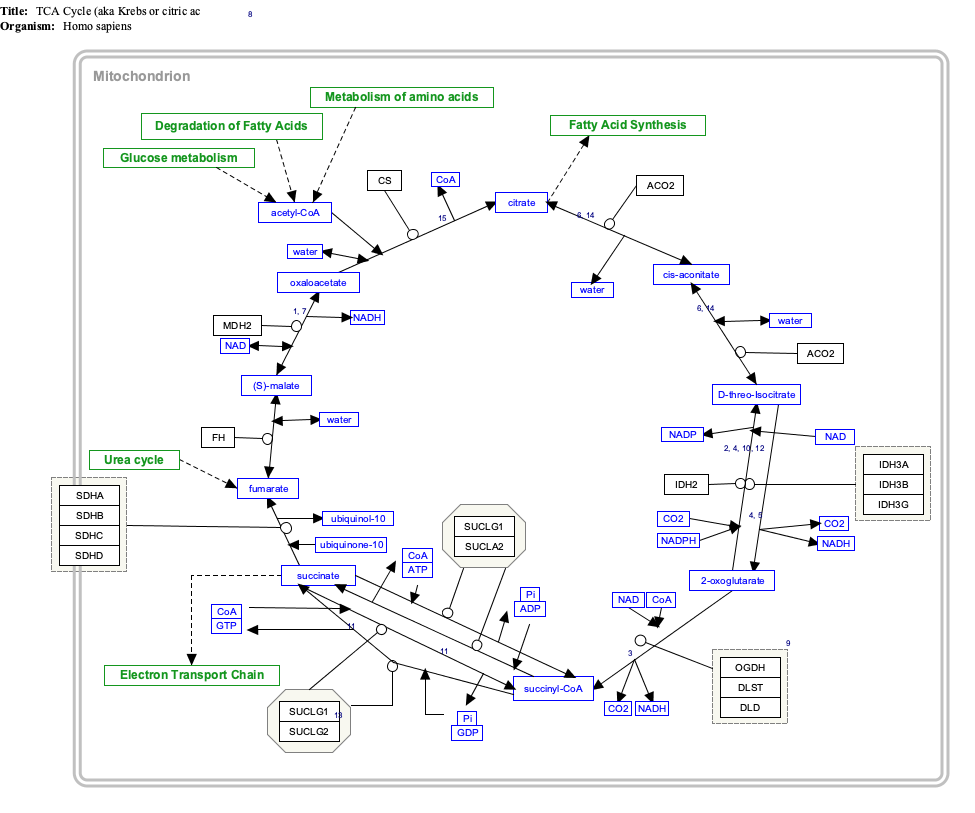
Interactive pathway map

Click on genes, proteins and metabolites below to link to respective articles. [§ 1]

[[File:
TCACycle WP78.png Go to articleGo to articleGo to articleGo to articleGo to articleGo to articleGo to articleGo to articleGo to articleGo to articleGo to articleGo to articleGo to articleGo to articleGo to articleGo to articleGo to articleGo to articleGo to articleGo to articleGo to articleGo to articleGo to articleGo to articleGo to articleGo to articleGo to articleGo to articleGo to articleGo to articleGo to articleGo to articleGo to articleGo to articleGo to articleGo to articleGo to articleGo to articleGo to articleGo to articleGo to articleGo to HMDBGo to articleGo to articleGo to articleGo to articleGo to articleGo to articleGo to articleGo to articleGo to articleGo to article
[[
]]
[[
]]
[[
]]
[[
]]
[[
]]
[[
]]
[[
]]
[[
]]
[[
]]
[[
]]
[[
]]
[[
]]
[[
]]
[[
]]
[[
]]
[[
]]
[[
]]
[[
]]
[[
]]
[[
]]
[[
]]
[[
]]
[[
]]
[[
]]
[[
]]
[[
]]
[[
]]
[[
]]
[[
]]
[[
]]
[[
]]
[[
]]
[[
]]
[[
]]
[[
]]
[[
]]
[[
]]
[[
]]
[[
]]
[[
]]
[[
]]
[[
]]
[[
]]
[[
]]
[[
]]
[[
]]
[[
]]
[[
]]
[[
]]
[[
]]
[[
]]
[[
]]
[[
]]
[[
]]
[[
]]
[[
]]
[[
]]
[[
]]
[[
]]
[[
]]
[[
]]
[[
]]
[[
]]
[[
]]
TCACycle WP78.png Go to articleGo to articleGo to articleGo to articleGo to articleGo to articleGo to articleGo to articleGo to articleGo to articleGo to articleGo to articleGo to articleGo to articleGo to articleGo to articleGo to articleGo to articleGo to articleGo to articleGo to articleGo to articleGo to articleGo to articleGo to articleGo to articleGo to articleGo to articleGo to articleGo to articleGo to articleGo to articleGo to articleGo to articleGo to articleGo to articleGo to articleGo to articleGo to articleGo to articleGo to articleGo to HMDBGo to articleGo to articleGo to articleGo to articleGo to articleGo to articleGo to articleGo to articleGo to articleGo to article
|alt=TCACycle_WP78 edit]]
TCACycle_WP78 edit
  1. The interactive pathway map can be edited at WikiPathways: "TCACycle_WP78".

Related Research Articles

<span class="mw-page-title-main">Oxidative phosphorylation</span> Metabolic pathway

Oxidative phosphorylation or electron transport-linked phosphorylation or terminal oxidation is the metabolic pathway in which cells use enzymes to oxidize nutrients, thereby releasing chemical energy in order to produce adenosine triphosphate (ATP). In eukaryotes, this takes place inside mitochondria. Almost all aerobic organisms carry out oxidative phosphorylation. This pathway is so pervasive because it releases more energy than alternative fermentation processes such as anaerobic glycolysis.

<span class="mw-page-title-main">Electron transport chain</span> Energy-producing metabolic pathway

An electron transport chain (ETC) is a series of protein complexes and other molecules that transfer electrons from electron donors to electron acceptors via redox reactions (both reduction and oxidation occurring simultaneously) and couples this electron transfer with the transfer of protons (H+ ions) across a membrane. The electrons that transferred from NADH and FADH2 to the ETC involves 4 multi-subunit large enzymes complexes and 2 mobile electron carriers. Many of the enzymes in the electron transport chain are membrane-bound.

<span class="mw-page-title-main">Respiratory complex I</span>

Respiratory complex I, EC 7.1.1.2 is the first large protein complex of the respiratory chains of many organisms from bacteria to humans. It catalyzes the transfer of electrons from NADH to coenzyme Q10 (CoQ10) and translocates protons across the inner mitochondrial membrane in eukaryotes or the plasma membrane of bacteria.

<span class="mw-page-title-main">Coenzyme Q – cytochrome c reductase</span> Class of enzymes

The coenzyme Q : cytochrome c – oxidoreductase, sometimes called the cytochrome bc1 complex, and at other times complex III, is the third complex in the electron transport chain, playing a critical role in biochemical generation of ATP. Complex III is a multisubunit transmembrane protein encoded by both the mitochondrial and the nuclear genomes. Complex III is present in the mitochondria of all animals and all aerobic eukaryotes and the inner membranes of most eubacteria. Mutations in Complex III cause exercise intolerance as well as multisystem disorders. The bc1 complex contains 11 subunits, 3 respiratory subunits, 2 core proteins and 6 low-molecular weight proteins.

<span class="mw-page-title-main">Succinic acid</span> Dicarboxylic acid

Succinic acid is a dicarboxylic acid with the chemical formula (CH2)2(CO2H)2. The name derives from Latin succinum, meaning amber. In living organisms, succinic acid takes the form of an anion, succinate, which has multiple biological roles as a metabolic intermediate being converted into fumarate by the enzyme succinate dehydrogenase in complex 2 of the electron transport chain which is involved in making ATP, and as a signaling molecule reflecting the cellular metabolic state. It is marketed as food additive E363. Succinate is generated in mitochondria via the tricarboxylic acid cycle (TCA). Succinate can exit the mitochondrial matrix and function in the cytoplasm as well as the extracellular space, changing gene expression patterns, modulating epigenetic landscape or demonstrating hormone-like signaling. As such, succinate links cellular metabolism, especially ATP formation, to the regulation of cellular function. Dysregulation of succinate synthesis, and therefore ATP synthesis, happens in some genetic mitochondrial diseases, such as Leigh syndrome, and Melas syndrome, and degradation can lead to pathological conditions, such as malignant transformation, inflammation and tissue injury.

<span class="mw-page-title-main">Succinate dehydrogenase</span> Enzyme

Succinate dehydrogenase (SDH) or succinate-coenzyme Q reductase (SQR) or respiratory complex II is an enzyme complex, found in many bacterial cells and in the inner mitochondrial membrane of eukaryotes. It is the only enzyme that participates in both the citric acid cycle and the electron transport chain. Histochemical analysis showing high succinate dehydrogenase in muscle demonstrates high mitochondrial content and high oxidative potential.

<span class="mw-page-title-main">SDHD</span> Protein-coding gene in the species Homo sapiens

Succinate dehydrogenase [ubiquinone] cytochrome b small subunit, mitochondrial (CybS), also known as succinate dehydrogenase complex subunit D (SDHD), is a protein that in humans is encoded by the SDHD gene. Names previously used for SDHD were PGL and PGL1. Succinate dehydrogenase is an important enzyme in both the citric acid cycle and the electron transport chain.

<span class="mw-page-title-main">Succinate dehydrogenase complex subunit C</span> Protein-coding gene in the species Homo sapiens

Succinate dehydrogenase complex subunit C, also known as succinate dehydrogenase cytochrome b560 subunit, mitochondrial, is a protein that in humans is encoded by the SDHC gene. This gene encodes one of four nuclear-encoded subunits that comprise succinate dehydrogenase, also known as mitochondrial complex II, a key enzyme complex of the tricarboxylic acid cycle and aerobic respiratory chains of mitochondria. The encoded protein is one of two integral membrane proteins that anchor other subunits of the complex, which form the catalytic core, to the inner mitochondrial membrane. There are several related pseudogenes for this gene on different chromosomes. Mutations in this gene have been associated with pheochromocytomas and paragangliomas. Alternatively spliced transcript variants have been described.

<span class="mw-page-title-main">Paraganglioma</span> Medical condition

A paraganglioma is a rare neuroendocrine neoplasm that may develop at various body sites. When the same type of tumor is found in the adrenal gland, they are referred to as a pheochromocytoma. They are rare tumors, with an overall estimated incidence of 1/300,000. There is no test that determines benign from malignant tumors; long-term follow-up is therefore recommended for all individuals with paraganglioma.

<span class="mw-page-title-main">SDHA</span> Protein-coding gene in the species Homo sapiens

Succinate dehydrogenase complex, subunit A, flavoprotein variant is a protein that in humans is encoded by the SDHA gene. This gene encodes a major catalytic subunit of succinate-ubiquinone oxidoreductase, a complex of the mitochondrial respiratory chain. The complex is composed of four nuclear-encoded subunits and is localized in the mitochondrial inner membrane. SDHA contains the FAD binding site where succinate is deprotonated and converted to fumarate. Mutations in this gene have been associated with a form of mitochondrial respiratory chain deficiency known as Leigh Syndrome. A pseudogene has been identified on chromosome 3q29. Alternatively spliced transcript variants encoding different isoforms have been found for this gene.

<span class="mw-page-title-main">MT-ND6</span> Mitochondrial gene coding for a protein involved in the respiratory chain

MT-ND6 is a gene of the mitochondrial genome coding for the NADH-ubiquinone oxidoreductase chain 6 protein (ND6). The ND6 protein is a subunit of NADH dehydrogenase (ubiquinone), which is located in the mitochondrial inner membrane and is the largest of the five complexes of the electron transport chain. Variations in the human MT-ND6 gene are associated with Leigh's syndrome, Leber's hereditary optic neuropathy (LHON) and dystonia.

<span class="mw-page-title-main">ETFB</span> Protein-coding gene in humans

The human ETFB gene encodes the Electron-transfer-flavoprotein, beta subunit, also known as ETF-β. Together with Electron-transfer-flavoprotein, alpha subunit, encoded by the 'ETFA' gene, it forms the heterodimeric Electron transfer flavoprotein (ETF). The native ETF protein contains one molecule of FAD and one molecule of AMP, respectively.

<span class="mw-page-title-main">NDUFS1</span> Protein-coding gene in the species Homo sapiens

NADH-ubiquinone oxidoreductase 75 kDa subunit, mitochondrial (NDUFS1) is an enzyme that in humans is encoded by the NDUFS1 gene. The encoded protein, NDUFS1, is the largest subunit of complex I, located on the inner mitochondrial membrane, and is important for mitochondrial oxidative phosphorylation. Mutations in this gene are associated with complex I deficiency.

<span class="mw-page-title-main">NDUFA5</span> Protein-coding gene in the species Homo sapiens

NADH dehydrogenase [ubiquinone] 1 alpha subcomplex subunit 5 is an enzyme that in humans is encoded by the NDUFA5 gene. The NDUFA5 protein is a subunit of NADH dehydrogenase (ubiquinone), which is located in the mitochondrial inner membrane and is the largest of the five complexes of the electron transport chain.

<span class="mw-page-title-main">NDUFB6</span> Protein-coding gene in the species Homo sapiens

NADH dehydrogenase [ubiquinone] 1 beta subcomplex subunit 6, also known as complex I-B17, is a protein that in humans is encoded by the NDUFB6 gene. NADH dehydrogenase (ubiquinone) 1 beta subcomplex subunit 6, is an accessory subunit of the NADH dehydrogenase (ubiquinone) complex, located in the mitochondrial inner membrane. It is also known as Complex I and is the largest of the five complexes of the electron transport chain.

<span class="mw-page-title-main">NDUFA10</span> Protein-coding gene in the species Homo sapiens

NADH dehydrogenase [ubiquinone] 1 alpha subcomplex subunit 10 is an enzyme that in humans is encoded by the NDUFA10 gene. The NDUFA10 protein is a subunit of NADH dehydrogenase (ubiquinone), which is located in the mitochondrial inner membrane and is the largest of the five complexes of the electron transport chain. Mutations in subunits of NADH dehydrogenase (ubiquinone), also known as Complex I, frequently lead to complex neurodegenerative diseases such as Leigh's syndrome. Furthermore, reduced NDUFA10 expression levels due to FOXM1-directed hypermethylation are associated with human squamous cell carcinoma and may be related to other forms of cancer.

<span class="mw-page-title-main">NDUFB11</span> Protein-coding gene in the species Homo sapiens

NADH dehydrogenase [ubiquinone] 1 beta subcomplex subunit 11, mitochondrial is an enzyme that in humans is encoded by the NDUFB11 gene. NADH dehydrogenase (ubiquinone) 1 beta subcomplex subunit 11 is an accessory subunit of the NADH dehydrogenase (ubiquinone) complex, located in the mitochondrial inner membrane. It is also known as Complex I and is the largest of the five complexes of the electron transport chain. NDUFB11 mutations have been associated with linear skin defects with multiple congenital anomalies 3 and mitochondrial complex I deficiency.

<span class="mw-page-title-main">Carney's triad</span>

Carney triad (CT) is characterized by the coexistence of three types of neoplasms, mainly in young women, including gastric gastrointestinal stromal tumor, pulmonary chondroma, and extra-adrenal paraganglioma. The underlying genetic defect remains elusive. CT is distinct from Carney complex, and the Carney-Stratakis syndrome.

<span class="mw-page-title-main">SDHAF2</span> Protein-coding gene in the species Homo sapiens

Succinate dehydrogenase complex assembly factor 2, formerly known as SDH5 and also known as SDH assembly factor 2 or SDHAF2 is a protein that in humans is encoded by the SDHAF2 gene. This gene encodes a mitochondrial protein needed for the flavination of a succinate dehydrogenase complex subunit required for activity of the complex. Mutations in this gene are associated with pheochromocytoma and paraganglioma.

<span class="mw-page-title-main">SDHAF1</span> Protein-coding gene in the species Homo sapiens

Succinate dehydrogenase complex assembly factor 1 (SDHAF1), also known as LYR motif-containing protein 8 (LYRM8), is a protein that in humans is encoded by the SDHAF1, or LYRM8, gene. SDHAF1 is a chaperone protein involved in the assembly of the succinate dehydrogenase (SDH) complex. Mutations in this gene are associated with SDH-defective infantile leukoencephalopathy and mitochondrial complex II deficiency.

References

  1. 1 2 3 GRCh38: Ensembl release 89: ENSG00000117118 - Ensembl, May 2017
  2. 1 2 3 GRCm38: Ensembl release 89: ENSMUSG00000009863 - Ensembl, May 2017
  3. "Human PubMed Reference:". National Center for Biotechnology Information, U.S. National Library of Medicine.
  4. "Mouse PubMed Reference:". National Center for Biotechnology Information, U.S. National Library of Medicine.
  5. 1 2 "Entrez Gene: succinate dehydrogenase complex".
  6. Kita K, Oya H, Gennis RB, Ackrell BA, Kasahara M (January 1990). "Human complex II (succinate-ubiquinone oxidoreductase): cDNA cloning of iron sulfur (Ip) subunit of liver mitochondria". Biochem. Biophys. Res. Commun. 166 (1): 101–8. doi:10.1016/0006-291X(90)91916-G. PMID   2302193.
  7. Au HC, Ream-Robinson D, Bellew LA, Broomfield PL, Saghbini M, Scheffler IE (July 1995). "Structural organization of the gene encoding the human iron-sulfur subunit of succinate dehydrogenase". Gene. 159 (2): 249–53. doi:10.1016/0378-1119(95)00162-Y. PMID   7622059.
  8. Zong NC, Li H, Li H, Lam MP, Jimenez RC, Kim CS, Deng N, Kim AK, Choi JH, Zelaya I, Liem D, Meyer D, Odeberg J, Fang C, Lu HJ, Xu T, Weiss J, Duan H, Uhlen M, Yates JR, Apweiler R, Ge J, Hermjakob H, Ping P (Oct 2013). "Integration of cardiac proteome biology and medicine by a specialized knowledgebase". Circulation Research. 113 (9): 1043–53. doi:10.1161/CIRCRESAHA.113.301151. PMC   4076475 . PMID   23965338.
  9. "SDHB - Succinate dehydrogenase [ubiquinone] iron-sulfur subunit, mitochondrial". Cardiac Organellar Protein Atlas Knowledgebase (COPaKB). Archived from the original on 2018-07-19. Retrieved 2018-07-18.
  10. Sun, F; Huo, X; Zhai, Y; Wang, A; Xu, J; Su, D; Bartlam, M; Rao, Z (1 July 2005). "Crystal structure of mitochondrial respiratory membrane protein complex II". Cell. 121 (7): 1043–57. doi: 10.1016/j.cell.2005.05.025 . PMID   15989954. S2CID   16697879.
  11. Horsefield, R; Yankovskaya, V; Sexton, G; Whittingham, W; Shiomi, K; Omura, S; Byrne, B; Cecchini, G; Iwata, S (17 March 2006). "Structural and computational analysis of the quinone-binding site of complex II (succinate-ubiquinone oxidoreductase): a mechanism of electron transfer and proton conduction during ubiquinone reduction". The Journal of Biological Chemistry. 281 (11): 7309–16. doi: 10.1074/jbc.m508173200 . PMID   16407191.
  12. 1 2 Neumann HP, Pawlu C, Peczkowska M, Bausch B, McWhinney SR, Muresan M, Buchta M, Franke G, Klisch J, Bley TA, Hoegerle S, Boedeker CC, Opocher G, Schipper J, Januszewicz A, Eng C (August 2004). "Distinct clinical features of paraganglioma syndromes associated with SDHB and SDHD gene mutations". JAMA. 292 (8): 943–51. doi: 10.1001/jama.292.8.943 . PMID   15328326.
  13. 1 2 Brouwers FM, Eisenhofer G, Tao JJ, Kant JA, Adams KT, Linehan WM, Pacak K (November 2006). "High frequency of SDHB germline mutations in patients with malignant catecholamine-producing paragangliomas: implications for genetic testing". J. Clin. Endocrinol. Metab. 91 (11): 4505–9. doi: 10.1210/jc.2006-0423 . PMID   16912137.
  14. Conference: National Institute of Health (U.S.A.), "SDHB-related Pheochromocytoma: Recent Discoveries & Current Diagnostic and Therapeutic Approaches", September 29, 2006
  15. Gottlieb E, Tomlinson IP (November 2005). "Mitochondrial tumour suppressors: a genetic and biochemical update". Nat. Rev. Cancer. 5 (11): 857–66. doi:10.1038/nrc1737. PMID   16327764. S2CID   20851047.
  16. Selak MA, Armour SM, MacKenzie ED, Boulahbel H, Watson DG, Mansfield KD, Pan Y, Simon MC, Thompson CB, Gottlieb E (January 2005). "Succinate links TCA cycle dysfunction to oncogenesis by inhibiting HIF-alpha prolyl hydroxylase". Cancer Cell. 7 (1): 77–85. doi: 10.1016/j.ccr.2004.11.022 . PMID   15652751.
  17. MacKenzie ED, Selak MA, Tennant DA, Payne LJ, Crosby S, Frederiksen CM, Watson DG, Gottlieb E (May 2007). "Cell-permeating alpha-ketoglutarate derivatives alleviate pseudohypoxia in succinate dehydrogenase-deficient cells". Mol. Cell. Biol. 27 (9): 3282–9. doi:10.1128/MCB.01927-06. PMC   1899954 . PMID   17325041.
  18. Lee S, Nakamura E, Yang H, Wei W, Linggi MS, Sajan MP, Farese RV, Freeman RS, Carter BD, Kaelin WG, Schlisio S (August 2005). "Neuronal apoptosis linked to EglN3 prolyl hydroxylase and familial pheochromocytoma genes: developmental culling and cancer". Cancer Cell. 8 (2): 155–67. doi: 10.1016/j.ccr.2005.06.015 . PMID   16098468.
  19. Baysal BE (2007). "A recurrent stop-codon mutation in succinate dehydrogenase subunit B gene in normal peripheral blood and childhood T-cell acute leukemia". PLOS ONE. 2 (5): e436. Bibcode:2007PLoSO...2..436B. doi: 10.1371/journal.pone.0000436 . PMC   1855983 . PMID   17487275. Open Access logo PLoS transparent.svg
  20. Baysal BE, De Jong K, Liu B, Wang J, Patnaik SK, Wallace PK, Taggart RT (2013). "Hypoxia-inducible C-to-U coding RNA editing downregulates SDHB in monocytes". PeerJ. 1: e152. doi:10.7717/peerj.152. PMC   3775634 . PMID   24058882.

Further reading