This article is about fractures of nonacetabular portions of the pelvis. For fractures of the proximal femur, see Hip fracture. For fractures of the acetabulum, see Acetabular fracture.
Common causes include falls, motor vehicle collisions, a vehicle hitting a pedestrian, or a direct crush injury.[2] In younger people significant trauma is typically required while in older people less significant trauma can result in a fracture.[1] They are divided into two types: stable and unstable.[1] Unstable fractures are further divided into anterior posterior compression, lateral compression, vertical shear, and combined mechanism fractures.[2][1] Diagnosis is suspected based on symptoms and examination with confirmation by X-rays or CT scan.[1] If a person is fully awake and has no pain of the pelvis medical imaging is not needed.[2]
Pelvic fractures make up around 3% of adult fractures.[1] Stable fractures generally have a good outcome.[1] The risk of death with an unstable fracture is about 15%, while those who also have low blood pressure have a risk of death approaching 50%.[2][4] Unstable fractures are often associated with injuries to other parts of the body.[3]
Signs and symptoms
Symptoms include pain, particularly with movement.[1]
Bruising of the perineum, scrotum, or labia, known as Coopernail's sign, is also suggestive of pelvic fracture.[5]
Complications
Complications are likely to result in cases of excess blood loss or puncture to certain organs, possibly leading to shock.[6][7] Swelling and bruising may result, more so in high-impact injuries.[7] Pain in the affected areas may differ where severity of impact increases its likelihood and may radiate if symptoms are aggravated when one moves around.[citation needed]
The bony pelvis consists of the ilium (i.e., iliac wings), ischium, and pubis, which form an anatomic ring with the sacrum. Disruption of this ring requires significant energy. When it comes to the stability and the structure of the pelvis, or pelvic girdle, understanding its function as support for the trunk and legs helps to recognize the effect a pelvic fracture has on someone.[8] The pubic bone, the ischium and the ilium make up the pelvic girdle, fused together as one unit. They attach to both sides of the spine and circle around to create a ring and sockets to place hipjoints. Attachment to the spine is important to direct force into the trunk from the legs as movement occurs, extending to one's back. This requires the pelvis to be strong enough to withstand pressure and energy. Various muscles play important roles in pelvic stability. Because of the forces involved, pelvic fractures frequently involve injury to organs contained within the bony pelvis. In addition, trauma to extra-pelvic organs is common. Pelvic fractures are often associated with severe hemorrhage due to the extensive blood supply to the region. The veins of the presacral pelvic plexus are particularly vulnerable. Greater than 85 percent of bleeding due to pelvic fractures is venous or from the open surfaces of the bone.[citation needed]
Diagnosis
If a person is fully awake and has no pain in the pelvis, medical imaging of the pelvis is not needed.[2]
Classification
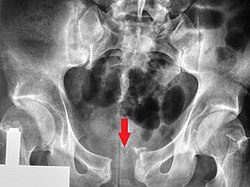
Fractures of the superior (in two places) and inferior pubic rami on the person's right, in a person who has had prior hip replacements
Pelvic fractures are most commonly described using one of two classification systems. The different forces on the pelvis result in different fractures. Sometimes they are determined based on stability or instability.[9]
In type A injuries, the sacroiliac complex is intact. The pelvic ring has a stable fracture that can be managed nonoperatively. Type B injuries are caused by either external or internal rotational forces resulting in partial disruption of the posterior sacroiliac complex. These are often unstable. Type C injuries are characterized by complete disruption of the posterior sacroiliac complex and are both rotationally and vertically unstable. These injuries are the result of great force, usually from a motor vehicle crash, fall from a height or severe compression.[citation needed]
Young-Burgess classification
Superior view, Pelvic Fracture Types (2006). Force and break are shown by matching color: Anteroposterior compression type I (orange), Anteroposterior compression type II (green), Anteroposterior compression type III (blue); Lateral compression type I (red), Lateral compression type II (purple), F. Lateral compression type III (black). Increased force and breaks are shown by increasing size. This fracture is best viewed anteriorly, while the other fractures are viewed superiorly. The arrow indicates where the force is coming from, and the colored lines indicate where the break occurs.
The Young-Burgess classification system is based on mechanism of injury: anteroposterior compression type I, II and III, lateral compression types I, II and III, and vertical shear,[6] or a combination of forces.
Lateral compression (LC) fractures involve transverse fractures of the pubic rami, either ipsilateral or contralateral to a posterior injury.
Grade I– Associated sacral compression on side of impact
Grade II– Associated posterior iliac ("crescent") fracture on side of impact
The most common force type, lateral compression (LC) forces, from side-impact automobile accidents and pedestrian injuries, can result in an internal rotation.[10] The superior and inferior pubic rami may fracture anteriorly, for example. Injuries from shear forces, like falls from above, can result in disruption of ligaments or bones. When multiple forces occur, it is called combined mechanical injury (CMI). The best imaging modality to use for this classification is probably a pelvic CT scan.[11]
Open book fracture
One specific kind of pelvic fracture is known as an 'open book' fracture. This is often the result of a heavy impact to the groin (pubis), a common motorcycling accident injury. In this kind of injury, the left and right halves of the pelvis are separated at front and rear, the front opening more than the rear, i.e. like an open book that falls to the ground and splits in the middle. Depending on the severity, this may require surgical reconstruction before rehabilitation.[12] Forces from an anterior or posterior direction, like head-on car accidents, usually cause external rotation of the hemipelvis, an “open-book” injury. Open fractures have an increased risk of infection and hemorrhaging from vessel injury, leading to higher mortality.[13]
Prevention
As the human body ages, the bones become weaker and brittle and are therefore more susceptible to fractures. Certain precautions are crucial in order to lower the risk of getting pelvic fractures. The most damaging is one from a car accident, cycling accident, or falling from a high building which can result in a high energy injury.[14] This can be very dangerous because the pelvis supports many internal organs and can damage these organs. Falling is one of the most common causes of pelvic fracture. Therefore, proper precautions should be taken to prevent this from happening.[citation needed]
Treatment
An example of pelvic binding using a sheet and cable ties
A pelvic fracture is often complicated and treatment can be a long and painful process. Depending on the severity, pelvic fractures can be treated with or without surgery.[15]
Initial
A high index of suspicion should be held for pelvic injuries in anyone with major trauma. The pelvis should be stabilized with a pelvic binder.[16] This can be a purpose-made device, but improvised pelvic binders have also been used around the world to good effect.[17] Stabilisation of the pelvic ring reduces blood loss from the pelvic vessels and reduced the risk of death.
Surgery
Surgery is often required for pelvic fractures. Many methods of pelvic stabilization are used including external fixation or internal fixation and traction.[18][19] There are often other injuries associated with a pelvic fracture so the type of surgery involved must be thoroughly planned.[20]
Rehabilitation
Pelvic fractures that are treatable without surgery are treated with bed rest. Once the fracture has healed enough, rehabilitation can be started with first standing upright with the help of a physical therapist, followed by starting to walk using a walker and eventually progressing to a cane.[citation needed]
Prognosis
Mortality rates in people with pelvic fractures are between 10 and 16 percent.[21] However, death is typically due to associated trauma affecting other organs, such as the brain. Death rates due to complications directly related to pelvic fractures, such as bleeding, are relatively low.[21]
Epidemiology
In the United States of America, about 10 percent of people that seek treatment at a level 1 trauma center after a blunt force injury have a pelvic fracture.[21] Motorcycle injuries are the most common cause of pelvic fractures, followed by injuries to pedestrians caused by motor vehicles, large falls (over 15 feet), and motor vehicle crashes.[21]
↑ Dimon, Theodore Jr. (2010). The body in motion: its evolution and design. Berkeley, Calif.: North Atlantic Books. pp.49–56. ISBN978-1556439704.
↑ Young, JW; Resnik, CS (December 1990). "Fracture of the Pelvis: Current Concepts of Classification". AJR. American Journal of Roentgenology. 155 (6): 1169–75. doi:10.2214/ajr.155.6.2122661. PMID2122661.
↑ Davis DD, Foris LA, Kane SM, etal. (January 2021). "Pelvic Fracture". National Center for Biotechnology Information, U.S. National Library of Medicine. PMID28613485. Retrieved 2 October 2021.
↑ Mirghasemi A, Mohamadi A, Ara AM, Gabaran NR, Sadat MM (2009). "Completely displaced S-1/S-2 growth plate fracture in an adolescent: case report and review of literature". J Orthop Trauma. 23 (10): 734–8. doi:10.1097/BOT.0b013e3181a23d8b. PMID19858983. S2CID6651435.
↑ Taguchi, T; Kawai, S; Kaneko, K; Yugue, D (1999). "Operative management of displaced fractures of the sacrum". Journal of Orthopaedic Science. 4 (5): 347–52. doi:10.1007/s007760050115. PMID10542038. S2CID46282760.
↑ Hancharenka, V.; Tuzikov, A.; Arkhipau, V.; Kryvanos, A. (March 2009). "Preoperative planning of pelvic and lower limbs surgery by CT image processing". Pattern Recognition and Image Analysis. 19 (1): 109–113. doi:10.1134/S1054661809010209. S2CID34590414.
1 2 3 4 Vincent, Jean-Louis (2011). Textbook of Critical Care (6thed.). Philadelphia, PA: Elsevier/Saunders. p.1523. ISBN9781437713671.
This page is based on this Wikipedia article Text is available under the CC BY-SA 4.0 license; additional terms may apply. Images, videos and audio are available under their respective licenses.