Maisonneuve fracture

Last updated
Maisonneuve fracture
Maisonneuve fracture Fibula.JPG
Radiograph showing a Maisonneuve fracture of the proximal fibula
Specialty Orthopedics
Symptoms Swelling around medial and lateral sides of ankle joint, pain during external rotation of foot
Complications Osteoarthritis, peroneal nerve palsy
CausesForceful, external rotation of the foot
Risk factors Sporting injuries, falls, motor vehicle accidents
Diagnostic method Physical examination, radiography, X-ray, CT, MRI, arthroscopy
Differential diagnosis Isolated tibiofibular syndesmosis injury, isolated fibula fracture
Treatment Orthopedic casting, ORIF, CRIF

The Maisonneuve fracture is a spiral fracture of the proximal third of the fibula associated with a tear of the distal tibiofibular syndesmosis and the interosseous membrane. There is an associated fracture of the medial malleolus or rupture of the deep deltoid ligament of the ankle. This type of injury can be difficult to detect. [1] [2]

Contents

The Maisonneuve fracture is typically a result of excessive, external rotative force being applied to the deltoid and syndesmotic ligaments. Due to this, the Maisonneuve fracture is described as a pronation-external rotation injury according to the Lauge-Hansen classification system. [3] It is also classified as a Type C ankle fracture according to the Danis-Weber classification system. [4]

The Maisonneuve fracture is similar to the Galeazzi fracture in the sense that there is an important ligamentous disruption in association with the fracture. [5] The fracture is named after the surgeon Jules Germain François Maisonneuve. [6]

Cause

Forceful, external rotation of the ankle joint is the main cause of a Maisonneuve fracture. [4] [7] Engaging in high-intensity sports or falling over can increase the risk of tearing the deltoid ligament or cause an avulsion fracture of the medial malleolus from external rotation of the foot. [4] [6] In some cases, motor vehicle accidents can also result in a Maisonneuve fracture. [8]

Signs and symptoms

Fracture of the medial malleolus seen on X-ray scan (left ankle) Fracture of Medial Malleolus.jpg
Fracture of the medial malleolus seen on X-ray scan (left ankle)

Common symptoms of a Maisonneuve fracture are pain, swelling, tenderness, and bruising around the ankle joint and inferior (or distal) tibiofibular joint. More specifically, as a pronation-external rotation injury, pain during external rotation of the ankle joint is expected. Additionally, there is a reduced range of motion of the foot and an inability to weight-bear due to ankle pain. [4] [7] Pain may also be felt around the medial and lateral aspects of the ankle, and more rarely around the superior (or proximal) tibiofibular joint. [9] Damage to the deltoid ligament or interosseous membrane can cause haemorrhaging around the surrounding tissues, resulting in a localised oedema. [8]

As the syndesmotic ligaments are responsible for stabilising the ankle mortise and tibiotalar joint, disruption to this syndesmosis can cause a reduction of the space between the distal tibia, fibula, and talus. A long-term effect of this is painful ankle osteoarthritis due to the direct contact between the tibia and talus. [10] [11]

If a Maisonneuve fracture is left untreated, instability of the tibiotalar joint and deltoid ligament can cause a valgus deformity of the ankle. This leaves the ankle joint in a state of chronic pronation, characterised by a protrusion of the medial malleolus into the subcutaneous tissue. [11]

Pathophysiology

Fracture of the lateral malleolus seen on X-ray scan (left ankle) Left lateral malleolus avulsion fracture detail.jpg
Fracture of the lateral malleolus seen on X-ray scan (left ankle)

The Maisonneuve fracture generally follows a specific pattern of injury. The following are described as subsequent events that result in a Maisonneuve fracture: [3] [4] [12]

In cases where the anterior aspect of the tibiofibular syndesmosis can resist mechanical stress, only an oblique fracture of the lateral malleolus is produced. Diastasis of the lateral malleolus may also occur, in which it is posterolaterally displaced from the tibia. [9]

Although most Maisonneuve cases report a pronation-external rotation mechanism of injury, clinical studies have recorded instances of supination-external rotation being the mechanism of injury. [6] Slight or high degrees of plantarflexion prior to supination-external rotation of the foot have been identified in patients with proximal fibular fractures. [8]

Diagnosis

A digital radiography machine Digital radiography Machine 01.jpg
A digital radiography machine

Diagnosing a Maisonneuve fracture requires a combination of medical history, physical examination, and radiographic imaging. [11] Patients generally do not report pain near the proximal fibula, so physical examination such as palpation along the fibula is effective for differentiating a Maisonneuve fracture from an isolated syndesmotic injury. [4] Feeling pain near the proximal fibula during palpation is a positive indication of a Maisonneuve fracture. [12] Ankle instability is often associated with a damaged proximal fibula in a Maisonneuve fracture, so patients are typically asked about the mechanism of injury. Mortise stability is examined to rule out the possibility of an isolated fibular fracture. [6]

Ankle radiographs are used to detect widening of the tibiofibular syndesmosis or medial clear space. The medial clear space is the area between the talus of the ankle and the medial malleolus. Damage to the deltoid ligament and syndesmotic ligaments result in mortise instability, causing the talus to laterally shift and widen the medial clear space. [4] [12] A clinical study, conducted in 2006 and published in the Journal of Bone and Joint Surgery , found that the medial clear space size of a normal ankle and an injured ankle measured at 4 millimetres and 5.4 millimetres in length respectively. [11] To confirm diagnosis, full-leg radiographs are used to inspect for fractures of the proximal fibula and widening of the interosseous clear space (or tibiofibular clear space). The interosseous clear space is the area between the medial side of the fibula and lateral side of the tibia. A peer-reviewed study, published in Injury in 2004, found that an interosseous clear space greater than 10 millimetres indicates diastasis of the syndesmotic ligaments. [4]

If necessary, computed tomography (CT) or magnetic resonance imaging (MRI) may also be used to clarify diagnosis. MRI scans can check for interosseous membrane or tibial tubercle damage if high instability of the ankle is diagnosed. [8] [11] Arthroscopy may be used to diagnose a syndesmotic lesion but is often not recommended due to operative difficulty. [13] Stress radiographs of the ankle are used to assess the integrity of the deltoid ligament and tibiofibular syndesmosis. [9] [13] The size of the medial clear space can also be measured using stress radiography. [11]

Classification

Types of bone fractures Types of fracture.jpg
Types of bone fractures

A Maisonneuve fracture may be a simple fracture or comminuted fracture: [8]

X-ray, CT, or MRI scans can be used to diagnose the extent of the Maisonneuve fracture's damage and determine whether it is a simple or comminution fracture. [8] During diagnosis, a supination-external rotation pattern of injury may also be concluded if there is an isolated fracture of the posterior tubercle of the tibia. [9]

Treatment

Treatment can be achieved by either non-operative (or conservative) or operative means. The main operative treatments for a Maisonneuve fracture are open-reduction surgery and closed-reduction surgery, both of which usually preceding internal fixation of the injury. These procedures are known as Open Reduction Internal Fixation (ORIF) and Closed Reduction Internal Fixation (CRIF). [6] [13]

Internal fixators

Syndesmotic screws are the main, internal fixators used in surgeries for a Maisonneuve fracture. Two main types of syndesmotic screws are used: trans-syndesmotic screws (positioned at the level of the syndesmosis) and supra-syndesmotic screws (positioned above the syndesmosis). [14]

Based on several clinical results, syndesmotic screws are recommended to be fixed at least 1 centimetre proximal to the tibiofibular syndesmosis or 4 to 6 centimetres proximal to the tibiotalar joint line. [4] [15] Cadaveric analyses, from a comparative study published in Foot & Ankle International in 1997, suggest that screw fixation at 2 centimetres proximal to the tibiotalar joint line is also adequate. [16] Biodegradable implants such as bioabsorbable screws, which do not require postoperative removal, may be used as an alternative to metallic hardware. However, biodegradable implants still limit rotation of the ankle and dorsiflexion of the foot. [4] [6] [13]

Non-operative treatment

In cases where only the posterior ligaments of the tibiofibular syndesmosis are partially damaged, non-operative treatment such as long-leg casting for at least 6 weeks is recommended. [4] [17] Immobilisation techniques such as casting are often paired with non-weight bearing precautions. [11] [12] Gradually, physiotherapy rehabilitation programs allow patients to weight-bear after at least 8 weeks of postoperative casting. [12] Orthopaedic surgeons also administer these non-operative treatments for cases where the medial malleolus remains intact. [6]

Operative treatment

Open Reduction Internal Fixation (ORIF)

Distraction osteogenesis Ellipse Precice Banner Blue 04 PREVIEW.jpg
Distraction osteogenesis

Open-reduction surgery is typically not performed at the level of the proximal fibula, as dissection near the proximal end may risk severing the common peroneal nerve. Instead, reducing the proximal fibula at the level of the distal tibiofibular syndesmosis is recommended. [6] [12] A hook test is performed, using a curved hook, to assess the stability of the fibula. If instability is detected, further distraction of the fibula can be done to repair the full bone. The fibula can then be guided into the fibular notch located on the tibia, effectively restoring its length. Internal rotation of the foot may then be used to correct anatomical alignment. [12] [13]

Following open-reduction, internal fixation is usually performed to stabilise the ankle mortise. To account for the distal fibula being slightly posterior to the distal tibia, drill holes are angled at 30° from the anteromedial aspect of the tibia to the posterolateral aspect of the fibula. [4] [12] [15] Trans-syndesmotic screws can be inserted in this way to ensure tibia fixation. Additional supra-syndesmotic screws may be temporarily inserted, for approximately 3 to 6 months, if instability is still present after fibular reduction. To reduce the fibula and restore the ankle mortise to its proper anatomical configuration, partial dorsiflexion of the foot is maintained prior to intraoperative screw fixation. This is because, in a neutral or maximally dorsiflexed position of the foot, the trochlear surface of the talus may reduce maximal postoperative dorsiflexion due to rigidity after screw fixation. [9] [12]

Assessing the severity of syndesmotic lesions can be performed with fluoroscopic screening. [13] Guidance under fluoroscopy can also assist with syndesmotic screw fixation. [4] Restoration of the anteromedial joint capsule of the ankle can be achieved with suturing techniques. [7] [9]

Closed Reduction Internal Fixation (CRIF)

Short-leg cast Short leg cast.jpg
Short-leg cast

Closed-reduction surgery requires no dissection or incisions being made into the leg to operate. It is most commonly applied in cases where the Maisonneuve fracture has only extensively damaged the anterior portion of the syndesmotic ligaments. [6] That is, the posterior hinge of the ankle is still stable, and the foot can be internally rotated using traction to restore fibular bone length. [4] Long-leg casting or short-leg casting is applied postoperatively to maintain this alignment. [12]

It is generally recommended that medial malleolar fractures do not require surgical intervention if closed reduction is sufficient enough for the restoration of bone length. Otherwise, large medial malleolar fractures can be fixed using trans-syndesmotic screws, figure-of-8 wires, or Kirschner wires. [6] For smaller medial malleolar fractures, repair with a wire-tension band is sufficient enough. [9]

Complications

Delaying diagnosis and treatment can result in intraoperative complications. In one case of a clinical study, conducted in 2000 and published in Orthopedics , the insertion of a super-syndesmotic screw caused the lateral malleolus to be further shifted laterally; subsequent removal of the screw was necessary. [7]

Complications that may postoperatively occur include:

Surgical hardware complications

Incorrectly positioned screws can potentially make contact with articular surfaces, which can cause calcification around the affected area. Screw breakage can also cause pain in these areas. [4]

Postoperative hardware removal can cause problems such as infection, joint rigidity, or diastasis if fixation was not sufficiently long enough. [13] In areas where residual stiffness has persisted, patients may report feeling pain or a mild aching sensation. [9] Generally, it is recommended that hardware removal should be done anywhere from 6 weeks to 12 weeks after internal fixation to allow the tibiofibular syndesmosis to properly heal. [6] [12] Syndesmotic screws should be removed prior to rehabilitative training; bearing weight without prior hardware removal may result in ankle stiffness due to reduced dorsiflexion of the foot and potential screw breakage. [4]

Postoperative follow-ups

Postoperative follow-ups are done to ensure that treatment has produced satisfactory results, such as checking if malreduction of any of the associated structures in a Maisonneuve fracture has occurred. [13] Follow-ups may be performed from 6 months to 2 years after the surgery and are applicable for both non-operative and operative treatments. [4] [9]

Epidemiology

Psoriatic arthritis affecting the foot Psoriatic arthritis2010.JPG
Psoriatic arthritis affecting the foot

Exact incidence rates are unknown, but it is believed that the Maisonneuve fracture accounts for 5% of all ankle injuries treated in surgery. [9] [17] The Maisonneuve fracture has been reported in patients as young as 17–19 years old, and up to 42–70 years old. [7] [9] [11] The injury is mostly seen in male patients. A clinical article on studies conducted between 2014 and 2019, published in Orthopaedic Surgery in 2020, reported that 78% of admitted patients with a Maisonneuve fracture were male. [8]

Sporting injuries are the most common risk factor of causing a Maisonneuve fracture. Sport-related injuries were associated with 50% of all Maisonneuve fracture cases examined in one clinical study. [4] Comorbidity of the Maisonneuve fracture and other health conditions, such as hypertension, obesity, and psoriatic arthritis, have been identified in patient medical histories. [4] [6]

Related Research Articles

<span class="mw-page-title-main">Foot</span> Anatomical structure found in vertebrates

The foot is an anatomical structure found in many vertebrates. It is the terminal portion of a limb which bears weight and allows locomotion. In many animals with feet, the foot is a separate organ at the terminal part of the leg made up of one or more segments or bones, generally including claws and/or nails.

<span class="mw-page-title-main">Human leg</span> Lower extremity or limb of the human body (foot, lower leg, thigh and hip)

The leg is the entire lower limb of the human body, including the foot, thigh or sometimes even the hip or buttock region. The major bones of the leg are the femur, tibia, and adjacent fibula. The thigh is between the hip and knee, while the calf (rear) and shin (front) are between the knee and foot.

<span class="mw-page-title-main">Tibia</span> Leg bone in vertebrates

The tibia, also known as the shinbone or shankbone, is the larger, stronger, and anterior (frontal) of the two bones in the leg below the knee in vertebrates ; it connects the knee with the ankle. The tibia is found on the medial side of the leg next to the fibula and closer to the median plane. The tibia is connected to the fibula by the interosseous membrane of leg, forming a type of fibrous joint called a syndesmosis with very little movement. The tibia is named for the flute tibia. It is the second largest bone in the human body, after the femur. The leg bones are the strongest long bones as they support the rest of the body.

<span class="mw-page-title-main">Fibula</span> Leg bone in vertebrates

The fibula or calf bone is a leg bone on the lateral side of the tibia, to which it is connected above and below. It is the smaller of the two bones and, in proportion to its length, the most slender of all the long bones. Its upper extremity is small, placed toward the back of the head of the tibia, below the knee joint and excluded from the formation of this joint. Its lower extremity inclines a little forward, so as to be on a plane anterior to that of the upper end; it projects below the tibia and forms the lateral part of the ankle joint.

<span class="mw-page-title-main">Ankle</span> Region where the foot and the leg meet

The ankle, the talocrural region or the jumping bone (informal) is the area where the foot and the leg meet. The ankle includes three joints: the ankle joint proper or talocrural joint, the subtalar joint, and the inferior tibiofibular joint. The movements produced at this joint are dorsiflexion and plantarflexion of the foot. In common usage, the term ankle refers exclusively to the ankle region. In medical terminology, "ankle" can refer broadly to the region or specifically to the talocrural joint.

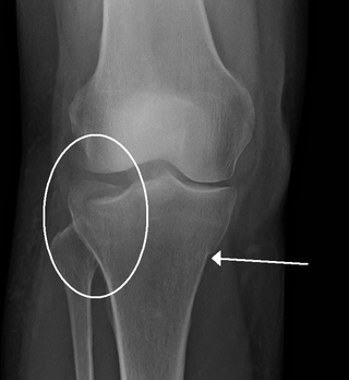
Pott's fracture, also known as Pott's syndrome I and Dupuytren fracture, is an archaic term loosely applied to a variety of bimalleolar ankle fractures. The injury is caused by a combined abduction external rotation from an eversion force. This action strains the sturdy medial (deltoid) ligament of the ankle, often tearing off the medial malleolus due to its strong attachment. The talus then moves laterally, shearing off the lateral malleolus or, more commonly, breaking the fibula superior to the tibiofibular syndesmosis. If the tibia is carried anteriorly, the posterior margin of the distal end of the tibia is also sheared off by the talus. A fractured fibula in addition to detaching the medial malleolus will tear the tibiofibular syndesmosis. The combined fracture of the medial malleolus, lateral malleolus, and the posterior margin of the distal end of the tibia is known as a "trimalleolar fracture".

<span class="mw-page-title-main">Syndesmosis</span> Type of fibrous joint

A syndesmosis is a type of fibrous joint in which two parallel bones are united to each other by fibrous connective tissue. The gap between the bones may be narrow, with the bones joined by ligaments, or the gap may be wide and filled in by a broad sheet of connective tissue called an interosseous membrane. The syndesmoses found in the forearm and leg serve to unite parallel bones and prevent their separation.

<span class="mw-page-title-main">Talus bone</span> One of the foot bones that forms the tarsus

The talus, talus bone, astragalus, or ankle bone is one of the group of foot bones known as the tarsus. The tarsus forms the lower part of the ankle joint. It transmits the entire weight of the body from the lower legs to the foot.

<span class="mw-page-title-main">Ankle fracture</span> Medical condition

An ankle fracture is a break of one or more of the bones that make up the ankle joint. Symptoms may include pain, swelling, bruising, and an inability to walk on the injured leg. Complications may include an associated high ankle sprain, compartment syndrome, stiffness, malunion, and post-traumatic arthritis.

<span class="mw-page-title-main">Inferior tibiofibular joint</span>

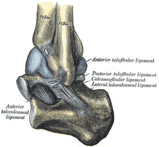
The inferior tibiofibular joint, also known as the distal tibiofibular joint, is formed by the rough, convex surface of the medial side of the distal end of the fibula, and a rough concave surface on the lateral side of the tibia.

<span class="mw-page-title-main">Superior tibiofibular joint</span> Joint in the knee

The superior tibiofibular articulation is an arthrodial joint between the lateral condyle of tibia and the head of the fibula.

<span class="mw-page-title-main">Anterior tibiofibular ligament</span>

The anterior ligament of the lateral malleolus is a flat, trapezoidal band of fibers, broader below than above, which extends obliquely downward and lateralward between the adjacent margins of the tibia and fibula, on the front aspect of the syndesmosis.

<span class="mw-page-title-main">Fibrous joint</span> Fixed joints between bones held together by dense, fibrous tissue

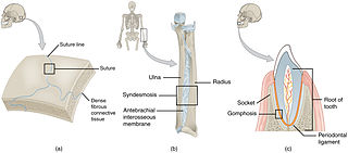
In anatomy, fibrous joints are joints connected by fibrous tissue, consisting mainly of collagen. These are fixed joints where bones are united by a layer of white fibrous tissue of varying thickness. In the skull, the joints between the bones are called sutures. Such immovable joints are also referred to as synarthroses.

<span class="mw-page-title-main">Malleolus</span> Ankle bone protrusion

A malleolus is the bony prominence on each side of the human ankle.

<span class="mw-page-title-main">Trimalleolar fracture</span> Medical condition

A trimalleolar fracture is a fracture of the ankle that involves the lateral malleolus, the medial malleolus, and the distal posterior aspect of the tibia, which can be termed the posterior malleolus. The trauma is sometimes accompanied by ligament damage and dislocation.

A high ankle sprain, also known as a syndesmotic ankle sprain (SAS), is a sprain of the syndesmotic ligaments that connect the tibia and fibula in the lower leg, thereby creating a mortise and tenon joint for the ankle. High ankle sprains are described as high because they are located above the ankle. They comprise approximately 15% of all ankle sprains. Unlike the common lateral ankle sprains, when ligaments around the ankle are injured through an inward twisting, high ankle sprains are caused when the lower leg and foot externally rotates.

<span class="mw-page-title-main">Danis–Weber classification</span> Method to classify an ankle fracture

The Danis–Weber classification is a method of describing ankle fractures. It has three categories:

<span class="mw-page-title-main">Crus fracture</span>

A crus fracture is a fracture of the lower legs bones meaning either or both of the tibia and fibula.

<span class="mw-page-title-main">Syndesmotic screw</span> Type of screw

A syndesmotic screw is a metal screw designed to replace the syndesmosis of the human body, usually temporarily. If the syndosmosis is torn apart as result of bone fracture, surgeons will sometimes fix the relevant bones together with a syndesmotic screw, temporarily replacing the normal articulation.

<span class="mw-page-title-main">Medial knee injuries</span> Medical condition

Medial knee injuries are the most common type of knee injury. The medial ligament complex of the knee consists of:

References

  1. Wilson, F. C. (2000). Fractures of the ankle: pathogenesis and treatment. J South Orthop Assoc. 9(2):105-115.
  2. Thordarson, D. B. (1996). Detecting and treating common foot and ankle fractures: Part 1: the ankle and hindfoot. Phys Sportsmed. 24(9): 29-38.
  3. 1 2 Lauge-Hansen, N. (1950). Fractures of the ankle. II. Combined experimental-surgical and experimental-roentgenologic investigations. Arch Surg. 60(5): 957- 985.
  4. 1 2 3 4 5 6 7 8 9 10 11 12 13 14 15 16 17 18 19 Sproule, J. A., Khalid, M., O’Sullivan, M., & McCabe, J. P. (2004). Outcome after surgery for Maisonneuve fracture of the fibula. Injury. 35(8): 791-798.
  5. Atesok, K. I., Jupiter, J. B., & Weiss, A. P. (2011). Galeazzi fracture. J Am Acad Orthop Surg. 19(10): 623-633.
  6. 1 2 3 4 5 6 7 8 9 10 11 12 Stufkens, S. A., van den Bekerom, M. P. J., Doornberg, J. N., Niek van Dijk, C., & Kloen, P. (2011). Evidence-Based Treatment of Maisonneuve Fractures. J Foot Ankle Surg. 50(1): 62-67.
  7. 1 2 3 4 5 6 Babis, G. C., Papagelopoulos, P. J., Tsarouchas, J., Zoubos, A. B., Korres, D. S., & Nikiforidis, P. (2000). Operative treatment for maisonneuve fracture of the proximal fibula. Orthopedics. 23(7): 687-690.
  8. 1 2 3 4 5 6 7 He, J., Ma, X., Xin, J., Cao, H., Li, N., Sun, Z., Wang, G., Fu, X., Zhao, B., & Hu, F. (2020). Pathoanatomy and Injury Mechanism of Typical Maisonneuve Fracture. Orthopaedic Surgery. DOI: 10.1111/os.12733
  9. 1 2 3 4 5 6 7 8 9 10 11 Pankovich, A. M. (1976). Maisonneuve fracture of the fibula. J Bone Joint Surg Am. 58(3): 337-342.
  10. Ramsey, P. L. & Hamilton, W. (1976). Changes in tibiotalar area of contact caused by lateral talar shift. J Bone Joint Surg Am. 58(3): 356-357.
  11. 1 2 3 4 5 6 7 8 9 Levy, B. A., Vogt, K. J., Herrera, D. A., & Cole, P. A. (2006). Maisonneuve fracture equivalent with proximal tibiofibular dislocation. A case report and literature review. J Bone Joint Surg Am. 88(5): 1111-1116.
  12. 1 2 3 4 5 6 7 8 9 10 11 12 Duchesneau, S. & Fallat, L. M. (1995). The Maisonneuve Fracture. J Foot Ankle Surg. 34(5): 422-428.
  13. 1 2 3 4 5 6 7 8 Schnetzke, M., Vetter, S. Y., Beisemann, N., Swartman, B., Grützner, P. A., & Franke, J. (2016). Management of syndesmotic injuries: What is the evidence?. World J Orthop. 7(11): 718-725.
  14. Kukreti, S., Faraj, A., & Miles, J. N. V. (2005). Does position of syndesmotic screw affect functional and radiological outcome in ankle fractures?. Injury. 36(9): 1121-1124.
  15. 1 2 van den Bekerom, M. P. J. & Raven, E. E. J. (2007). Current concepts review: operative techniques for stabilizing the distal tibiofibular syndesmosis. Foot Ankle Int. 28(12): 1302-1308.
  16. McBryde A., Chiasson, B., Wilhelm, A., Donovan, F., Ray, T., & Bacilla, P. (1997). Syndesmotic screw placement: a biomechanical analysis. Foot Ankle Int. 18(5): 262-266.
  17. 1 2 Lock, T.R., Schaffer, J. J., & Manoli, A. (1987). Maisonneuve fracture: case of missed diagnosis. Ann Emerg Med. 16(7): 805-807.