Periodontal scaler

Last updated
Periodontal scaler Dental scaler.JPG
Periodontal scaler
Periodontal scalers have sharp tips to access tight embrasure spaces between teeth and are triangular in cross-section. Scaler - top.jpg
Periodontal scalers have sharp tips to access tight embrasure spaces between teeth and are triangular in cross-section.
A posterior scaler shown in relation to a posterior tooth on a typodont. Posterior scaler on teeth.jpg
A posterior scaler shown in relation to a posterior tooth on a typodont.

Periodontal scalers are dental instruments used in the prophylactic and periodontal care of teeth (most often human teeth), including scaling and root planing. The working ends come in a variety of shapes and sizes, but they are always narrow at the tip, so as to allow for access to narrow embrasure spaces between teeth. They differ from periodontal curettes, which possess a blunt tip.

Contents

Use

Together with periodontal curettes, periodontal scalers are used to remove calculus from teeth. While curettes are often universal in that they can be used on both supra- and sub-gingival calculus removals, scalers are restricted to supra-gingival use. [1] Use of a scaler below the gum line is likely to damage the gingiva (gums). [2]

The anterior scaler (yellow ring) is straight, while the posterior scaler (orange ring) has an angled terminal shank (highlighted in red) to allow for easy access to the surfaces of posterior teeth. Scalers - shank.jpg
The anterior scaler (yellow ring) is straight, while the posterior scaler (orange ring) has an angled terminal shank (highlighted in red) to allow for easy access to the surfaces of posterior teeth.

Scalers have scraping edges on both sides of their blades and thus are fit for both mesial and distal surfaces of any tooth in the area in which they are being used. [2]

Scalers are best used when their terminal shank, namely, the last portion of the functional shank closest to the working end, is angled slightly toward the surface of the tooth. [2]

Design and materials

The composition of hand instruments is continuously evolving, which is why it may be a challenge to find the proper instrument for the right clinical situation. [3] With the broad variation of instrument designs and materials, it allows dental professionals to implement periodontal therapy with reduced strain and increased comfort levels for both the clinician and the patient. [3] The following are some factors to consider with the design of periodontal scalers:

With the continuous, evolving technology of hand instruments, dental professionals are given the opportunity to implement dental treatment more effectively for the patient while improving their work surroundings simultaneously. [3]

Design characteristics

Generally, there are two cutting edges per working end: the outer cutting edge (farther from the instrument handle) and the inner cutting edge (closer to the instrument handle). [4] This way, both edges can adapt to both the interproximal surfaces of any tooth, to which they are being used. Typically, periodontal scalers have pointed backs, but some new scaler designs have working ends with rounded backs as well. [4] Additionally, they have triangular cross sections; this limits their instrumentation use to above the gingiva (supragingival) to prevent any tissue trauma. [4] Periodontal scalers also have pointed tips and faces perpendicular to the lower shank; this is why the terminal shank must be tilted towards the tooth in order to establish correct angulation since the cutting edges are at equal levels with each other. [4]

There are two types of periodontal scaler designs:

Techniques

When it comes to periodontal therapy, there are multiple steps that are required prior to activating a proper working stroke on the teeth. First, a modified pen grasp position must be achieved before starting periodontal instrumentation; this position involves the thumb and the index finger placed on the instrument handle, the middle finger resting on the shank, and the little finger neutral and relaxed near the ring finger. [4] By holding periodontal instruments in this manner, it allows for precise control of the instrument, effective detection of rough areas on the tooth structure (i.e. calculus or irregular tooth anatomy), and reduces musculoskeletal stress on the clinician. [4]

In addition, there are several characteristics of a calculus removal stroke that are vital to the effectiveness of periodontal instrumentation; [6] these include stabilization, adaptation, angulation, lateral pressure, characteristics, stroke direction, and stroke number. [4]

Once all these characteristics are understood, the clinician will activate the periodontal debridement strokes using the periodontal scalers.

When performing periodontal debridement, the instrumentation of anterior teeth and posterior teeth are applied with the following steps. First, the fulcrum finger rests on a tooth to support the clinician’s hand, then at the initiation of a stroke, the clinician will press down on the fulcrum finger to further gain control. [4] It is crucial to tilt the lower shank of the periodontal scaler slightly towards the tooth surface being worked on to obtain correct angulation. [4] This ensures that a 70º-80º angulation is achieved between the tooth surface and the face of the instrument. [4] The instrument is then walked across the surface being worked on with short, controlled, overlapping strokes while applying moderate pressure. [4] Overall, the working-end of the instrument is only moving a few millimeters at a time. [4] Simultaneously, the clinician must roll the handle of the instrument to maintain adaptation throughout, to prevent any soft tissue injury. [4] After completing a calculus removal stroke, the clinician may then utilize an assessment stroke, characterized by feather-light pressure, to judge the removal of the calculus deposit. [4] These steps are repeated until the complete removal of calculus is achieved throughout the entire dentition.

These instrumentation techniques are followed to achieve effective periodontal therapy using periodontal instruments, [6] such as periodontal scalers. Moreover, following these instrumentation principles would improve the quality of life for the clinician as well; this includes lowering the risk of musculoskeletal disorders (MSD) in their hands and/or forearm, conserving more effort than required, and increasing the efficiency of instrumentation. [6] These principles form a foundation to improve efficiency and effectiveness of periodontal therapy while protecting the proper ergonomics of the clinician. [6]

A healthy periodontium is achieved by completely removing the living bacteria present in both the biofilm and calculus. [7] This bacteria is responsible for periodontal disease, a term that encompasses both gingivitis and periodontitis. [7] It is also important to note that with different levels of periodontal disease, the clinician may apply sextant scaling or quadrant scaling to achieve best results for a healthy periodontium. [8] In this case, a sextant or a quadrant will be completed per appointment, which would require multiple appointments to complete debridement of the entire mouth.

Related Research Articles

<span class="mw-page-title-main">Periodontal disease</span> Medical condition

Periodontal disease, also known as gum disease, is a set of inflammatory conditions affecting the tissues surrounding the teeth. In its early stage, called gingivitis, the gums become swollen and red and may bleed. It is considered the main cause of tooth loss for adults worldwide. In its more serious form, called periodontitis, the gums can pull away from the tooth, bone can be lost, and the teeth may loosen or fall out. Bad breath may also occur.

<span class="mw-page-title-main">Calculus (dental)</span> Form of hardened dental plaque

In dentistry, calculus or tartar is a form of hardened dental plaque. It is caused by precipitation of minerals from saliva and gingival crevicular fluid (GCF) in plaque on the teeth. This process of precipitation kills the bacterial cells within dental plaque, but the rough and hardened surface that is formed provides an ideal surface for further plaque formation. This leads to calculus buildup, which compromises the health of the gingiva (gums). Calculus can form both along the gumline, where it is referred to as supragingival, and within the narrow sulcus that exists between the teeth and the gingiva, where it is referred to as subgingival.

<span class="mw-page-title-main">Toothache</span> Medical condition of the teeth

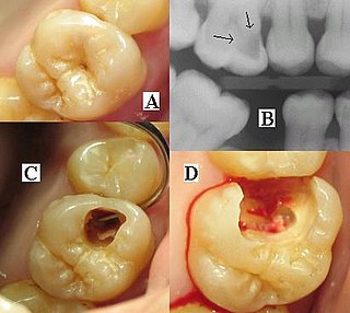
Toothache, also known as dental pain, is pain in the teeth or their supporting structures, caused by dental diseases or pain referred to the teeth by non-dental diseases. When severe it may impact sleep, eating, and other daily activities.

A dental explorer or sickle probe is an instrument in dentistry commonly used in the dental armamentarium. A sharp point at the end of the explorer is used to enhance tactile sensation.

Periodontology or periodontics is the specialty of dentistry that studies supporting structures of teeth, as well as diseases and conditions that affect them. The supporting tissues are known as the periodontium, which includes the gingiva (gums), alveolar bone, cementum, and the periodontal ligament. A periodontist is a dentist that specializes in the prevention, diagnosis and treatment of periodontal disease and in the placement of dental implants.

<span class="mw-page-title-main">Veterinary dentistry</span> Branch of veterinary medicine

Veterinary dentistry is the field of dentistry applied to the care of animals. It is the art and science of prevention, diagnosis, and treatment of conditions, diseases, and disorders of the oral cavity, the maxillofacial region, and its associated structures as it relates to animals.

<span class="mw-page-title-main">Dental instrument</span> Tools of the dental profession

Dental instruments are tools that dental professionals use to provide dental treatment. They include tools to examine, manipulate, treat, restore, and remove teeth and surrounding oral structures.

A dental auxiliary is any oral health practitioner other than a dentist & dental hygienist, including the supporting team assisting in dental treatment. They include dental assistants, dental therapists and oral health therapists, dental technologists, and orthodontic auxiliaries. The role of dental auxiliaries is usually set out in regional dental regulations, defining the treatment that can be performed.

<span class="mw-page-title-main">Gingival sulcus</span> Space between tooth and gums

The gingival sulcus is an area of potential space between a tooth and the surrounding gingival tissue and is lined by sulcular epithelium. The depth of the sulcus is bounded by two entities: apically by the gingival fibers of the connective tissue attachment and coronally by the free gingival margin. A healthy sulcular depth is three millimeters or less, which is readily self-cleansable with a properly used toothbrush or the supplemental use of other oral hygiene aids.

<span class="mw-page-title-main">Dental radiography</span> X-ray imaging in dentistry

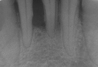
Dental radiographs, commonly known as X-rays, are radiographs used to diagnose hidden dental structures, malignant or benign masses, bone loss, and cavities.

<span class="mw-page-title-main">Scaling and root planing</span> Dental procedure

Scaling and root planing, also known as conventional periodontal therapy, non-surgical periodontal therapy or deep cleaning, is a procedure involving removal of dental plaque and calculus and then smoothing, or planing, of the (exposed) surfaces of the roots, removing cementum or dentine that is impregnated with calculus, toxins, or microorganisms, the agents that cause inflammation. It is a part of non-surgical periodontal therapy. This helps to establish a periodontium that is in remission of periodontal disease. Periodontal scalers and periodontal curettes are some of the tools involved.

<span class="mw-page-title-main">Root canal treatment</span> Dental treatment

Root canal treatment is a treatment sequence for the infected pulp of a tooth which is intended to result in the elimination of infection and the protection of the decontaminated tooth from future microbial invasion. Root canals, and their associated pulp chamber, are the physical hollows within a tooth that are naturally inhabited by nerve tissue, blood vessels and other cellular entities. Together, these items constitute the dental pulp.

The periodontal curette is a type of hand-activated instrument used in dentistry and dental hygiene for the purpose of scaling and root planing. The periodontal curette is considered a treatment instrument and is classified into two main categories: universal curettes and Gracey curettes. Periodontal curettes have one face, one or two cutting edges and a rounded back and rounded toe. They are typically the instrument of choice for subgingival calculus removal.

<span class="mw-page-title-main">Tooth polishing</span>

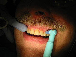
Tooth polishing is done to smooth the surfaces of teeth and restorations. The purpose of polishing is to remove extrinsic stains, remove dental plaque accumulation, increase aesthetics and to reduce corrosion of metallic restorations. Tooth polishing has little therapeutic value and is usually done as a cosmetic procedure after debridement and before fluoride application. Common practice is to use a prophy cup—a small motorized rubber cup—along with an abrasive polishing compound.

<span class="mw-page-title-main">Debridement (dental)</span> Removal of plaque and calculus from teeth

In dentistry, debridement refers to the removal by dental cleaning of accumulations of plaque and calculus (tartar) in order to maintain dental health. Debridement may be performed using ultrasonic instruments, which fracture the calculus, thereby facilitating its removal, as well as hand tools, including periodontal scaler and curettes, or through the use of chemicals such as hydrogen peroxide.

<span class="mw-page-title-main">Periodontal abscess</span> Medical condition

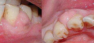
A periodontal abscess, is a localized collection of pus within the tissues of the periodontium. It is a type of dental abscess. A periodontal abscess occurs alongside a tooth, and is different from the more common periapical abscess, which represents the spread of infection from a dead tooth. To reflect this, sometimes the term "lateral (periodontal) abscess" is used. In contrast to a periapical abscess, periodontal abscesses are usually associated with a vital (living) tooth. Abscesses of the periodontium are acute bacterial infections classified primarily by location.

A phoenix abscess is an acute exacerbation of a chronic periapical lesion. It is a dental abscess that can occur immediately following root canal treatment. Another cause is due to untreated necrotic pulp. It is also the result of inadequate debridement during the endodontic procedure. Risk of occurrence of a phoenix abscess is minimised by correct identification and instrumentation of the entire root canal, ensuring no missed anatomy.

Chronic periodontitis is one of the seven categories of periodontitis as defined by the American Academy of Periodontology 1999 classification system. Chronic periodontitis is a common disease of the oral cavity consisting of chronic inflammation of the periodontal tissues that is caused by the accumulation of profuse amounts of dental plaque. Periodontitis initially begins as gingivitis and can progress onto chronic and subsequent aggressive periodontitis according to the 1999 classification.

Peri-implant mucositis is defined as an inflammatory lesion of the peri-implant mucosa in the absence of continuing marginal bone loss.

Periodontal surgery is a form of dental surgery that prevents or corrects anatomical, traumatic, developmental, or plaque-induced defects in the bone, gingiva, or alveolar mucosa. The objectives of this surgery include accessibility of instruments to root surface, elimination of inflammation, creation of an oral environment for plaque control, periodontal diseases control, oral hygiene maintenance, maintain proper embrasure space, address gingiva-alveolar mucosa problems, and esthetic improvement. The surgical procedures include crown lengthening, frenectomy, and mucogingival flap surgery.

References

  1. Nield-Gehrig 2008, p. 287, 309.
  2. 1 2 3 Nield-Gehrig 2008, p. 288.
  3. 1 2 3 4 5 6 7 Bennet, Barbara (November 2007). "All about hand instruments". Dimensions of Dental Hygiene. 5 (11): 20–23. ISSN   1542-7838 via CINAHL Plus with Full Text.
  4. 1 2 3 4 5 6 7 8 9 10 11 12 13 14 15 16 17 18 19 20 Nield-Gehrig, Jill S. (2013). Fundamentals of periodontal instrumentation and advanced root instrumentation (7th ed.). Philadelphia: Lippincott Williams & Wilkins. ISBN   9781609133313.
  5. 1 2 3 4 5 Gehrig, Jill S. (2016). Fundamentals of periodontal instrumentation and advanced root instrumentation (8th ed.). Wolters Kluwer Health Adis. ISBN   9781496345530.
  6. 1 2 3 4 Matsuda, Stacy A. (November 2008). "Anatomy of a stroke: Building the foundation for effective therapy and good ergonomics". Dimensions of Dental Hygiene. 6 (11): 22–26. ISSN   1542-7838 via CINAHL Plus with Full Text.
  7. 1 2 Cobbs, Charles M. (October 2008). "Microbes, inflammation, scaling and root planing, and the periodontal condition". Journal of Dental Hygiene. 3 (82): 4–9. ISSN   1043-254X via CINAHL Plus with Full Text.
  8. Newman, Michael G.; Takei, Henry H.; Klokkevoid, Perry R.; Carranza, Fermin A. (2015). Carranza's Clinical Periodontology (12th ed.). St. Louis, Missouri: Elsevier Saunders. ISBN   9780323188241.

Sources