Microscopy is the technical field of using microscopes to view objects and areas of objects that cannot be seen with the naked eye. There are three well-known branches of microscopy: optical, electron, and scanning probe microscopy, along with the emerging field of X-ray microscopy.

A microscope is an instrument used to see objects that are too small to be seen by the naked eye. Microscopy is the science of investigating small objects and structures using such an instrument. Microscopic means invisible to the eye unless aided by a microscope.

The optical microscope, often referred to as the light microscope, is a type of microscope that commonly uses visible light and a system of lenses to magnify images of small objects. Optical microscopes are the oldest design of microscope and were possibly invented in their present compound form in the 17th century. Basic optical microscopes can be very simple, although many complex designs aim to improve resolution and sample contrast. Often used in the classroom and at home unlike the electron microscope which is used for closer viewing.
Angular resolution or spatial resolution describes the ability of any image-forming device such as an optical or radio telescope, a microscope, a camera, or an eye, to distinguish small details of an object, thereby making it a major determinant of image resolution. In physics and geosciences, the term spatial resolution refers to the precision of a measurement with respect to space.

The resolution of an optical imaging system – a microscope, telescope, or camera – can be limited by factors such as imperfections in the lenses or misalignment. However, there is a principal limit to the resolution of any optical system, due to the physics of diffraction. An optical system with resolution performance at the instrument's theoretical limit is said to be diffraction-limited.

In optical engineering, the objective is the optical element that gathers light from the object being observed and focuses the light rays to produce a real image. Objectives can be a single lens or mirror, or combinations of several optical elements. They are used in microscopes, telescopes, cameras, slide projectors, CD players and many other optical instruments. Objectives are also called object lenses, object glasses, or objective glasses.
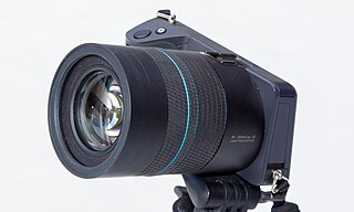
A light field camera, also known as plenoptic camera, captures information about the light field emanating from a scene; that is, the intensity of light in a scene, and also the direction that the light rays are traveling in space. This contrasts with a conventional camera, which records only light intensity.
Differential interference contrast (DIC) microscopy, also known as Nomarski interference contrast (NIC) or Nomarski microscopy, is an optical microscopy technique used to enhance the contrast in unstained, transparent samples. DIC works on the principle of interferometry to gain information about the optical path length of the sample, to see otherwise invisible features. A relatively complex optical system produces an image with the object appearing black to white on a grey background. This image is similar to that obtained by phase contrast microscopy but without the bright diffraction halo. The technique was developed by Polish physicist Georges Nomarski in 1952.

Dark-field microscopy describes microscopy methods, in both light and electron microscopy, which exclude the unscattered beam from the image. As a result, the field around the specimen is generally dark.

Bright-field microscopy is the simplest of all the optical microscopy illumination techniques. Sample illumination is transmitted white light, and contrast in the sample is caused by attenuation of the transmitted light in dense areas of the sample. Bright-field microscopy is the simplest of a range of techniques used for illumination of samples in light microscopes, and its simplicity makes it a popular technique. The typical appearance of a bright-field microscopy image is a dark sample on a bright background, hence the name.

Phase-contrast microscopy is an optical microscopy technique that converts phase shifts in light passing through a transparent specimen to brightness changes in the image. Phase shifts themselves are invisible, but become visible when shown as brightness variations.

August Karl Johann Valentin Köhler was a German professor and early staff member of Carl Zeiss AG in Jena, Germany. He is best known for his development of the microscopy technique of Köhler illumination, an important principle in optimizing microscopic resolution power by evenly illuminating the field of view. This invention revolutionized light microscope design and is widely used in traditional as well as modern digital imaging techniques today.

A petrographic microscope is a type of optical microscope used in petrology and optical mineralogy to identify rocks and minerals in thin sections. The microscope is used in optical mineralogy and petrography, a branch of petrology which focuses on detailed descriptions of rocks. The method is called "polarized light microscopy" (PLM). Depending on the grade of observation required, petrological microscopes are derived from conventional brightfield microscopes of similar basic capabilities by:
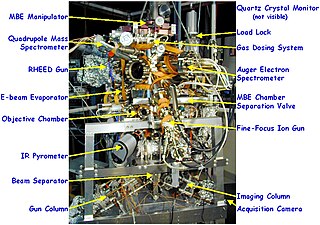
Low-energy electron microscopy, or LEEM, is an analytical surface science technique used to image atomically clean surfaces, atom-surface interactions, and thin (crystalline) films. In LEEM, high-energy electrons are emitted from an electron gun, focused using a set of condenser optics, and sent through a magnetic beam deflector. The “fast” electrons travel through an objective lens and begin decelerating to low energies near the sample surface because the sample is held at a potential near that of the gun. The low-energy electrons are now termed “surface-sensitive” and the near-surface sampling depth can be varied by tuning the energy of the incident electrons. The low-energy elastically backscattered electrons travel back through the objective lens, reaccelerate to the gun voltage, and pass through the beam separator again. However, now the electrons travel away from the condenser optics and into the projector lenses. Imaging of the back focal plane of the objective lens into the object plane of the projector lens produces a diffraction pattern at the imaging plane and recorded in a number of different ways. The intensity distribution of the diffraction pattern will depend on the periodicity at the sample surface and is a direct result of the wave nature of the electrons. One can produce individual images of the diffraction pattern spot intensities by turning off the intermediate lens and inserting a contrast aperture in the back focal plane of the objective lens, thus allowing for real-time observations of dynamic processes at surfaces. Such phenomena include : tomography, phase transitions, adsorption, reaction, segregation, thin film growth, etching, strain relief, sublimation, and magnetic microstructure. These investigations are only possible because of the accessibility of the sample; allowing for a wide variety of in situ studies over a wide temperature range. LEEM was invented by Ernst Bauer in 1962; however, not fully developed until 1985.
The optical properties of all liquid and solid materials change as a function of the wavelength of light used to measure them. This change as a function of wavelength is called the dispersion of the optical properties. The graph created by plotting the optical property of interest by the wavelength at which it is measured is called a dispersion curve.

Optical sectioning is the process by which a suitably designed microscope can produce clear images of focal planes deep within a thick sample. This is used to reduce the need for thin sectioning using instruments such as the microtome. Many different techniques for optical sectioning are used and several microscopy techniques are specifically designed to improve the quality of optical sectioning.
A conoscopic interference pattern or interference figure is a pattern of birefringent colours crossed by dark bands, which can be produced using a geological petrographic microscope for the purposes of mineral identification and investigation of mineral optical and chemical properties. The figures are produced by optical interference when diverging light rays travel through an optically non-isotropic substance - that is, one in which the substance's refractive index varies in different directions within it. The figure can be thought of as a "map" of how the birefringence of a mineral would vary with viewing angle away from perpendicular to the slide, where the central colour is the birefringence seen looking straight down, and the colours further from the centre equivalent to viewing the mineral at ever increasing angles from perpendicular. The dark bands correspond to positions where optical extinction would be seen. In other words, the interference figure presents all possible birefringence colours for the mineral at once.

Hoffman modulation contrast microscopy is an optical microscopy technique for enhancing the contrast in unstained biological specimens. The technique was invented by Robert Hoffman in 1975. Like differential interference contrast microscopy, contrast is increased by using components in the light path which convert phase gradients in the specimen into differences in light intensity that are rendered in an image that appears three-dimensional. The 3D appearance may be misleading, as a feature which appears to cast a shadow may not necessarily have a distinct physical geometry corresponding to the shadow. The technique is particularly suitable for optical sectioning at lower magnifications.
A condenser is an optical lens which renders a divergent beam from a point source into a parallel or converging beam to illuminate an object.