Two-photon excitation microscopy

Last updated
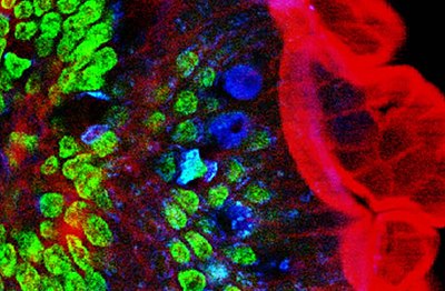
Two-photon excitation microscopy of mouse intestine. Red: actin. Green: cell nuclei. Blue: mucus of goblet cells. Obtained at 780 nm using a Ti-sapphire laser. MultiPhotonExcitation-Fig10-doi10.1186slash1475-925X-5-36-clipping.JPEG
Two-photon excitation microscopy of mouse intestine. Red: actin. Green: cell nuclei. Blue: mucus of goblet cells. Obtained at 780 nm using a Ti-sapphire laser.

Two-photon excitation microscopy (TPEF or 2PEF) is a fluorescence imaging technique that is particularly well-suited to image scattering living tissue of up to about one millimeter in thickness. Unlike traditional fluorescence microscopy, where the excitation wavelength is shorter than the emission wavelength, two-photon excitation requires simultaneous excitation by two photons with longer wavelength than the emitted light. The laser is focused onto a specific location in the tissue and scanned across the sample to sequentially produce the image. Due to the non-linearity of two-photon excitation, mainly fluorophores in the micrometer-sized focus of the laser beam are excited, which results in the spatial resolution of the image. This contrasts with confocal microscopy, where the spatial resolution is produced by the interaction of excitation focus and the confined detection with a pinhole.

Contents

Two-photon excitation microscopy typically uses near-infrared (NIR) excitation light which can also excite fluorescent dyes. Using infrared light minimizes scattering in the tissue because infrared light is scattered less in typical biological tissues. Due to the multiphoton absorption, the background signal is strongly suppressed. Both effects lead to an increased penetration depth for this technique. Two-photon excitation can be a superior alternative to confocal microscopy due to its deeper tissue penetration, efficient light detection, and reduced photobleaching. [1] [2]

Two-photon fluorescence image (green) of a cross section of rhizome colored with lily of the valley. The excitement is at 840 nm, and the red and blue colors represent other channels of multiphoton techniques which have been superimposed. Convallaria rhizom.jpg
Two-photon fluorescence image (green) of a cross section of rhizome colored with lily of the valley. The excitement is at 840 nm, and the red and blue colors represent other channels of multiphoton techniques which have been superimposed.

Concept

Schematic representation of the energy levels (Jablonski diagrams) of the fluorescence process, example of a fluorescent dye that emits light at 460 nm. One (purple, 1PEF), two (light red, 2PEF) or three (dark red, 3PEF) photons are absorbed to emit a photon of fluorescence (turquoise). MultiPhotonExcitation-Fig1-doi10.1186slash1475-925X-5-36.JPEG
Schematic representation of the energy levels (Jabłoński diagrams) of the fluorescence process, example of a fluorescent dye that emits light at 460 nm. One (purple, 1PEF), two (light red, 2PEF) or three (dark red, 3PEF) photons are absorbed to emit a photon of fluorescence (turquoise).
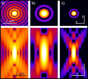
Optical response from a point source. From left to right: calculated intensity distributions xy (top) and rz (bottom), with logarithmic scale, for a point source imaged by means of a wide field (a), 2PEF (b) and confocal microscopy (c). The 2PEF and confocal forms have a better signal-to-noise ratio than the wide field. The 2PEF distribution is larger due to the fact that a wavelength twice as long as in the case of a wide or confocal field is responsible for the intensity distribution. These intensity distributions are also known as point spread functions. Optical conditions: the excitation wavelengths are 488 nm and 900 nm respectively for 1PEF and 2PEF; the emission wavelength is 520 nm; the numerical aperture is 1.3 with an oil immersion objective. MultiPhotonExcitation-Fig7-doi10.1186slash1475-925X-5-36.JPEG
Optical response from a point source. From left to right: calculated intensity distributions xy (top) and rz (bottom), with logarithmic scale, for a point source imaged by means of a wide field (a), 2PEF (b) and confocal microscopy (c). The 2PEF and confocal forms have a better signal-to-noise ratio than the wide field. The 2PEF distribution is larger due to the fact that a wavelength twice as long as in the case of a wide or confocal field is responsible for the intensity distribution. These intensity distributions are also known as point spread functions. Optical conditions: the excitation wavelengths are 488 nm and 900 nm respectively for 1PEF and 2PEF; the emission wavelength is 520 nm; the numerical aperture is 1.3 with an oil immersion objective.

Two-photon excitation employs two-photon absorption, a concept first described by Maria Goeppert Mayer (1906–1972) in her doctoral dissertation in 1931, [3] and first observed in 1961 in a CaF2:Eu2+ crystal using laser excitation by Wolfgang Kaiser. [4] Isaac Abella showed in 1962 in caesium vapor that two-photon excitation of single atoms is possible. [5]

Two-photon excitation fluorescence microscopy has similarities to other confocal laser microscopy techniques such as laser scanning confocal microscopy and Raman microscopy. These techniques use focused laser beams scanned in a raster pattern to generate images, and both have an optical sectioning effect. Unlike confocal microscopes, multiphoton microscopes do not contain pinhole apertures that give confocal microscopes their optical sectioning quality. The optical sectioning produced by multiphoton microscopes is a result of the point spread function of the excitation. The concept of two-photon excitation is based on the idea that two photons, of comparably lower photon energy than needed for one-photon excitation, can also excite a fluorophore in one quantum event. Each photon carries approximately half the energy necessary to excite the molecule. The emitted photon is at a higher energy (shorter wavelength) than either of the two exciting photons. The probability of the near-simultaneous absorption of two photons is extremely low. Therefore, a high peak flux of excitation photons is typically required, usually generated by femtosecond pulsed laser. For example, the same average laser power but without pulsing results in no detectable fluorescence compared to fluorescence generated by the pulsed laser via the two-photon effect. The longer wavelength, lower energy (typically infrared) excitation lasers of multiphoton microscopes are well-suited to use in imaging live cells as they cause less damage than the short-wavelength lasers typically used for single-photon excitation, so living tissues may be observed for longer periods with fewer toxic effects.

The most commonly used fluorophores have excitation spectra in the 400500 nm range, whereas the laser used to excite the two-photon fluorescence lies in the ~7001100 nm (infrared) range produced by Ti-sapphire lasers. If the fluorophore absorbs two infrared photons simultaneously, it will absorb enough energy to be raised into the excited state. The fluorophore will then emit a single photon with a wavelength that depends on the type of fluorophore used (typically in the visible spectrum). Because two photons are absorbed during the excitation of the fluorophore, the probability of fluorescent emission from the fluorophores increases quadratically with the excitation intensity. Therefore, much more two-photon fluorescence is generated where the laser beam is tightly focused than where it is more diffuse. Effectively, excitation is restricted to the tiny focal volume (~1 femtoliter), resulting in a high degree of rejection of out-of-focus objects. This localization of excitation is the key advantage compared to single-photon excitation microscopes, which need to employ elements such as pinholes to reject out-of-focus fluorescence. The fluorescence from the sample is then collected by a high-sensitivity detector, such as a photomultiplier tube. This observed light intensity becomes one pixel in the eventual image; the focal point is scanned throughout a desired region of the sample to form all the pixels of the image.

Development

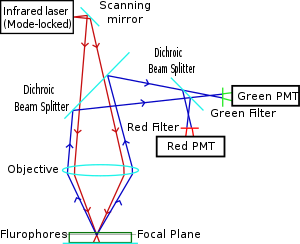
A diagram of a two-photon microscope. Diagram of a two-photon excitation microscope en.svg
A diagram of a two-photon microscope.

Two-photon microscopy was pioneered and patented by Winfried Denk and James Strickler in the lab of Watt W. Webb at Cornell University in 1990. They combined the idea of two-photon absorption with the use of a laser scanner. [1] [6] In two-photon excitation microscopy an infrared laser beam is focused through an objective lens. The Ti-sapphire laser normally used has a pulse width of approximately 100 femtoseconds (fs) and a repetition rate of about 80 MHz, allowing the high photon density and flux required for two-photon absorption, and is tunable across a wide range of wavelengths.

The use of infrared light to excite fluorophores in light-scattering tissue has added benefits. [7] Longer wavelengths are scattered to a lesser degree than shorter ones, which is a benefit to high-resolution imaging. In addition, these lower-energy photons are less likely to cause damage outside the focal volume. Compared to a confocal microscope, photon detection is much more effective since even scattered photons contribute to the usable signal. These benefits for imaging in scattering tissues were only recognized several years after the invention of two-photon excitation microscopy. [8]

There are several caveats to using two-photon microscopy: The pulsed lasers needed for two-photon excitation are much more expensive than the continuous wave (CW) lasers used in confocal microscopy. The two-photon absorption spectrum of a molecule may vary significantly from its one-photon counterpart. Higher-order photodamage becomes a problem and bleaching scales with the square of the laser power, whereas it is linear for single-photon (confocal). For very thin objects such as isolated cells, single-photon (confocal) microscopes can produce images with higher optical resolution due to their shorter excitation wavelengths. In scattering tissue, on the other hand, the superior optical sectioning and light detection capabilities of the two-photon microscope result in better performance.

Applications

Main

Two-photon microscopy has been involved in numerous fields including: physiology, neurobiology, embryology and tissue engineering. Even thin, nearly transparent tissues (such as skin cells) have been visualized with clear detail due to this technique. [9] Two-photon microscopy's high speed imaging capabilities may also be utilized in noninvasive optical biopsy. [10] Two-photon microscopy has been aptly used for producing localized chemical reactions, [8] and effect that has been used also for two-photon-based lithography. Using two-photon fluorescence and second-harmonic generation–based microscopy, it was shown that organic porphyrin-type molecules can have different transition dipole moments for two-photon fluorescence and second harmonic generation, [11] which are otherwise thought to occur from the same transition dipole moment. [12] Non-degenerative two-photon excitation, or using 2 photons of unequal wavelengths, was shown to increase the fluorescence of all tested small molecules and fluorescent proteins. [13]

Cancer research

2PEF was also proven to be very valuable for characterizing skin cancer. [14] It had also been shown to reveal tumor cell arrest, tumor cell-platelet interaction, tumor cell-leukocyte interaction and metastatic colonization processes. [15]

Embryonic research

2PEF has shown to be advantageous over other techniques, such as confocal microscopy when it comes to long-term live-cell imaging of mammalian embryos. [16]

Kidney research

2PEF has also been used in visualization of difficult-to-access cell types, especially in regards to kidney cells. [17] It has been used in better understanding fluid dynamics and filtration. [18]

Diagram of in vivo brain function imaging. Shows the general schematic for imaging, along with neuronal and vascular images. Imaging was performed using various fluorescent dyes. Two-photon microscopy of in vivo brain function.jpg
Diagram of in vivo brain function imaging. Shows the general schematic for imaging, along with neuronal and vascular images. Imaging was performed using various fluorescent dyes.

Neurosciences

2PEF as well as the extension of this method to 3PEF are used to characterize intact neural tissues in the brain of living and even behaving animals. In particular, the method is advantageous for calcium imaging of a neuron or populations of neurons, [19] for photopharmacology including localized uncaging of components such as glutamate [20] or isomerization of photoswitchable drugs, [21] [22] and for the imaging of other genetically encoded sensors that report the concentration of neurotransmitters. [23]

Currently, two-photon microscopy is widely used to image the live firing of neurons in model organisms including fruit flies ( Drosophila melanogaster), rats, songbirds, primates, ferrets, mice ( Mus musculus), zebrafish. [24] [25] [26] The animals are typically head-fixed due to the size of the microscope and scan devices, but also miniatured microscopes are being developed that enable imaging of neurons in the moving and freely behaving animals. [27] [28]

Higher-order excitation

Simultaneous absorption of three or more photons is also possible, allowing for higher-order multiphoton excitation microscopy. [29] So-called "three-photon excitation fluorecence microscopy" (3PEF) is the most used technique after 2PEF, to which it is complementary. Localized isomerization of photoswitchable drugs in vivo using three-photon excitation has also been reported. [30]

Dyes and fluorescent proteins for two-photon excitation microscopy

In general, all commonly used fluorescent proteins (CFP, GFP, YFP, RFP) and dyes can be excited in two-photon mode. Two-photon excitation spectra are often considerably broader, making it more difficult to excite fluorophores selectively by switching excitation wavelengths.[ citation needed ]

Several green, red and NIR emitting dyes (probes and reactive labels) with extremely high 2-photon absorption cross-sections have been reported. [31] Due to the donor-acceptor-donor type structure, squaraine dyes such as Seta-670, Seta-700 and Seta-660 exhibit very high 2-photon absorption (2PA) efficiencies in comparison to other dyes, [31] [32] [33] SeTau-647 and SeTau-665, a new type of squaraine-rotaxane, exhibit extremely high two-photon action cross-sections of up to 10,000 GM in the near IR region, unsurpassed by any other class of organic dyes. [31]

See also

Sources

Related Research Articles

<span class="mw-page-title-main">Microscopy</span> Viewing of objects which are too small to be seen with the naked eye

Microscopy is the technical field of using microscopes to view objects and areas of objects that cannot be seen with the naked eye. There are three well-known branches of microscopy: optical, electron, and scanning probe microscopy, along with the emerging field of X-ray microscopy.
<span class="mw-page-title-main">Fluorophore</span> Agents that emit light after excitation by light

A fluorophore is a fluorescent chemical compound that can re-emit light upon light excitation. Fluorophores typically contain several combined aromatic groups, or planar or cyclic molecules with several π bonds.

A total internal reflection fluorescence microscope (TIRFM) is a type of microscope with which a thin region of a specimen, usually less than 200 nanometers can be observed.

<span class="mw-page-title-main">Fluorescence microscope</span> Optical microscope that uses fluorescence and phosphorescence

A fluorescence microscope is an optical microscope that uses fluorescence instead of, or in addition to, scattering, reflection, and attenuation or absorption, to study the properties of organic or inorganic substances. "Fluorescence microscope" refers to any microscope that uses fluorescence to generate an image, whether it is a simple set up like an epifluorescence microscope or a more complicated design such as a confocal microscope, which uses optical sectioning to get better resolution of the fluorescence image.

<span class="mw-page-title-main">Confocal microscopy</span> Optical imaging technique

Confocal microscopy, most frequently confocal laser scanning microscopy (CLSM) or laser scanning confocal microscopy (LSCM), is an optical imaging technique for increasing optical resolution and contrast of a micrograph by means of using a spatial pinhole to block out-of-focus light in image formation. Capturing multiple two-dimensional images at different depths in a sample enables the reconstruction of three-dimensional structures within an object. This technique is used extensively in the scientific and industrial communities and typical applications are in life sciences, semiconductor inspection and materials science.

Chemical imaging is the analytical capability to create a visual image of components distribution from simultaneous measurement of spectra and spatial, time information. Hyperspectral imaging measures contiguous spectral bands, as opposed to multispectral imaging which measures spaced spectral bands.

<span class="mw-page-title-main">Second-harmonic imaging microscopy</span>

Second-harmonic imaging microscopy (SHIM) is based on a nonlinear optical effect known as second-harmonic generation (SHG). SHIM has been established as a viable microscope imaging contrast mechanism for visualization of cell and tissue structure and function. A second-harmonic microscope obtains contrasts from variations in a specimen's ability to generate second-harmonic light from the incident light while a conventional optical microscope obtains its contrast by detecting variations in optical density, path length, or refractive index of the specimen. SHG requires intense laser light passing through a material with a noncentrosymmetric molecular structure, either inherent or induced externally, for example by an electric field.

Winfried Denk is a German physicist. He built the first two-photon microscope while he was a graduate student in Watt W. Webb's lab at Cornell University, in 1989.

<span class="mw-page-title-main">Fluorescence in the life sciences</span> Scientific investigative technique

Fluorescence is used in the life sciences generally as a non-destructive way of tracking or analysing biological molecules. Some proteins or small molecules in cells are naturally fluorescent, which is called intrinsic fluorescence or autofluorescence. Alternatively, specific or general proteins, nucleic acids, lipids or small molecules can be "labelled" with an extrinsic fluorophore, a fluorescent dye which can be a small molecule, protein or quantum dot. Several techniques exist to exploit additional properties of fluorophores, such as fluorescence resonance energy transfer, where the energy is passed non-radiatively to a particular neighbouring dye, allowing proximity or protein activation to be detected; another is the change in properties, such as intensity, of certain dyes depending on their environment allowing their use in structural studies.

<span class="mw-page-title-main">Optical sectioning</span> Imaging of focal planes within a thick sample

Optical sectioning is the process by which a suitably designed microscope can produce clear images of focal planes deep within a thick sample. This is used to reduce the need for thin sectioning using instruments such as the microtome. Many different techniques for optical sectioning are used and several microscopy techniques are specifically designed to improve the quality of optical sectioning.

Super-resolution microscopy is a series of techniques in optical microscopy that allow such images to have resolutions higher than those imposed by the diffraction limit, which is due to the diffraction of light. Super-resolution imaging techniques rely on the near-field or on the far-field. Among techniques that rely on the latter are those that improve the resolution only modestly beyond the diffraction-limit, such as confocal microscopy with closed pinhole or aided by computational methods such as deconvolution or detector-based pixel reassignment, the 4Pi microscope, and structured-illumination microscopy technologies such as SIM and SMI.

Photo-activated localization microscopy and stochastic optical reconstruction microscopy (STORM) are widefield fluorescence microscopy imaging methods that allow obtaining images with a resolution beyond the diffraction limit. The methods were proposed in 2006 in the wake of a general emergence of optical super-resolution microscopy methods, and were featured as Methods of the Year for 2008 by the Nature Methods journal. The development of PALM as a targeted biophysical imaging method was largely prompted by the discovery of new species and the engineering of mutants of fluorescent proteins displaying a controllable photochromism, such as photo-activatible GFP. However, the concomitant development of STORM, sharing the same fundamental principle, originally made use of paired cyanine dyes. One molecule of the pair, when excited near its absorption maximum, serves to reactivate the other molecule to the fluorescent state.

Endomicroscopy is a technique for obtaining histology-like images from inside the human body in real-time, a process known as ‘optical biopsy’. It generally refers to fluorescence confocal microscopy, although multi-photon microscopy and optical coherence tomography have also been adapted for endoscopic use. Commercially available clinical and pre-clinical endomicroscopes can achieve a resolution on the order of a micrometre, have a field-of-view of several hundred µm, and are compatible with fluorophores which are excitable using 488 nm laser light. The main clinical applications are currently in imaging of the tumour margins of the brain and gastro-intestinal tract, particularly for the diagnosis and characterisation of Barrett’s Esophagus, pancreatic cysts and colorectal lesions. A number of pre-clinical and transnational applications have been developed for endomicroscopy as it enables researchers to perform live animal imaging. Major pre-clinical applications are in gastro-intestinal tract, toumour margin detection, uterine complications, ischaemia, live imaging of cartilage and tendon and organoid imaging.
<span class="mw-page-title-main">Light sheet fluorescence microscopy</span> Fluorescence microscopy technique

Light sheet fluorescence microscopy (LSFM) is a fluorescence microscopy technique with an intermediate-to-high optical resolution, but good optical sectioning capabilities and high speed. In contrast to epifluorescence microscopy only a thin slice of the sample is illuminated perpendicularly to the direction of observation. For illumination, a laser light-sheet is used, i.e. a laser beam which is focused only in one direction. A second method uses a circular beam scanned in one direction to create the lightsheet. As only the actually observed section is illuminated, this method reduces the photodamage and stress induced on a living sample. Also the good optical sectioning capability reduces the background signal and thus creates images with higher contrast, comparable to confocal microscopy. Because light sheet fluorescence microscopy scans samples by using a plane of light instead of a point, it can acquire images at speeds 100 to 1,000 times faster than those offered by point-scanning methods.

<span class="mw-page-title-main">Live-cell imaging</span> Study of living cells using time-lapse microscopy

Live-cell imaging is the study of living cells using time-lapse microscopy. It is used by scientists to obtain a better understanding of biological function through the study of cellular dynamics. Live-cell imaging was pioneered in the first decade of the 21st century. One of the first time-lapse microcinematographic films of cells ever made was made by Julius Ries, showing the fertilization and development of the sea urchin egg. Since then, several microscopy methods have been developed to study living cells in greater detail with less effort. A newer type of imaging using quantum dots have been used, as they are shown to be more stable. The development of holotomographic microscopy has disregarded phototoxicity and other staining-derived disadvantages by implementing digital staining based on cells’ refractive index.

Wide-field multiphoton microscopy refers to an optical non-linear imaging technique tailored for ultrafast imaging in which a large area of the object is illuminated and imaged without the need for scanning. High intensities are required to induce non-linear optical processes such as two-photon fluorescence or second harmonic generation. In scanning multiphoton microscopes the high intensities are achieved by tightly focusing the light, and the image is obtained by beam scanning. In wide-field multiphoton microscopy the high intensities are best achieved using an optically amplified pulsed laser source to attain a large field of view (~100 µm). The image in this case is obtained as a single frame with a CCD without the need of scanning, making the technique particularly useful to visualize dynamic processes simultaneously across the object of interest. With wide-field multiphoton microscopy the frame rate can be increased up to a 1000-fold compared to multiphoton scanning microscopy. Wide-field multiphoton microscopes are not yet commercially available, but working prototypes exist in several optics laboratories.

<span class="mw-page-title-main">Photoacoustic microscopy</span>

Photoacoustic microscopy is an imaging method based on the photoacoustic effect and is a subset of photoacoustic tomography. Photoacoustic microscopy takes advantage of the local temperature rise that occurs as a result of light absorption in tissue. Using a nanosecond pulsed laser beam, tissues undergo thermoelastic expansion, resulting in the release of a wide-band acoustic wave that can be detected using a high-frequency ultrasound transducer. Since ultrasonic scattering in tissue is weaker than optical scattering, photoacoustic microscopy is capable of achieving high-resolution images at greater depths than conventional microscopy methods. Furthermore, photoacoustic microscopy is especially useful in the field of biomedical imaging due to its scalability. By adjusting the optical and acoustic foci, lateral resolution may be optimized for the desired imaging depth.

Pump–probe microscopy is a non-linear optical imaging modality used in femtochemistry to study chemical reactions. It generates high-contrast images from endogenous non-fluorescent targets. It has numerous applications, including materials science, medicine, and art restoration.

Three-photon microscopy (3PEF) is a high-resolution fluorescence microscopy based on nonlinear excitation effect. Different from two-photon excitation microscopy, it uses three exciting photons. It typically uses 1300 nm or longer wavelength lasers to excite the fluorescent dyes with three simultaneously absorbed photons. The fluorescent dyes then emit one photon whose energy is three times the energy of each incident photon. Compared to two-photon microscopy, three-photon microscopy reduces the fluorescence away from the focal plane by , which is much faster than that of two-photon microscopy by . In addition, three-photon microscopy employs near-infrared light with less tissue scattering effect. This causes three-photon microscopy to have higher resolution than conventional microscopy.

<span class="mw-page-title-main">Non-degenerate two-photon absorption</span> Simultaneous absorption of two photons of differing energies by a molecule

In atomic physics, non-degenerate two-photon absorption or two-color two-photon excitation is a type of two-photon absorption (TPA) where two photons with different energies are (almost) simultaneously absorbed by a molecule, promoting a molecular electronic transition from a lower energy state to a higher energy state. The sum of the energies of the two photons is equal to, or larger than, the total energy of the transition.
References

  1. 1 2 Denk, Winifried; Strickler, James H.; Webb, Watt W. (6 April 1990). "Two-Photon Laser Scanning Fluorescence Microscopy". Science. 248 (4951): 73–76. Bibcode:1990Sci...248...73D. doi:10.1126/science.2321027. PMID   2321027. S2CID   18431535.
  2. Stockert, Juan Carlos; Blazquez-Castro, Alfonso (2017). "Non-linear Optics". Fluorescence Microscopy in Life Sciences. pp. 642–686. doi:10.2174/9781681085180117010023. ISBN   978-1-68108-518-0.
  3. Goeppert-Mayer M. (1931). "Über Elementarakte mit zwei Quantensprüngen". Annals of Physics. 9 (3): 273–95. Bibcode:1931AnP...401..273G. doi: 10.1002/andp.19314010303 .
  4. Kaiser, W.; Garrett, C. (September 1961). "Two-Photon Excitation in CaF2:Eu2+". Physical Review Letters. 7 (6): 229–231. Bibcode:1961PhRvL...7..229K. doi:10.1103/PhysRevLett.7.229.
  5. Abella, I. D. (December 1962). "Optical Double-Photon Absorption in Cesium Vapor". Physical Review Letters. 9 (11): 453–455. Bibcode:1962PhRvL...9..453A. doi:10.1103/PhysRevLett.9.453.
  6. US 5034613 "Two-photon laser microscopy."
  7. Helmchen F.; Denk W. (2005). "Deep tissue two-photon microscopy". Nat Methods. 2 (12): 932–40. doi:10.1038/nmeth818. PMID   16299478. S2CID   3339971.
  8. 1 2 Denk W.; Delaney K. (1994). "Anatomical and functional imaging of neurons using 2-photon laser scanning microscopy". J Neurosci Methods. 54 (2): 151–62. doi:10.1016/0165-0270(94)90189-9. PMID   7869748. S2CID   3772937.
  9. Masters BR.; So PTC; Gratton E. (1997). "Multiphoton excitation fluorescence microscopy and spectroscopy of in vivo human skin". Biophysical Journal. 72 (6): 2405–2412. Bibcode:1997BpJ....72.2405M. doi:10.1016/s0006-3495(97)78886-6. PMC   1184440 . PMID   9168018.
  10. Bewersdorf, Jörg; Pick, Rainer; Hell, Stefan W. (1 May 1998). "Multifocal multiphoton microscopy". Optics Letters. 23 (9): 655–657. Bibcode:1998OptL...23..655B. doi:10.1364/ol.23.000655. PMID   18087301. S2CID   17549598.
  11. Khadria A, Coene Y, Gawel P, Roche C, Clays K, Anderson HL (2017). "Push–pull pyropheophorbides for nonlinear optical imaging". Organic and Biomolecular Chemistry. 15 (4): 947–956. doi:10.1039/C6OB02319C. PMID   28054076. S2CID   3540505.
  12. Reeve JE, Corbett AD, Boczarow I, Wilson T, Bayley H, Anderson HL (2012). "Probing the Orientational Distribution of Dyes in Membranes through Multiphoton Microscopy". Biophysical Journal. 103 (5): 907–917. Bibcode:2012BpJ...103..907R. doi:10.1016/j.bpj.2012.08.003. PMC   3433607 . PMID   23009840.
  13. Sadegh, Sanaz; Yang, Mu-Han; Ferri, Christopher G. L.; Thunemann, Martin; Saisan, Payam A.; Wei, Zhe; Rodriguez, Erik A.; Adams, Stephen R.; Kiliç, Kivilcim; Boas, David A.; Sakadžić, Sava; Devor, Anna; Fainman, Yeshaiahu (18 September 2019). "Efficient non-degenerate two-photon excitation for fluorescence microscopy". Optics Express. 27 (20): 28022–28035. Bibcode:2019OExpr..2728022S. doi:10.1364/OE.27.028022. PMC   6825618 . PMID   31684560.
  14. Paoli, John; Smedh, Maria; Ericson, Marica B. (September 2009). "Multiphoton Laser Scanning Microscopy—A Novel Diagnostic Method for Superficial Skin Cancers". Seminars in Cutaneous Medicine and Surgery. 28 (3): 190–195. doi:10.1016/j.sder.2009.06.007. PMID   19782943.
  15. Tanaka, Koji; Toiyama, Yuji; Okugawa, Yoshinaga; Okigami, Masato; Inoue, Yasuhiro; Uchida, Keiichi; Araki, Toshimitsu; Mohri, Yasuhiko; Mizoguchi, Akira; Kusunoki, Masato (15 May 2014). "In vivo optical imaging of cancer metastasis using multiphoton microscopy: a short review". American Journal of Translational Research. 6 (3): 179–187. PMC   4058302 . PMID   24936213.
  16. Squirrell, Jayne M.; Wokosin, David L.; White, John G.; Bavister, Barry D. (August 1999). "Long-term two-photon fluorescence imaging of mammalian embryos without compromising viability". Nature Biotechnology. 17 (8): 763–767. doi:10.1038/11698. ISSN   1546-1696. PMC   5087329 . PMID   10429240.
  17. Diaspro, Alberto; Chirico, Giuseppe; Collini, Maddalena (May 2005). "Two-photon fluorescence excitation and related techniques in biological microscopy". Quarterly Reviews of Biophysics. 38 (2): 97–166. doi:10.1017/S0033583505004129. ISSN   1469-8994. PMID   16478566. S2CID   33514441.
  18. Ashworth, S. L.; Sandoval, R. M.; Tanner, G. A.; Molitoris, B. A. (2007-08-02). "Two-photon microscopy: Visualization of kidney dynamics". Kidney International. 72 (4): 416–421. doi: 10.1038/sj.ki.5002315 . ISSN   0085-2538. PMID   17538570.
  19. Grienberger, Christine; Konnerth, Arthur (2012). "Imaging calcium in neurons". Neuron. 73 (5): 862–885. doi: 10.1016/j.neuron.2012.02.011 . PMID   22405199.
  20. Svoboda, Karel; Yasuda, Ryohei (June 2006). "Principles of Two-Photon Excitation Microscopy and Its Applications to Neuroscience". Neuron. 50 (6): 823–839. doi: 10.1016/j.neuron.2006.05.019 . PMID   16772166. S2CID   7278880.
  21. Izquierdo-Serra, Mercè; Gascón-Moya, Marta; Hirtz, Jan J.; Pittolo, Silvia; Poskanzer, Kira E.; Ferrer, Èric; Alibés, Ramon; Busqué, Félix; Yuste, Rafael; Hernando, Jordi; Gorostiza, Pau (2014-06-18). "Two-Photon Neuronal and Astrocytic Stimulation with Azobenzene-Based Photoswitches". Journal of the American Chemical Society. 136 (24): 8693–8701. doi:10.1021/ja5026326. ISSN   0002-7863. PMC   4096865 . PMID   24857186.
  22. Pittolo, Silvia; Lee, Hyojung; Lladó, Anna; Tosi, Sébastien; Bosch, Miquel; Bardia, Lídia; Gómez-Santacana, Xavier; Llebaria, Amadeu; Soriano, Eduardo; Colombelli, Julien; Poskanzer, Kira E.; Perea, Gertrudis; Gorostiza, Pau (2019-07-02). "Reversible silencing of endogenous receptors in intact brain tissue using 2-photon pharmacology". Proceedings of the National Academy of Sciences. 116 (27): 13680–13689. doi:10.1073/pnas.1900430116. ISSN   0027-8424. PMC   6613107 . PMID   31196955.
  23. Sabatini, Bernardo; Tian, Lin (2020). "Neuromodulator Dynamics In Vivo with Genetically Encoded Indicators". Neuron. 108 (1): 17–32. doi: 10.1016/j.neuron.2020.09.036 . PMID   33058762.
  24. Benninger, Richard K.P.; Piston, David W. (June 2013). "Two‐Photon Excitation Microscopy for the Study of Living Cells and Tissues". Current Protocols in Cell Biology. 59 (1): Unit 4.11.1–24. doi:10.1002/0471143030.cb0411s59. PMC   4004770 . PMID   23728746.
  25. Huang, Cheng; Maxey, Jessica R.; Sinha, Supriyo; Savall, Joan; Gong, Yiyang; Schnitzer, Mark J. (December 2018). "Long-term optical brain imaging in live adult fruit flies". Nature Communications. 9 (1): 872. Bibcode:2018NatCo...9..872H. doi:10.1038/s41467-018-02873-1. PMC   5830414 . PMID   29491443.
  26. Demas, Jeffrey; Manley, Jason; Tejera, Frank; Barber, Kevin; Kim, Hyewon; Traub, Francisca Martínez; Chen, Brandon; Vaziri, Alipasha (September 2021). "High-speed, cortex-wide volumetric recording of neuroactivity at cellular resolution using light beads microscopy". Nature Methods. 18 (9): 1103–1111. doi:10.1038/s41592-021-01239-8. PMC   8958902 . PMID   34462592. S2CID   237366015.
  27. Helmchen, Fritjof; Fee, Michale; Tank, David; Denk, Winfried (2001). "A Miniature Head-Mounted Two-Photon Microscope: High-Resolution Brain Imaging in Freely Moving Animals". Neuron. 31 (6): 903–912. doi: 10.1016/S0896-6273(01)00421-4 . PMID   11580892.
  28. Zong W, Obenhaus HA, Skytøen ER, Eneqvist H, de Jong NL, Vale R; et al. (2022). "Large-scale two-photon calcium imaging in freely moving mice". Cell. 185 (7): 1240–1256.e30. doi:10.1016/j.cell.2022.02.017. PMC   8970296 . PMID   35305313.{{cite journal}}: CS1 maint: multiple names: authors list (link)
  29. Xu, C; Zipfel, W; Shear, J B; Williams, R M; Webb, W W (1 October 1996). "Multiphoton fluorescence excitation: new spectral windows for biological nonlinear microscopy". Proceedings of the National Academy of Sciences of the United States of America. 93 (20): 10763–10768. Bibcode:1996PNAS...9310763X. doi: 10.1073/pnas.93.20.10763 . PMC   38229 . PMID   8855254.
  30. Sortino, Rosalba; Cunquero, Marina; Castro-Olvera, Gustavo; Gelabert, Ricard; Moreno, Miquel; Riefolo, Fabio; Matera, Carlo; Fernàndez-Castillo, Noèlia; Agnetta, Luca; Decker, Michael; Lluch, José Maria; Hernando, Jordi; Loza-Alvarez, Pablo; Gorostiza, Pau (2023-10-12). "Three‐Photon Infrared Stimulation of Endogenous Neuroreceptors in Vivo". Angewandte Chemie International Edition. doi: 10.1002/anie.202311181 . hdl: 2445/203764 . ISSN   1433-7851.
  31. 1 2 3 Podgorski, Kaspar; Terpetschnig, Ewald; Klochko, Oleksii P.; Obukhova, Olena M.; Haas, Kurt (14 December 2012). "Ultra-Bright and -Stable Red and Near-Infrared Squaraine Fluorophores for In Vivo Two-Photon Imaging". PLOS ONE. 7 (12): e51980. Bibcode:2012PLoSO...751980P. doi: 10.1371/journal.pone.0051980 . PMC   3522634 . PMID   23251670.
  32. Liu, Lingzhi; Shao, Mei; Dong, Xiaohu; Yu, Xuefeng; Liu, Zhihong; He, Zhike; Wang, Ququan (15 October 2008). "Homogeneous Immunoassay Based on Two-Photon Excitation Fluorescence Resonance Energy Transfer". Analytical Chemistry. 80 (20): 7735–7741. doi:10.1021/ac801106w. PMID   18800850.
  33. Przhonska, Olga V.; Webster, Scott; Padilha, Lazaro A.; Hu, Honghua; Kachkovski, Alexey D.; Hagan, David J.; Van Stryland, Eric W. (2010). "Two-Photon Absorption in Near-IR Conjugated Molecules: Design Strategy and Structure–Property Relations". Advanced Fluorescence Reporters in Chemistry and Biology I. Springer Series on Fluorescence. Vol. 8. pp. 105–147. doi:10.1007/978-3-642-04702-2_4. ISBN   978-3-642-04700-8.