
Meiosis is a special type of cell division of germ cells in sexually-reproducing organisms used to produce the gametes, such as sperm or egg cells. It involves two rounds of division that ultimately result in four cells with only one copy of each chromosome (haploid). Additionally, prior to the division, genetic material from the paternal and maternal copies of each chromosome is crossed over, creating new combinations of code on each chromosome. Later on, during fertilisation, the haploid cells produced by meiosis from a male and female will fuse to create a cell with two copies of each chromosome again, the zygote.

In cell biology, mitosis is a part of the cell cycle in which replicated chromosomes are separated into two new nuclei. Cell division by mitosis gives rise to genetically identical cells in which the total number of chromosomes is maintained. Therefore, mitosis is also known as equational division. In general, mitosis is preceded by S phase of interphase and is often followed by telophase and cytokinesis; which divides the cytoplasm, organelles and cell membrane of one cell into two new cells containing roughly equal shares of these cellular components. The different stages of mitosis altogether define the mitotic (M) phase of an animal cell cycle—the division of the mother cell into two daughter cells genetically identical to each other.

Cell division is the process by which a parent cell divides, when a mother cell divides into two or more daughter cells. Cell division usually occurs as part of a larger cell cycle. In eukaryotes, there are two distinct types of cell division; a vegetative division, whereby each daughter cell is genetically identical to the parent cell (mitosis), and a reproductive cell division, whereby the number of chromosomes in the daughter cells is reduced by half to produce haploid gametes (meiosis). In cell biology, mitosis (/maɪˈtoʊsɪs/) is a part of the cell cycle, in which, replicated chromosomes are separated into two new nuclei. Cell division gives rise to genetically identical cells in which the total number of chromosomes is maintained. In general, mitosis is preceded by the S stage of interphase and is often followed by telophase and cytokinesis; which divides the cytoplasm, organelles, and cell membrane of one cell into two new cells containing roughly equal shares of these cellular components. The different stages of mitosis all together define the mitotic (M) phase of animal cell cycle—the division of the mother cell into two genetically identical daughter cells. Meiosis results in four haploid daughter cells by undergoing one round of DNA replication followed by two divisions. Homologous chromosomes are separated in the first division, and sister chromatids are separated in the second division. Both of these cell division cycles are used in the process of sexual reproduction at some point in their life cycle. Both are believed to be present in the last eukaryotic common ancestor.

Cytokinesis is the part of the cell division process during which the cytoplasm of a single eukaryotic cell divides into two daughter cells. Cytoplasmic division begins during or after the late stages of nuclear division in mitosis and meiosis. During cytokinesis the spindle apparatus partitions and transports duplicated chromatids into the cytoplasm of the separating daughter cells. It thereby ensures that chromosome number and complement are maintained from one generation to the next and that, except in special cases, the daughter cells will be functional copies of the parent cell. After the completion of the telophase and cytokinesis, each daughter cell enters the interphase of the cell cycle.

In cell biology, the cleavage furrow is the indentation of the cell's surface that begins the progression of cleavage, by which animal and some algal cells undergo cytokinesis, the final splitting of the membrane, in the process of cell division. The same proteins responsible for muscle contraction, actin and myosin, begin the process of forming the cleavage furrow, creating an actomyosin ring. Other cytoskeletal proteins and actin binding proteins are involved in the procedure.

Telophase is the final stage in both meiosis and mitosis in a eukaryotic cell. During telophase, the effects of prophase and prometaphase are reversed. As chromosomes reach the cell poles, a nuclear envelope is re-assembled around each set of chromatids, the nucleoli reappear, and chromosomes begin to decondense back into the expanded chromatin that is present during interphase. The mitotic spindle is disassembled and remaining spindle microtubules are depolymerized. Telophase accounts for approximately 2% of the cell cycle's duration.
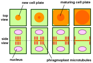
The phragmoplast is a plant cell specific structure that forms during late cytokinesis. It serves as a scaffold for cell plate assembly and subsequent formation of a new cell wall separating the two daughter cells. The phragmoplast can only be observed in Phragmoplastophyta, a clade that includes the Coleochaetophyceae, Zygnematophyceae, Mesotaeniaceae, and Embryophyta. Some algae use another type of microtubule array, a phycoplast, during cytokinesis.
In developmental biology, cleavage is the division of cells in the early embryo, following fertilization. The zygotes of many species undergo rapid cell cycles with no significant overall growth, producing a cluster of cells the same size as the original zygote. The different cells derived from cleavage are called blastomeres and form a compact mass called the morula. Cleavage ends with the formation of the blastula.

Preprophase is an additional phase during mitosis in plant cells that does not occur in other eukaryotes such as animals or fungi. It precedes prophase and is characterized by two distinct events:
- The formation of the preprophase band, a dense microtubule ring underneath the plasma membrane.
- The initiation of microtubule nucleation at the nuclear envelope.
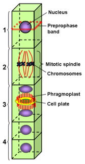
The preprophase band is a microtubule array found in plant cells that are about to undergo cell division and enter the preprophase stage of the plant cell cycle. Besides the phragmosome, it is the first microscopically visible sign that a plant cell is about to enter mitosis. The preprophase band was first observed and described by Jeremy Pickett-Heaps and Donald Northcote at Cambridge University in 1966.

Aurora kinase B is a protein that functions in the attachment of the mitotic spindle to the centromere.

Tetraspora is a genus of green algae in the family Tetrasporaceae of the order Chlamydomonadales, division Chlorophyta. Species of Tetraspora are unicellular green algae that exist in arrangements of four and consist of cells being packaged together in a gelatinous envelope that creates macroscopic colonies. These are primarily freshwater organisms, although there have been few cases where they have been found inhabiting marine environments and even contaminated water bodies. Tetraspora species can be found all around the globe, with the exception of Antarctica. Despite the ubiquitous presence, greatest growth of the genera's species is seen in the polar climatic zones.

Citron Rho-interacting kinase is an enzyme that in humans is encoded by the CIT gene.

An aster is a cellular structure shaped like a star, consisting of a centrosome and its associated microtubules during the early stages of mitosis in an animal cell. Asters do not form during mitosis in plants. Astral rays, composed of microtubules, radiate from the centrosphere and look like a cloud. Astral rays are one variant of microtubule which comes out of the centrosome; others include kinetochore microtubules and polar microtubules.

Binucleated cells are cells that contain two nuclei. This type of cell is most commonly found in cancer cells and may arise from a variety of causes. Binucleation can be easily visualized through staining and microscopy. In general, binucleation has negative effects on cell viability and subsequent mitosis.

Kinesin family member 15 is a protein that in humans is encoded by the KIF15 gene.
Centralspindlin is a motor complex implicated in cell division. It contributes to virtually every step in cytokinesis, It is highly conserved in animal cells as a component of the spindle midzone and midbody. Centralspindlin is required for the assembly of the mitotic spindle as well as for microtubule bundling and anchoring of midbody microtubules to the plasma membrane. This complex is also implicated in tethering the spindle apparatus to the plasma membrane during cytokinesis This interaction permits cleavage furrow ingression. In addition, centralspindlin's interaction with the ESCRT III allows for abscission to occur.
The central spindle is a microtubule based structure, which forms in between segregating chromosomes during anaphase where the two sets of microtubules, emanating from opposite halves of the cell, overlap, and become arranged into antiparallel bundles by various microtubule associated proteins (MAPs) and motor proteins. The central spindle is widely regarded as a key regulating center for cytokinesis, recruiting proteins for successful cleavage furrow positioning and membrane abscission. For these important roles to be achieved successfully the central spindle has to be carefully regulated to control the size of the overlap region, the alignment of those overlaps and the overall length and symmetry of the structure. Without this regulation, signaling faults in cytokinesis can occur, resulting in unequal chromosome segregation or polyploid cells, greatly increasing the risk of cancer.

In molecular biology, an actomyosin contractile ring is a prominent structure during cytokinesis. It forms perpendicular to the axis of the spindle apparatus towards the end of telophase, in which sister chromatids are identically separated at the opposite sides of the spindle forming nuclei. The actomyosin ring follows an orderly sequence of events: identification of the active division site, formation of the ring, constriction of the ring, and disassembly of the ring. It is composed of actin and myosin II bundles, thus the term actomyosin. The actomyosin ring operates in contractile motion, although the mechanism on how or what triggers the constriction is still an evolving topic. Other cytoskeletal proteins are also involved in maintaining the stability of the ring and driving its constriction. Apart from cytokinesis, in which the ring constricts as the cells divide, actomyosin ring constriction has also been found to activate during wound closure. During this process, actin filaments are degraded, preserving the thickness of the ring. After cytokinesis is complete, one of the two daughter cells inherits a remnant known as the midbody ring.

Vampyrella is a genus of amoebae belonging to the vampyrellid cercozoans usually ranging from 30-60 µm. Members of the genus alternate between two life stages: a free-living trophozoite stage and a cyst stage in which mitosis occurs. This taxon has received a great deal of attention due to their peculiar feeding behaviour of perforating the cell wall of algal cells and drawing out the contents for nourishment.
