Protein inhibitor of activated STAT

Last updated

PIAS and the JAK-STAT pathway. Upon stimulation by IL-6, PIAS3 can inhibit transcription activation by activated STAT3. Jakstat pathway PIAS.svg
PIAS and the JAK-STAT pathway. Upon stimulation by IL-6, PIAS3 can inhibit transcription activation by activated STAT3.

Protein inhibitor of activated STAT (PIAS), also known as E3 SUMO-protein ligase PIAS, is a protein that regulates transcription in mammals. PIAS proteins act as transcriptional co-regulators with at least 60 different proteins in order to either activate or repress transcription. The transcription factors STAT, NF-κB, p73, and p53 are among the many proteins that PIAS interacts with.

Contents

The seven proteins that belong to the mammalian PIAS family are encoded by four genes: PIAS1 , PIAS2 (PIASx), PIAS3 , and PIAS4 (PIASy). Apart from PIAS1, each gene encodes two protein isoforms. Homologues of PIAS proteins have been found in other eukaryotes, including Zimp/dPIAS in Drosophila melanogaster and zfPIAS4a in zebrafish. SIZ1 and SIZ2 were two homologues identified in yeast.

PIAS proteins contain each conserved domain and motif of the PIAS protein family, with a few exceptions. The known functions of these domains and motifs are similar among all PIAS protein family members. These functions include acting as E3 SUMO-protein ligases during SUMOylation, which is an important process in transcriptional regulation. Presently, less is known about the higher order structure of PIAS proteins. The three-dimensional protein structures of PIAS2, PIAS3, and SIZ1 have only recently been solved.

PIAS proteins have potential applications in cancer treatment and prevention. They may also play an important role in regulating immune system responses.

Discovery

The discovery of PIAS3 was first published in 1997. The discovery was made while the JAK-STAT pathway was being studied. [1] The discovery of other PIAS proteins, including PIAS1, PIASxα, PIASxβ, and PIASy, was published the following year. [2] The interaction between STATs and PIASs was characterized by the yeast two-hybrid assay. [1] [2] PIAS proteins were named based on their ability to inhibit STAT. For example, PIAS1 inhibited STAT1, [2] and PIAS3 inhibited STAT3. [1]

When it was discovered that PIAS proteins did far more than simply inhibit STATs, it was proposed that the PIAS acronym should stand for Pleiotropic Interactors Associated with SUMO based on their association with SUMO proteins. [3] Additionally, E3 SUMO-protein ligase PIAS is an alternative name for PIAS proteins. [4]

The discovery of PIAS3L, an isoform of PIAS3, was published in 2003. [5] In addition, the discovery of PIASyE6- was published in 2004. It is an isoform of PIASy that doesn't contain exon 6. [6]

Types of PIAS proteins

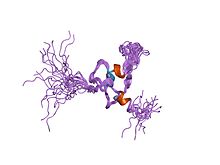
PHD zinc finger domain of SIZ1. SIZ1 is a PIAS protein homologue found in yeast. PDB 1wew EBI.jpg
PHD zinc finger domain of SIZ1. SIZ1 is a PIAS protein homologue found in yeast.

The table below lists the seven known proteins that belong to the mammalian PIAS protein family. [3] [7] Due to alternative splicing, some PIAS protein-encoding genes encode multiple protein products called isoforms. [8] PIAS1 is the only gene of this family that does not encode any isoforms. [3]

GeneEncoded Protein(s)
PIAS1PIAS1
PIAS2 (PIASx)PIASxα, PIASxβ
PIAS3PIAS3, PIAS3L (also known as PIAS3β)
PIAS4 (PIASy)PIASy, PIASyE6-

Homologues

Homologues of PIAS proteins have been found in other eukaryotes, and several are listed below:

Function

PIAS proteins contribute to the control of gene expression, and may be considered transcriptional co-regulators. [14] While PIAS proteins interact with at least 60 different proteins involved in transcription, [15] they are known to act as E3 SUMO-protein ligases. [14] In essence, the RING-finger-like zinc-binding domain of the PIAS protein assists in the attachment of a SUMO protein to the target transcription factor. Attachment of a SUMO protein to the target allows for protein–protein interaction between PIAS and the transcription factor. This interaction can either upregulate or downregulate transcription. [3] [16] For example, the activity of transcription factor p53 was stimulated after it was SUMOylated by PIASy. [17] In contrast, the activity of transcription factor p73 was repressed after it was SUMOylated by PIAS1. [18] One function of PIAS proteins is to relocate transcriptional regulators to different compartments within the nucleus of the cell. [14]

PIAS proteins also play a key role in double-stranded break DNA repair. [19] Exposure to UV light, chemicals, and ionizing radiation can cause DNA damage, and the most detrimental type of DNA damage is a double-stranded break. [19] PIAS1, PIAS3, and PIAS4 have been shown to recruit proteins to the site of the damage and promote repair. [19] [20]

Additionally, PIAS proteins are important transcriptional co-regulators of the JAK/STAT signaling pathway. PIAS protein's interaction with STAT signaling requires tyrosine phosphorylation of STAT proteins. [21] Additionally, PIAS1 binds preferentially to un-methylated STAT1. [21] Although the exact mechanism isn't clear, PIAS1 and PIASy both inhibit STAT1 signaling. [2] [22] PIAS3 was found to specifically inhibit STAT3 signaling after stimulation by the cytokine IL-6. [1] Also, it is known that PIAS1 can inhibit NF-κB activity upon stimulation by the cytokine TNF and the LPS endotoxin. [15]

Structure

The domains (SAP, RLD, AD, S/T) and motifs (PINIT, SIM) found in most protein inhibitors of activated STAT (PIAS) Protein inhibitors of activated STAT (PIAS) domains and motifs.svg
The domains (SAP, RLD, AD, S/T) and motifs (PINIT, SIM) found in most protein inhibitors of activated STAT (PIAS)

The three-dimensional protein structures of PIAS2, [23] PIAS3, [24] and PIAS-like protein SIZ1 [25] were recently solved using X-ray crystallography. The structures of PIAS2 and PIAS3 were listed in the Structural Genomics Consortium in 2012 and 2013, respectively, by A. Dong et al. Details of the SIZ1 structure were published by Ali A. Yunus and Christopher D. Lima in 2009.

Four PIAS domains and two PIAS motifs have been identified. They include the N-terminal scaffold attachment factor-A/B, acinus and PIAS (SAP) domain, the Pro-Ile-Asn-Ile-Thr (PINIT) motif, the RING-finger-like zinc-binding domain (RLD), the highly acidic domain (AD), the SUMO-interacting motif (SIM), and the serine/threonine-rich C-terminal region (S/T). [3] [7] [15] [26]

PIAS protein regions
NameAbbreviationFunction(s)
N-terminal scaffold attachment factor-A/B, acinus and PIAS domainSAPBinds to DNA matrix-attachment regions, proteins (i.e.: p53, nuclear receptors) [3] [7] [15] [27]
Pro-Ile-Asn-Ile-Thr motifPINITnuclear retention [5]
RING-finger-like zinc-binding domainRLDSUMOylation; interaction with other proteins [3]
Highly acidic domainADunknown [7]
SUMO-interacting motifSIMrecognition and interaction with SUMO proteins [3]
Serine/threonine-rich C-terminal regionS/Tunknown [7]

SAP

p53 binding domain of PIAS-1. PDB 1v66 EBI.jpg
p53 binding domain of PIAS-1.

The N-terminal scaffold attachment factor-A/B, acinus and PIAS (SAP) domain is found in all PIAS proteins. [15] It is composed of four alpha helices. [27] It binds to areas of chromatin that are rich in adenine (A) and thymine (T). These A/T rich regions are known as matrix-attachment regions. [28] Once bound, the matrix-attachment regions anchor loops of chromatin to the nuclear matrix. The nuclear matrix is a structure within the nucleus where it is thought that transcription regulation takes place. [7] [15] SAP also binds to p53. [27]

Each SAP domain contains an LXXLL amino acid motif. [15] L = leucine, and X = any amino acid. This motif is used to bind to nuclear receptors. Nuclear receptors are transcription factors that regulate transcription upon ligand binding. [29]

PINIT

The Pro-Ile-Asn-Ile-Thr (PINIT) motif was discovered in PIAS3L, an isoform of PIAS3. PIAS proteins tend to go back and forth between the nucleus and cytosol as they carry out their activities. PINIT is needed to localize PIAS3 and PIAS3L to the nucleus. [5]

PIASy has a slight difference in its PINIT motif: leucine is in place of the second isoleucine (PINLT). Furthermore, the PINIT motif is not found in PIASy isoform PIASyE6-. This isoform, lacking exon 6, is still retained in the nucleus despite lacking the PINIT motif. The reason for this is unknown. [6]

RLD

The RING-finger-like zinc-binding domain is present in all PIAS proteins. RLD is essential for PIAS proteins to function as E3 SUMO-protein ligases. It is also needed for successful interaction with other proteins. Its three dimensional structure is thought to be similar to typical RING finger domains. It contains one histidine residue and five cysteine residues [3]

AD and SIM

The highly acidic domain (AD), present in all PIAS proteins, contains a SUMO-interacting motif (SIM). [15] The SIM motif may be needed for PIAS proteins to accurately recognize and interact with other SUMO proteins. However, it is not needed for E3 SUMO-protein ligase activity to occur. [3] The function of the highly acidic domain is unknown. [7]

S/T

The Serine/threonine-rich C-terminal (S/T) region is not found in all PIAS proteins. PIASy and PIASyE6- are the only members of the PIAS protein family that lack this region. [15] Furthermore, the length of this region varies among PIAS protein isoforms. [3] The function of the S/T region is unknown. [7]

Structural differences between PIAS proteins
Type [3] [7] Amino acid length [3] Protein regions [3] [7]
PIAS1651SAP, PINIT, RLD, AD, SIM, S/T
PIASxα572SAP, PINIT, RLD, AD, SIM, S/T
PIASxβ621SAP, PINIT, RLD, AD, SIM, S/T
PIAS3593SAP, PINIT, RLD, AD, SIM, S/T
PIAS3L628SAP, PINIT, RLD, AD, SIM, S/T
PIASy510SAP, PINIT, RLD, AD
PIASyE6-467SAP, RLD, AD

Potential applications

Defects in the DNA repair system lead to a predisposition for developing cancer. At least some of the PIAS proteins are implicated in DNA repair, and specifically in enhancing repair of double-stranded breaks. In cell culture, overexpression of PIAS3 demonstrated an increased resistance of HeLa cells to ionizing radiation. [19] This indicates a significant role for PIAS3 in DNA repair. [19] Additionally, overexpression of PIAS3 inhibited human lung cancer cell growth in vitro and rendered cancer cells up to twelve times more sensitive to chemotherapeutic drugs. [30] While inhibition of PIAS by siRNAs led cancer cells to accelerate cell proliferation and demonstrate higher levels of resistance to chemotherapy drugs. In a study of human brain tissue samples from glioblastoma multiforme patients, PIAS3 expression was found to be reduced compared to the control brain tissue. [31] Inhibition of PIAS3 resulted in increased glioblastoma propagation, while PIAS3 overexpression inhibited STAT-3 signaling and cell proliferation . Furthermore, patients with higher levels of BRCA1, PIAS1, and PIAS4 survived for a longer period of time in a retrospective study of advanced gastric cancer patients. [32]

Continuous activation of the JAK-STAT pathway can cause cancer in humans as well as less complex organisms such as Drosophila. [33] Given the preliminary evidence and their effects on important signaling pathways involved in cancer, PIAS proteins may be interesting targets for the development of treatments for cancers or as sensitizers for chemotherapeutic drugs and radiation in BRCA-deficient cancers. [19] [30]

In addition to its importance in various cancers, the JAK-STAT signaling pathway plays an important part in the human immune response and in particular with regards to adaptive immunity. [34] Clinical proof of concept for the use of JAK inhibitors for treatment of autoimmune and inflammatory disease has been demonstrated by Pfizer's tofacitinib, a JAK inhibitor recently approved in the US for the treatment of rheumatoid arthritis. [35] Additionally, tofacitinib is currently being studied for the treatment of ankylosing spondylitis, psoriatic arthritis, psoriasis, atopic dermatitis, and inflammatory bowel disease. [36]

Furthermore, STAT1 and STAT2 are essential factors in the cellular antiviral and adaptive immune defenses. [37] PIAS proteins and other regulators are necessary for homeostasis and for fine tuning the immune response. [38] PIAS proteins regulate STAT transcription through several mechanisms, and genetic studies in rodents have shown that PIAS1 plays an important physiological role in STAT1 regulation. Many of the 60 proteins that PIAS protein family is believed to interact with are immune regulatory factors. [15]

Related Research Articles

<span class="mw-page-title-main">Philadelphia chromosome</span> Genetic abnormality in leukemia cancer cells

The Philadelphia chromosome or Philadelphia translocation (Ph) is a specific genetic abnormality in chromosome 22 of leukemia cancer cells. This chromosome is defective and unusually short because of reciprocal translocation, t(9;22)(q34;q11), of genetic material between chromosome 9 and chromosome 22, and contains a fusion gene called BCR-ABL1. This gene is the ABL1 gene of chromosome 9 juxtaposed onto the breakpoint cluster region BCR gene of chromosome 22, coding for a hybrid protein: a tyrosine kinase signaling protein that is "always on", causing the cell to divide uncontrollably by interrupting the stability of the genome and impairing various signaling pathways governing the cell cycle.

<span class="mw-page-title-main">Paracrine signaling</span>

Paracrine signaling is a form of cell signaling, a type of cellular communication in which a cell produces a signal to induce changes in nearby cells, altering the behaviour of those cells. Signaling molecules known as paracrine factors diffuse over a relatively short distance, as opposed to cell signaling by endocrine factors, hormones which travel considerably longer distances via the circulatory system; juxtacrine interactions; and autocrine signaling. Cells that produce paracrine factors secrete them into the immediate extracellular environment. Factors then travel to nearby cells in which the gradient of factor received determines the outcome. However, the exact distance that paracrine factors can travel is not certain.

The JAK-STAT signaling pathway is a chain of interactions between proteins in a cell, and is involved in processes such as immunity, cell division, cell death, and tumour formation. The pathway communicates information from chemical signals outside of a cell to the cell nucleus, resulting in the activation of genes through the process of transcription. There are three key parts of JAK-STAT signalling: Janus kinases (JAKs), signal transducer and activator of transcription proteins (STATs), and receptors. Disrupted JAK-STAT signalling may lead to a variety of diseases, such as skin conditions, cancers, and disorders affecting the immune system.
<span class="mw-page-title-main">STAT protein</span> Family of intracellular transcription factors

Members of the signal transducer and activator of transcription (STAT) protein family are intracellular transcription factors that mediate many aspects of cellular immunity, proliferation, apoptosis and differentiation. They are primarily activated by membrane receptor-associated Janus kinases (JAK). Dysregulation of this pathway is frequently observed in primary tumors and leads to increased angiogenesis which enhances the survival of tumors and immunosuppression. Gene knockout studies have provided evidence that STAT proteins are involved in the development and function of the immune system and play a role in maintaining immune tolerance and tumor surveillance.

The prolactin receptor (PRLR) is a type I cytokine receptor encoded in humans by the PRLR gene on chromosome 5p13-14. It is the receptor for prolactin (PRL). The PRLR can also bind to and be activated by growth hormone (GH) and human placental lactogen (hPL). The PRLR is expressed in the mammary glands, pituitary gland, and other tissues. It plays an important role in lobuloalveolar development of the mammary glands during pregnancy and in lactation.
<span class="mw-page-title-main">SUMO protein</span> Family of proteins which attach to other proteins to modify them

In molecular biology, SUMOproteins are a family of small proteins that are covalently attached to and detached from other proteins in cells to modify their function. This process is called SUMOylation. SUMOylation is a post-translational modification involved in various cellular processes, such as nuclear-cytosolic transport, transcriptional regulation, apoptosis, protein stability, response to stress, and progression through the cell cycle.

<span class="mw-page-title-main">Mothers against decapentaplegic homolog 7</span> Protein-coding gene in the species Homo sapiens

Mothers against decapentaplegic homolog 7 or SMAD7 is a protein that in humans is encoded by the SMAD7 gene.

<span class="mw-page-title-main">STAT5</span> Protein family

Signal transducer and activator of transcription 5 (STAT5) refers to two highly related proteins, STAT5A and STAT5B, which are part of the seven-membered STAT family of proteins. Though STAT5A and STAT5B are encoded by separate genes, the proteins are 90% identical at the amino acid level. STAT5 proteins are involved in cytosolic signalling and in mediating the expression of specific genes. Aberrant STAT5 activity has been shown to be closely connected to a wide range of human cancers, and silencing this aberrant activity is an area of active research in medicinal chemistry.

<span class="mw-page-title-main">STAT3</span> Protein-coding gene in the species Homo sapiens

Signal transducer and activator of transcription 3 (STAT3) is a transcription factor which in humans is encoded by the STAT3 gene. It is a member of the STAT protein family.

<span class="mw-page-title-main">STAT1</span> Transcription factor and coding gene in humans

Signal transducer and activator of transcription 1 (STAT1) is a transcription factor which in humans is encoded by the STAT1 gene. It is a member of the STAT protein family.

<span class="mw-page-title-main">STAT4</span> Protein-coding gene in the species Homo sapiens

Signal transducer and activator of transcription 4 (STAT4) is a transcription factor belonging to the STAT protein family, composed of STAT1, STAT2, STAT3, STAT4, STAT5A, STAT5B, STAT6. STAT proteins are key activators of gene transcription which bind to DNA in response to cytokine gradient. STAT proteins are a common part of Janus kinase (JAK)- signalling pathways, activated by cytokines.STAT4 is required for the development of Th1 cells from naive CD4+ T cells and IFN-γ production in response to IL-12. There are two known STAT4 transcripts, STAT4α and STAT4β, differing in the levels of interferon-gamma production downstream.

<span class="mw-page-title-main">Promyelocytic leukemia protein</span> Protein-coding gene in the species Homo sapiens

Promyelocytic leukemia protein (PML) is the protein product of the PML gene. PML protein is a tumor suppressor protein required for the assembly of a number of nuclear structures, called PML-nuclear bodies, which form amongst the chromatin of the cell nucleus. These nuclear bodies are present in mammalian nuclei, at about 1 to 30 per cell nucleus. PML-NBs are known to have a number of regulatory cellular functions, including involvement in programmed cell death, genome stability, antiviral effects and controlling cell division. PML mutation or loss, and the subsequent dysregulation of these processes, has been implicated in a variety of cancers.

<span class="mw-page-title-main">STAT5A</span> Protein-coding gene in the species Homo sapiens

Signal transducer and activator of transcription 5A is a protein that in humans is encoded by the STAT5A gene. STAT5A orthologs have been identified in several placentals for which complete genome data are available.

<span class="mw-page-title-main">PIAS4</span> Protein-coding gene in the species Homo sapiens

E3 SUMO-protein ligase PIAS4 is one of several protein inhibitor of activated STAT (PIAS) proteins. It is also known as protein inhibitor of activated STAT protein gamma, and is an enzyme that in humans is encoded by the PIAS4 gene.

<span class="mw-page-title-main">PIAS1</span> Protein-coding gene in the species Homo sapiens

E3 SUMO-protein ligase PIAS1 is an enzyme that in humans is encoded by the PIAS1 gene.
<span class="mw-page-title-main">Protein inhibitor of activated STAT2</span> Protein-coding gene in the species Homo sapiens

E3 SUMO-protein ligase PIAS2 is an enzyme that in humans is encoded by the PIAS2 gene.

<span class="mw-page-title-main">PIAS3</span> Protein-coding gene in the species Homo sapiens

E3 SUMO-protein ligase PIAS3 is an enzyme that in humans is encoded by the PIAS3 gene.

<span class="mw-page-title-main">IRF5</span> Protein-coding gene in the species Homo sapiens

Interferon regulatory factor 5 is a protein that in humans is encoded by the IRF5 gene. The IRF family is a group of transcription factors that are involved in signaling for virus responses in mammals along with regulation of certain cellular functions.

<span class="mw-page-title-main">RNF14</span> Protein-coding gene in the species Homo sapiens

E3 ubiquitin-protein ligase RNF14 is an enzyme that in humans is encoded by the RNF14 gene.

<span class="mw-page-title-main">RNF128</span> Protein-coding gene in the species Homo sapiens

E3 ubiquitin-protein ligase RNF128 is an enzyme that in humans is encoded by the RNF128 gene.

References

  1. 1 2 3 4 Chung CD, Liao J, Liu B, Rao X, Jay P, Berta P, Shuai K (December 1997). "Specific inhibition of Stat3 signal transduction by PIAS3". Science. 278 (5344): 1803–5. Bibcode:1997Sci...278.1803C. doi:10.1126/science.278.5344.1803. PMID   9388184.
  2. 1 2 3 4 Liu B, Liao J, Rao X, Kushner SA, Chung CD, Chang DD, Shuai K (September 1998). "Inhibition of Stat1-mediated gene activation by PIAS1". Proceedings of the National Academy of Sciences of the United States of America. 95 (18): 10626–31. Bibcode:1998PNAS...9510626L. doi: 10.1073/pnas.95.18.10626 . PMC   27945 . PMID   9724754.
  3. 1 2 3 4 5 6 7 8 9 10 11 12 13 14 Rytinki MM, Kaikkonen S, Pehkonen P, Jääskeläinen T, Palvimo JJ (September 2009). "PIAS proteins: pleiotropic interactors associated with SUMO". Cellular and Molecular Life Sciences. 66 (18): 3029–41. doi:10.1007/s00018-009-0061-z. PMID   19526197. S2CID   5619331.
  4. Van Itallie CM, Mitic LL, Anderson JM (July 2012). "SUMOylation of claudin-2". Annals of the New York Academy of Sciences. 1258 (1): 60–4. Bibcode:2012NYASA1258...60V. doi:10.1111/j.1749-6632.2012.06541.x. PMID   22731716. S2CID   45684330.
  5. 1 2 3 Duval D, Duval G, Kedinger C, Poch O, Boeuf H (November 2003). "The 'PINIT' motif, of a newly identified conserved domain of the PIAS protein family, is essential for nuclear retention of PIAS3L". FEBS Letters. 554 (1–2): 111–8. doi: 10.1016/s0014-5793(03)01116-5 . PMID   14596924. S2CID   23261716.
  6. 1 2 Wong KA, Kim R, Christofk H, Gao J, Lawson G, Wu H (June 2004). "Protein inhibitor of activated STAT Y (PIASy) and a splice variant lacking exon 6 enhance sumoylation but are not essential for embryogenesis and adult life". Molecular and Cellular Biology. 24 (12): 5577–86. doi:10.1128/MCB.24.12.5577-5586.2004. PMC   419860 . PMID   15169916.
  7. 1 2 3 4 5 6 7 8 9 10 Shuai K, Liu B (August 2005). "Regulation of gene-activation pathways by PIAS proteins in the immune system". Nature Reviews. Immunology. 5 (8): 593–605. doi:10.1038/nri1667. PMID   16056253. S2CID   7466028.
  8. University, James D. Watson, Cold Spring Harbor Laboratory, Tania A. Baker, Massachusetts Institute of Technology, Alexander Gann, Cold Spring Harbor Laboratory, Michael Levine, University of California, Berkeley, Richard Losik, Harvard (2014). Molecular biology of the gene (Seventh ed.). Boston: Pearson/CSH Press. p. 469. ISBN   978-0321762436.{{cite book}}: CS1 maint: multiple names: authors list (link)
  9. Mohr SE, Boswell RE (March 1999). "Zimp encodes a homologue of mouse Miz1 and PIAS3 and is an essential gene in Drosophila melanogaster". Gene. 229 (1–2): 109–16. doi:10.1016/s0378-1119(99)00033-5. PMID   10095110.
  10. Betz A, Lampen N, Martinek S, Young MW, Darnell JE (August 2001). "A Drosophila PIAS homologue negatively regulates stat92E". Proceedings of the National Academy of Sciences of the United States of America. 98 (17): 9563–8. Bibcode:2001PNAS...98.9563B. doi: 10.1073/pnas.171302098 . PMC   55492 . PMID   11504941.
  11. Xiong R, Nie L, Xiang LX, Shao JZ (March 2012). "Characterization of a PIAS4 homologue from zebrafish: insights into its conserved negative regulatory mechanism in the TRIF, MAVS, and IFN signaling pathways during vertebrate evolution". Journal of Immunology. 188 (6): 2653–68. doi: 10.4049/jimmunol.1100959 . PMID   22345667. S2CID   207425842.
  12. Johnson ES, Gupta AA (September 2001). "An E3-like factor that promotes SUMO conjugation to the yeast septins". Cell. 106 (6): 735–44. doi: 10.1016/s0092-8674(01)00491-3 . PMID   11572779. S2CID   14375183.
  13. Takahashi Y, Kikuchi Y (October 2005). "Yeast PIAS-type Ull1/Siz1 is composed of SUMO ligase and regulatory domains". The Journal of Biological Chemistry. 280 (43): 35822–8. doi: 10.1074/jbc.M506794200 . PMID   16109721. S2CID   24493405.
  14. 1 2 3 Sharrocks AD (April 2006). "PIAS proteins and transcriptional regulation—more than just SUMO E3 ligases?". Genes & Development. 20 (7): 754–8. doi: 10.1101/gad.1421006 . PMID   16600908.
  15. 1 2 3 4 5 6 7 8 9 10 Shuai K (February 2006). "Regulation of cytokine signaling pathways by PIAS proteins". Cell Research. 16 (2): 196–202. doi: 10.1038/sj.cr.7310027 . PMID   16474434. S2CID   755228.
  16. Geiss-Friedlander R, Melchior F (December 2007). "Concepts in sumoylation: a decade on". Nature Reviews. Molecular Cell Biology. 8 (12): 947–56. doi:10.1038/nrm2293. PMID   18000527. S2CID   30462190.
  17. Bischof O, Schwamborn K, Martin N, Werner A, Sustmann C, Grosschedl R, Dejean A (June 2006). "The E3 SUMO ligase PIASy is a regulator of cellular senescence and apoptosis". Molecular Cell. 22 (6): 783–94. doi:10.1016/j.molcel.2006.05.016. PMID   16793547.
  18. Munarriz E, Barcaroli D, Stephanou A, Townsend PA, Maisse C, Terrinoni A, Neale MH, Martin SJ, Latchman DS, Knight RA, Melino G, De Laurenzi V (December 2004). "PIAS-1 is a checkpoint regulator which affects exit from G1 and G2 by sumoylation of p73". Molecular and Cellular Biology. 24 (24): 10593–610. doi:10.1128/MCB.24.24.10593-10610.2004. PMC   533962 . PMID   15572666.
  19. 1 2 3 4 5 6 Liu S, Fan Z, Geng Z, Zhang H, Ye Q, Jiao S, Xu X (October 2013). "PIAS3 promotes homology-directed repair and distal non-homologous end joining". Oncology Letters. 6 (4): 1045–1048. doi:10.3892/ol.2013.1472. PMC   3796434 . PMID   24137461.
  20. Galanty Y, Belotserkovskaya R, Coates J, Polo S, Miller KM, Jackson SP (December 2009). "Mammalian SUMO E3-ligases PIAS1 and PIAS4 promote responses to DNA double-strand breaks". Nature. 462 (7275): 935–9. Bibcode:2009Natur.462..935G. doi:10.1038/nature08657. PMC   2904806 . PMID   20016603.
  21. 1 2 Heinrich PC, Behrmann I, Haan S, Hermanns HM, Müller-Newen G, Schaper F (August 2003). "Principles of interleukin (IL)-6-type cytokine signalling and its regulation". The Biochemical Journal. 374 (Pt 1): 1–20. doi:10.1042/BJ20030407. PMC   1223585 . PMID   12773095.
  22. Liu B, Gross M, ten Hoeve J, Shuai K (March 2001). "A transcriptional corepressor of Stat1 with an essential LXXLL signature motif". Proceedings of the National Academy of Sciences of the United States of America. 98 (6): 3203–7. Bibcode:2001PNAS...98.3203L. doi: 10.1073/pnas.051489598 . PMC   30631 . PMID   11248056.
  23. Dong, A. "Human protein inhibitor of activated STAT, 2 (E3 SUMO ligase)". Structural Genomics Consortium (SGC). Retrieved 5 May 2014.
  24. Dong, A. "Human protein inhibitor of activated STAT, 3". Structural Genomics Consortium (SGC). Retrieved 5 May 2014.
  25. Yunus AA, Lima CD (September 2009). "Structure of the Siz/PIAS SUMO E3 ligase Siz1 and determinants required for SUMO modification of PCNA". Molecular Cell. 35 (5): 669–82. doi:10.1016/j.molcel.2009.07.013. PMC   2771690 . PMID   19748360.
  26. Palvimo JJ (December 2007). "PIAS proteins as regulators of small ubiquitin-related modifier (SUMO) modifications and transcription". Biochemical Society Transactions. 35 (Pt 6): 1405–8. doi:10.1042/BST0351405. PMID   18031232.
  27. 1 2 3 Okubo S, Hara F, Tsuchida Y, Shimotakahara S, Suzuki S, Hatanaka H, Yokoyama S, Tanaka H, Yasuda H, Shindo H (July 2004). "NMR structure of the N-terminal domain of SUMO ligase PIAS1 and its interaction with tumor suppressor p53 and A/T-rich DNA oligomers". The Journal of Biological Chemistry. 279 (30): 31455–61. doi: 10.1074/jbc.M403561200 . PMID   15133049. S2CID   9187033.
  28. Aravind L, Koonin EV (March 2000). "SAP – a putative DNA-binding motif involved in chromosomal organization". Trends in Biochemical Sciences. 25 (3): 112–4. doi:10.1016/s0968-0004(99)01537-6. PMID   10694879.
  29. Glass CK, Rosenfeld MG (January 2000). "The coregulator exchange in transcriptional functions of nuclear receptors". Genes & Development. 14 (2): 121–41. doi: 10.1101/gad.14.2.121 . PMID   10652267. S2CID   12793980.
  30. 1 2 Ogata Y, Osaki T, Naka T, Iwahori K, Furukawa M, Nagatomo I, Kijima T, Kumagai T, Yoshida M, Tachibana I, Kawase I (October 2006). "Overexpression of PIAS3 suppresses cell growth and restores the drug sensitivity of human lung cancer cells in association with PI3-K/Akt inactivation". Neoplasia. 8 (10): 817–25. doi:10.1593/neo.06409. PMC   1715929 . PMID   17032498.
  31. Brantley EC, Nabors LB, Gillespie GY, Choi YH, Palmer CA, Harrison K, Roarty K, Benveniste EN (August 2008). "Loss of protein inhibitors of activated STAT-3 expression in glioblastoma multiforme tumors: implications for STAT-3 activation and gene expression". Clinical Cancer Research. 14 (15): 4694–704. doi:10.1158/1078-0432.CCR-08-0618. PMC   3886729 . PMID   18676737.
  32. Wei J, Costa C, Ding Y, Zou Z, Yu L, Sanchez JJ, Qian X, Chen H, Gimenez-Capitan A, Meng F, Moran T, Benlloch S, Taron M, Rosell R, Liu B (October 2011). "mRNA expression of BRCA1, PIAS1, and PIAS4 and survival after second-line docetaxel in advanced gastric cancer". Journal of the National Cancer Institute. 103 (20): 1552–6. doi:10.1093/jnci/djr326. PMID   21862729.
  33. Amoyel M, Anderson AM, Bach EA (April 2014). "JAK/STAT pathway dysregulation in tumors: a Drosophila perspective". Seminars in Cell & Developmental Biology. 28: 96–103. doi:10.1016/j.semcdb.2014.03.023. PMC   4037387 . PMID   24685611.
  34. Liongue C, O'Sullivan LA, Trengove MC, Ward AC (2012). "Evolution of JAK-STAT pathway components: mechanisms and role in immune system development". PLOS ONE. 7 (3): e32777. Bibcode:2012PLoSO...732777L. doi: 10.1371/journal.pone.0032777 . PMC   3296744 . PMID   22412924.
  35. "FDA Press Release Regarding Approval of Tofacitinib". Food and Drug Administration . Retrieved 6 May 2014.
  36. "Pfizer's Product Pipeline" . Retrieved 6 May 2014.
  37. Au-Yeung N, Mandhana R, Horvath CM (July 2013). "Transcriptional regulation by STAT1 and STAT2 in the interferon JAK-STAT pathway". JAK-STAT. 2 (3): e23931. doi:10.4161/jkst.23931. PMC   3772101 . PMID   24069549.
  38. Morales JK, Falanga YT, Depcrynski A, Fernando J, Ryan JJ (December 2010). "Mast cell homeostasis and the JAK-STAT pathway". Genes and Immunity. 11 (8): 599–608. doi:10.1038/gene.2010.35. PMC   3099592 . PMID   20535135.