Interferon regulatory factors

Last updated
Interferon regulatory factor transcription factor
PDB 1irf EBI.jpg
interferon regulatory factor-2 dna binding domain, nmr, minimized average structure
Identifiers
SymbolIRF
Pfam PF00605
InterPro IPR001346
SCOP2 1if1 / SCOPe / SUPFAM
Available protein structures:
Pfam   structures / ECOD  
PDB RCSB PDB; PDBe; PDBj
PDBsum structure summary

Interferon regulatory factors (IRF) are proteins which regulate transcription of interferons (see regulation of gene expression). [1] Interferon regulatory factors contain a conserved N-terminal region of about 120 amino acids, which folds into a structure that binds specifically to the IRF-element (IRF-E) motifs, which is located upstream of the interferon genes. [2] Some viruses have evolved defense mechanisms that regulate and interfere with IRF functions to escape the host immune system. [3] For instance, the remaining parts of the interferon regulatory factor sequence vary depending on the precise function of the protein. [2] The Kaposi sarcoma herpesvirus, KSHV, [4] is a cancer virus that encodes four different IRF-like genes; [5] including vIRF1, [6] which is a transforming oncoprotein that inhibits type 1 interferon activity. [7] In addition, the expression of IRF genes is under epigenetic regulation by promoter DNA methylation. [8]

Contents

Role in IFN signaling

IRFs primarily regulate type I IFNs in the host after pathogen invasion and are considered the crucial mediators of an antiviral response. Following a viral infection, pathogens are detected by Pattern Recognition Receptors (PRRs), including various types of Toll-like Receptors (TLR) and cytosolic PRRs, in the host cell. [3] The downstream signaling pathways from PRR activation phosphorylate ubiquitously expressed IRFs (IRF1, IRF3, and IRF7) through IRF kinases, such as TANK-binding kinase 1 (TBK1). [9] Phosphorylated IRFs are translocated to the nucleus where they bind to IRF-E motifs and activate the transcription of Type I IFNs. In addition to IFNs, IRF1 and IRF5 has been found to induce transcription of pro-inflammatory cytokines.

Some IFNs like IRF2 and IRF4 regulate the activation of IFNs and pro-inflammatory cytokines through inhibition. IRF2 contains a repressor region that downregulates expression of type I IFNs. IRF4 competes with IRF5, and inhibits its sustained activity. [3]

Role in immune cell development

In addition to the signal transduction functions of IRFs in innate immune responses, multiple IRFs (IRF1, IRF2, IRF4, and IRF8) play essential roles in the development of immune cells, including dendritic, myeloid, natural killer (NK), B, and T cells. [3]

Dendritic cells (DC) are a group of heterogeneous cells that can be divided into different subsets with distinct functions and developmental programs. IRF4 and IRF8 specify and direct the differentiation of different subsets of DCs by stimulating subset-specific gene expression. [3] For example, IRF4 is required for the generation of CD4 + DCs, whereas IRF8 is essential for CD8α + DCs. In addition to IRF4 and IRF8, IRF1 and IRF2 are also involved in DC subset development.

IRF8 has also been implicated in the promotion of macrophage development from common myeloid progenitors (CMPs) and the inhibition of granulocytic differentiation during the divergence of granulocytes and monocytes.

IRF8 and IRF4 are also involved in the regulation of B and T-cell development at multiple stages. IRF8 and IRF4 function redundantly to drive common lymphoid progenitors (CLPs) to B-cell lineage. IRF8 and IRF4 are also required in the regulation of germinal center (GC) B cell differentiation.

Role in diseases

IRFs are critical regulators of immune responses and immune cell development, and abnormalities in IRF expression and function have been linked to numerous diseases. Due to their critical role in IFN type I activation, IRFs are implicated in autoimmune diseases that are linked to activation of IFN type I system, such as systemic lupus erythematosus (SLE). [10] Accumulating evidence also indicates that IRFs play a major role in the regulation of cellular responses linked to oncogenesis. [11] In addition to autoimmune diseases and cancers, IRFs are also found to be involved in the pathogenesis of metabolic, cardiovascular, and neurological diseases, such as hepatic steatosis, diabetes, cardiac hypertrophy, atherosclerosis, and stroke. [3]

Genes

See also

Related Research Articles

<span class="mw-page-title-main">Interferon</span> Signaling proteins released by host cells in response to the presence of pathogens

Interferons are a group of signaling proteins made and released by host cells in response to the presence of several viruses. In a typical scenario, a virus-infected cell will release interferons causing nearby cells to heighten their anti-viral defenses.

<span class="mw-page-title-main">T helper cell</span> Type of immune cell

The T helper cells (Th cells), also known as CD4+ cells or CD4-positive cells, are a type of T cell that play an important role in the adaptive immune system. They aid the activity of other immune cells by releasing cytokines. They are considered essential in B cell antibody class switching, breaking cross-tolerance in dendritic cells, in the activation and growth of cytotoxic T cells, and in maximizing bactericidal activity of phagocytes such as macrophages and neutrophils. CD4+ cells are mature Th cells that express the surface protein CD4. Genetic variation in regulatory elements expressed by CD4+ cells determines susceptibility to a broad class of autoimmune diseases.

<span class="mw-page-title-main">Kaposi's sarcoma-associated herpesvirus</span> Species of virus

Kaposi's sarcoma-associated herpesvirus (KSHV) is the ninth known human herpesvirus; its formal name according to the International Committee on Taxonomy of Viruses (ICTV) is Human gammaherpesvirus 8, or HHV-8 in short. Like other herpesviruses, its informal names are used interchangeably with its formal ICTV name. This virus causes Kaposi's sarcoma, a cancer commonly occurring in AIDS patients, as well as primary effusion lymphoma, HHV-8-associated multicentric Castleman's disease and KSHV inflammatory cytokine syndrome. It is one of seven currently known human cancer viruses, or oncoviruses. Even after many years since the discovery of KSHV/HHV8, there is no known cure for KSHV associated tumorigenesis.

<span class="mw-page-title-main">Primary effusion lymphoma</span> Medical condition

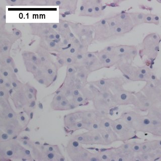
Primary effusion lymphoma (PEL) is classified as a diffuse large B cell lymphoma. It is a rare malignancy of plasmablastic cells that occurs in individuals that are infected with the Kaposi's sarcoma-associated herpesvirus. Plasmablasts are immature plasma cells, i.e. lymphocytes of the B-cell type that have differentiated into plasmablasts but because of their malignant nature do not differentiate into mature plasma cells but rather proliferate excessively and thereby cause life-threatening disease. In PEL, the proliferating plasmablastoid cells commonly accumulate within body cavities to produce effusions, primarily in the pleural, pericardial, or peritoneal cavities, without forming a contiguous tumor mass. In rare cases of these cavitary forms of PEL, the effusions develop in joints, the epidural space surrounding the brain and spinal cord, and underneath the capsule which forms around breast implants. Less frequently, individuals present with extracavitary primary effusion lymphomas, i.e., solid tumor masses not accompanied by effusions. The extracavitary tumors may develop in lymph nodes, bone, bone marrow, the gastrointestinal tract, skin, spleen, liver, lungs, central nervous system, testes, paranasal sinuses, muscle, and, rarely, inside the vasculature and sinuses of lymph nodes. As their disease progresses, however, individuals with the classical effusion-form of PEL may develop extracavitary tumors and individuals with extracavitary PEL may develop cavitary effusions.

The type III interferon group is a group of anti-viral cytokines, that consists of four IFN-λ (lambda) molecules called IFN-λ1, IFN-λ2, IFN-λ3, and IFN-λ4. They were discovered in 2003. Their function is similar to that of type I interferons, but is less intense and serves mostly as a first-line defense against viruses in the epithelium.

<span class="mw-page-title-main">STAT1</span> Transcription factor and coding gene in humans

Signal transducer and activator of transcription 1 (STAT1) is a transcription factor which in humans is encoded by the STAT1 gene. It is a member of the STAT protein family.

<span class="mw-page-title-main">IRF3</span> Protein-coding gene in the species Homo sapiens

Interferon regulatory factor 3, also known as IRF3, is an interferon regulatory factor.

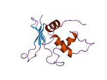
NSP1 (NS53), the product of rotavirus gene 5, is a nonstructural RNA-binding protein that contains a cysteine-rich region and is a component of early replication intermediates. RNA-folding predictions suggest that this region of the NSP1 mRNA can interact with itself, producing a stem-loop structure similar to that found near the 5'-terminus of the NSP1 mRNA.

<span class="mw-page-title-main">IRF2</span> Protein-coding gene in the species Homo sapiens

Interferon regulatory factor 2 is a protein that in humans is encoded by the IRF2 gene.

<span class="mw-page-title-main">RIG-I</span> Mammalian protein found in humans

RIG-I is a cytosolic pattern recognition receptor (PRR) that can mediate induction of a type-I interferon (IFN1) response. RIG-I is an essential molecule in the innate immune system for recognizing cells that have been infected with a virus. These viruses can include West Nile virus, Japanese Encephalitis virus, influenza A, Sendai virus, flavivirus, and coronaviruses.

<span class="mw-page-title-main">IRF1</span> Protein-coding gene in the species Homo sapiens

Interferon regulatory factor 1 is a protein that in humans is encoded by the IRF1 gene.

<span class="mw-page-title-main">Interferon alpha-1</span> Protein-coding gene in the species Homo sapiens

Interferon alpha-1 is a protein that in humans is encoded by the IFNA1 gene.

<span class="mw-page-title-main">IFNA2</span> Mammalian protein found in Homo sapiens

Interferon alpha-2 is a protein that in humans is encoded by the IFNA2 gene.

<span class="mw-page-title-main">IRF4</span> Protein-coding gene in the species Homo sapiens

Interferon regulatory factor 4 (IRF4) also known as MUM1 is a protein that in humans is encoded by the IRF4 gene. IRF4 functions as a key regulatory transcription factor in the development of human immune cells. The expression of IRF4 is essential for the differentiation of T lymphocytes and B lymphocytes as well as certain myeloid cells. Dysregulation of the IRF4 gene can result in IRF4 functioning either as an oncogene or a tumor-suppressor, depending on the context of the modification.

<span class="mw-page-title-main">IRF5</span> Protein-coding gene in the species Homo sapiens

Interferon regulatory factor 5 is a protein that in humans is encoded by the IRF5 gene. The IRF family is a group of transcription factors that are involved in signaling for virus responses in mammals along with regulation of certain cellular functions.

<span class="mw-page-title-main">PRDM1</span> Protein-coding gene in the species Homo sapiens

PR domain zinc finger protein 1, or B lymphocyte-induced maturation protein-1 (BLIMP-1), is a protein in humans encoded by the gene PRDM1 located on chromosome 6q21. BLIMP-1 is considered a 'master regulator' of hematopoietic stem cells, and plays a critical role in the development of plasma B cells, T cells, dendritic cells (DCs), macrophages, and osteoclasts. Pattern Recognition Receptors (PRRs) can activate BLIMP-1, both as a direct target and through downstream activation. BLIMP-1 is a transcription factor that triggers expression of many downstream signaling cascades. As a fine-tuned and contextual rheostat of the immune system, BLIMP-1 up- or down-regulates immune responses depending on the precise scenarios. BLIMP-1 is highly expressed in exhausted T-cells – clones of dysfunctional T-cells with diminished functions due to chronic immune response against cancer, viral infections, or organ transplant.

<span class="mw-page-title-main">IRF8</span> Protein-coding gene in the species Homo sapiens

Interferon regulatory factor 8 (IRF8) also known as interferon consensus sequence-binding protein (ICSBP), is a protein that in humans is encoded by the IRF8 gene. IRF8 is a transcription factor that plays critical roles in the regulation of lineage commitment and in myeloid cell maturation including the decision for a common myeloid progenitor (CMP) to differentiate into a monocyte precursor cell.

<span class="mw-page-title-main">CD200</span> Protein-coding gene in the species Homo sapiens

OX-2 membrane glycoprotein, also named CD200 is a human protein encoded by the CD200 gene. CD200 gene is in human located on chromosome 3 in proximity to genes encoding other B7 proteins CD80/CD86. In mice CD200 gene is on chromosome 16.

<span class="mw-page-title-main">Kaposi's sarcoma</span> Cancer of the skin, integumentary lymph nodes, or other organs

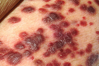
Kaposi's sarcoma (KS) is a type of cancer that can form masses in the skin, in lymph nodes, in the mouth, or in other organs. The skin lesions are usually painless, purple and may be flat or raised. Lesions can occur singly, multiply in a limited area, or may be widespread. Depending on the sub-type of disease and level of immune suppression, KS may worsen either gradually or quickly. Except for Classical KS where there is generally no immune suppression, KS is caused by a combination of immune suppression and infection by Human herpesvirus 8.

Type 1 regulatory cells or Tr1 (TR1) cells are a class of regulatory T cells participating in peripheral immunity as a subsets of CD4+ T cells. Tr1 cells regulate tolerance towards antigens of any origin. Tr1 cells are self or non-self antigen specific and their key role is to induce and maintain peripheral tolerance and suppress tissue inflammation in autoimmunity and graft vs. host disease.

References

  1. Paun A, Pitha PM (2007). "The IRF family, revisited". Biochimie. 89 (6–7): 744–53. doi:10.1016/j.biochi.2007.01.014. PMC   2139905 . PMID   17399883.
  2. 1 2 Weisz A, Marx P, Sharf R, Appella E, Driggers PH, Ozato K, Levi BZ (December 1992). "Human interferon consensus sequence binding protein is a negative regulator of enhancer elements common to interferon-inducible genes". The Journal of Biological Chemistry. 267 (35): 25589–96. doi: 10.1016/S0021-9258(19)74081-2 . PMID   1460054.
  3. 1 2 3 4 5 6 Zhao GN, Jiang DS, Li H (February 2015). "Interferon regulatory factors: at the crossroads of immunity, metabolism, and disease". Biochimica et Biophysica Acta (BBA) - Molecular Basis of Disease. 1852 (2): 365–78. doi: 10.1016/j.bbadis.2014.04.030 . PMID   24807060.
  4. Chang Y, Cesarman E, Pessin MS, Lee F, Culpepper J, Knowles DM, Moore PS (December 1994). "Identification of herpesvirus-like DNA sequences in AIDS-associated Kaposi's sarcoma". Science. 266 (5192): 1865–9. Bibcode:1994Sci...266.1865C. doi:10.1126/science.7997879. PMID   7997879. S2CID   29977325.
  5. Offermann MK (2007). "Kaposi Sarcoma Herpesvirus-Encoded Interferon Regulator Factors". Kaposi Sarcoma Herpesvirus: New Perspectives. Current Topics in Microbiology and Immunology. Vol. 312. pp. 185–209. doi:10.1007/978-3-540-34344-8_7. ISBN   978-3-540-34343-1. PMID   17089798.
  6. Moore PS, Boshoff C, Weiss RA, Chang Y (December 1996). "Molecular mimicry of human cytokine and cytokine response pathway genes by KSHV". Science. 274 (5293): 1739–44. Bibcode:1996Sci...274.1739M. doi:10.1126/science.274.5293.1739. PMID   8939871. S2CID   29713179.
  7. Gao SJ, Boshoff C, Jayachandra S, Weiss RA, Chang Y, Moore PS (October 1997). "KSHV ORF K9 (vIRF) is an oncogene which inhibits the interferon signaling pathway". Oncogene. 15 (16): 1979–85. doi: 10.1038/sj.onc.1201571 . PMID   9365244.
  8. Rotondo JC, Borghi A, Selvatici R, Magri E, Bianchini E, Montinari E, et al. (August 2016). "Hypermethylation-Induced Inactivation of the IRF6 Gene as a Possible Early Event in Progression of Vulvar Squamous Cell Carcinoma Associated With Lichen Sclerosus". JAMA Dermatology. 152 (8): 928–33. doi:10.1001/jamadermatol.2016.1336. PMID   27223861.
  9. Shah, Masaud; Choi, Sangdun (2016), "Interferon Regulatory Factor", in Choi, Sangdun (ed.), Encyclopedia of Signaling Molecules, New York, NY: Springer, pp. 1–10, doi: 10.1007/978-1-4614-6438-9_101496-1 , ISBN   978-1-4614-6438-9
  10. Santana-de Anda K, Gómez-Martín D, Díaz-Zamudio M, Alcocer-Varela J (December 2011). "Interferon regulatory factors: beyond the antiviral response and their link to the development of autoimmune pathology". Autoimmunity Reviews. 11 (2): 98–103. doi:10.1016/j.autrev.2011.08.006. PMID   21872684.
  11. Yanai H, Negishi H, Taniguchi T (November 2012). "The IRF family of transcription factors: Inception, impact and implications in oncogenesis". Oncoimmunology. 1 (8): 1376–1386. doi: 10.4161/onci.22475 . PMC   3518510 . PMID   23243601.
This article incorporates text from the public domain Pfam and InterPro: IPR001346