TBX2

Last updated
TBX2
Protein TBX2 PDB 1h6f.png
Identifiers
Aliases TBX2 , T-box 2, VETD, T-box transcription factor 2
External IDs OMIM: 600747 MGI: 98494 HomoloGene: 38123 GeneCards: TBX2
Orthologs
SpeciesHumanMouse
Entrez
Ensembl
UniProt
RefSeq (mRNA)

NM_005994

NM_009324

RefSeq (protein)

NP_005985

NP_033350

Location (UCSC) Chr 17: 61.4 – 61.41 Mb Chr 11: 85.72 – 85.73 Mb
PubMed search [3] [4]
Wikidata
View/Edit Human View/Edit Mouse

T-box transcription factor 2 Tbx2 is a transcription factor that is encoded by the Tbx2 gene on chromosome 17q21-22 in humans. [5] [6] [7] This gene is a member of a phylogenetically conserved family of genes that share a common DNA-binding domain, the T-box. Tbx2 and Tbx3 are the only T-box transcription factors that act as transcriptional repressors rather than transcriptional activators, and are closely related in terms of development and tumorigenesis. [8] This gene plays a significant role in embryonic and fetal development through control of gene expression, and also has implications in various cancers. Tbx2 is associated with numerous signaling pathways, BMP, TGFβ, Wnt, and FGF, which allow for patterning and proliferation during organogenesis in fetal development. [8]

Contents

Role in development

The molecule Tbx-2 is a transcription factor in the T box transcription factor family. Tbx2 helps form the outflow tract and atrioventricular canal. Tbx2 can repress genes as well as being competitors that take over binding sites. It also plays a role in cancer because it will suppress cell growth and supports invasiveness. In human melanoma, the expression of endogenous Tbx 2 is shown to help reduce the growth of melanomas. It has also been shown that overexpression of Tbx2 can lead to breast cancer. Tbx2 has shown septal defects of the outflow tract, and this has been shown using a knockout mouse. The knockout mouse is a mouse in which the gene is inactivated in order to study the role of genes. Tbx 2 also helps in regulating the cell cycle. This was first shown when Tbx2 was found in a chromosomal region that is often mutated in ovarian cancer and pancreatic cancer cells. [9]

During fetal development, the relationship of Tbx2 to FGF, BMP, and Wnt signaling pathways indicates its extensive control in development of various organ systems. It functions predominantly in the patterning of organ development rather than tissue proliferation. Tbx2 has implications in limb development, atrioventricular development of the heart, and development of the anterior brain tissues. [10] [11] [12]

During limb bud development, Shh and FGF signaling stimulate the outgrowth of the limb. At a certain point, Tbx2 concentrations are such that the signaling of Shh and FGF are terminated, halting further progression and outgrowth of the limb development. This occurs directly through Tbx2 repressing the expression of Grem1, creating a negative Grem1 zone, thereby disrupting the outgrowth signaling by Shh and FGF. [10]

Cardiac development is heavily regulated and requires the development of the four cardiac chambers, septum, and various valve components for outflow and inflow. In heart development, Tbx2 is up-regulated by BMP2 to stimulate atrioventricular development. [11] The development of a Tbx2 knockout mouse model allowed for the determination of specific roles of Tbx2 in cardiac development, and scientists determined Tbx2 and Tbx3 to be redundant in much of heart development. [11] Further, the use of these knockout models determined the significance of Tbx2 in the BMP signaling pathway for development of the atrioventricular canal, atrioventricular nodal phenotype, and atrioventricular cushion. [11]

The atrioventricular canal signaling cascade involves the atrial natriuretic factor gene (ANF). This gene is one of the first hallmarks of chamber formation in the developing myocardium. A small fragment within this gene can repress the promoter of cardiac troponin I (cTnI) selectively in the atrioventricular canal. T-box factor and NK2-homeobox factor binding element are involved in the repression of the atrioventricular canal without affecting its chamber activity. Tbx2 forms a complex with Nkx2.5 on the ANF gene to repress its promoter activity, so that the gene's expression is inhibited in the atrioventricular canal during chamber differentiation. [13] The atrioventricular canal is also the origin of the atrioventricular nodal axis and helps eventually coordinate the beating heart. The role of Tbx2 in cushion formation in the developing heart is by working with Tbx3 to trigger a feed-forward loop with BMP2 for the coordinated development of these cushions. [11] Tbx2 has also been found to temporally suppress the proliferation and differentiation a subset of the primary myocardial cells. [14]

Finally, during anterior brain development, BMP stimulates the expression of Tbx2, which suppresses FGF signaling. This suppression of FGF signaling further represses the expression of Flrt3, which is necessary for anterior brain development.

Tbx2 has been shown to be a master regulator in the differentiation of inner and outer hair cells. [15]

Associated congenital defects

It is known that Tbx2 functions in a dose-dependent manner; therefore, duplication or deletion of the region encompassing Tbx2 can cause various congenital defects, including: microcephaly, various ventricular-septal defects, and skeletal abnormalities. [16] [17] [18] Some specific abnormalities are discussed further below. Mutations in TBX2 cause predisposition to hernias. [19]

Abnormalities of the digits

During limb bud development, down-regulation of Tbx2 fails to inhibit Shh/FGF4 signaling; therefore, resulting in increased limb bud size and duplication of the 4th digit, polydactyly. [10] Opposite this, when Tbx2 is over expressed or duplicated, limb buds are smaller and can have reduced digit number because of the early termination of Shh and FGF4 signaling. [10]

Ventricular septal defects

This is a broad category encompassing many more specific congenital heart defects. Of those related to Tbx2, some are caused by duplication, or over expression, of Tbx2, and others are caused by deletion of the Tbx2 gene region. For example, patients with a duplication of the Tbx2 gene region have presented with atrioventricular abnormalities including: interventricular septal defect, patent foramen ovale, aortic coarctation, tricuspid valve insufficiency, and mitral valve stenosis. [18] Contrary, those with Tbx2 gene deletion have presented with pulmonary hypertension and other heart defects, but is less reported. [20] [17]

Role in tumorigenesis

Tbx2 has been implicated in cancers associated with the lung, breast, bone, pancreas, and melanoma. It is known to be over-expressed in this group of cancers, altering cell-signaling pathways leading to tumorigenesis. Several pathways have been suggested and studied using mouse knockout models of genes within the signaling pathways. Currently, research using the knockout model of Tbx2 for study of tumorigenesis is limited.

p14ARF/MDM2/p35/p21CIP1 Pathway. When up-regulated, Tbx2 inhibits p21CIP1 . p21CIP1 is necessary for tissue senescence, and when compromised, leaves the tissue vulnerable to tumor-promoting signals. [21]

Wnt/beta-catenin Pathway. The role of Tbx2 in Wnt signaling has yet to be confirmed; however, up-regulation of Tbx2 in the beta-catenin signaling pathway leads to loss of the adhesion molecule E-cadherin. [22] This returns cells to a mesenchymal state, and facilitates invasion of tumor cells.

EGR1 Signaling Pathway. Finally, Tbx2 up-regulation increases its interaction with EGR1. EGR1 represses NDGR1 to increase cell proliferation, resulting in metastasis or tumor development. [23]

Together, the up-regulation of Tbx2 on these signaling pathways can lead to development of malignant tumors.

Cancer treatment target

Understanding the signaling pathways, and the role of Tbx2 in tumorigenesis, can aid in developing gene-targeted cancer treatments. Because Tbx2 is up-regulated in various types of cancer cells in multiple organ systems, the potential for gene therapy is optimistic. Scientists are interested in targeting a small domain of Tbx2 and Tbx3 to reduce its expression, and utilize small peptides known to suppress tumor genes to inhibit proliferation. An in vitro study using a cell line of human prostate cancer blocked endogenous Tbx2 using Tbx2 dominant-negative retroviral vectors found reduced tumor cell proliferation. [24] Further, the same study suggests targeting WNT3A because of its role in cell-signaling with Tbx2, by utilizing a WNT antagonist such as SFRP-2. Because somatic cells have low expression of Tbx2, a targeted Tbx2 gene treatment would leave healthy somatic cells unharmed, thereby providing a treatment with low toxicity and negative side effects. [8] Much research is still required to determine the efficacy of these specific gene targets to anti-cancer treatments.

Related Research Articles

<span class="mw-page-title-main">Sonic hedgehog protein</span> Signaling molecule in animals

Sonic hedgehog protein(SHH) is encoded for by the SHH gene. The protein is named after the character Sonic the Hedgehog.

<span class="mw-page-title-main">Paracrine signaling</span>

Paracrine signaling is a form of cell signaling, a type of cellular communication in which a cell produces a signal to induce changes in nearby cells, altering the behaviour of those cells. Signaling molecules known as paracrine factors diffuse over a relatively short distance, as opposed to cell signaling by endocrine factors, hormones which travel considerably longer distances via the circulatory system; juxtacrine interactions; and autocrine signaling. Cells that produce paracrine factors secrete them into the immediate extracellular environment. Factors then travel to nearby cells in which the gradient of factor received determines the outcome. However, the exact distance that paracrine factors can travel is not certain.

The Wnt signaling pathways are a group of signal transduction pathways which begin with proteins that pass signals into a cell through cell surface receptors. The name Wnt is a portmanteau created from the names Wingless and Int-1. Wnt signaling pathways use either nearby cell-cell communication (paracrine) or same-cell communication (autocrine). They are highly evolutionarily conserved in animals, which means they are similar across animal species from fruit flies to humans.

<span class="mw-page-title-main">GLI2</span> Protein-coding gene in the species Homo sapiens

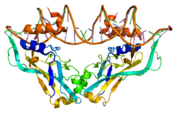
Zinc finger protein GLI2 also known as GLI family zinc finger 2 is a protein that in humans is encoded by the GLI2 gene. The protein encoded by this gene is a transcription factor.

<span class="mw-page-title-main">Lateral plate mesoderm</span>

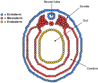
The lateral plate mesoderm is the mesoderm that is found at the periphery of the embryo. It is to the side of the paraxial mesoderm, and further to the axial mesoderm. The lateral plate mesoderm is separated from the paraxial mesoderm by a narrow region of intermediate mesoderm. The mesoderm is the middle layer of the three germ layers, between the outer ectoderm and inner endoderm.

The Hedgehog signaling pathway is a signaling pathway that transmits information to embryonic cells required for proper cell differentiation. Different parts of the embryo have different concentrations of hedgehog signaling proteins. The pathway also has roles in the adult. Diseases associated with the malfunction of this pathway include cancer.

<span class="mw-page-title-main">Apical ectodermal ridge</span>

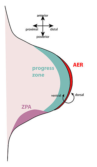
The apical ectodermal ridge (AER) is a structure that forms from the ectodermal cells at the distal end of each limb bud and acts as a major signaling center to ensure proper development of a limb. After the limb bud induces AER formation, the AER and limb mesenchyme—including the zone of polarizing activity (ZPA)—continue to communicate with each other to direct further limb development.

<span class="mw-page-title-main">Limb development</span>

Limb development in vertebrates is an area of active research in both developmental and evolutionary biology, with much of the latter work focused on the transition from fin to limb.

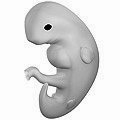
The limb bud is a structure formed early in vertebrate limb development. As a result of interactions between the ectoderm and underlying mesoderm, formation occurs roughly around the fourth week of development. In the development of the human embryo the upper limb bud appears in the third week and the lower limb bud appears four days later.

Gremlin is an inhibitor in the TGF beta signaling pathway. It primarily inhibits bone morphogenesis and is implicated in disorders of increased bone formation and several cancers.

<i>TBX5</i> (gene) Protein-coding gene that affects limb development and heart and bone function

T-box transcription factor TBX5, is a protein that in humans is encoded by the TBX5 gene. Abnormalities in the TBX5 gene can result in altered limb development, Holt-Oram syndrome, Tetra-amelia syndrome, and cardiac and skeletal problems.

<span class="mw-page-title-main">Fibroblast growth factor 8</span> Protein-coding gene in the species Homo sapiens

Fibroblast growth factor 8(FGF-8) is a protein that in humans is encoded by the FGF8 gene.

<span class="mw-page-title-main">FGF4</span> Fibroblast growth factor gene

Fibroblast growth factor 4 is a protein that in humans is encoded by the FGF4 gene.

<span class="mw-page-title-main">TBX3</span> Protein-coding gene in the species Homo sapiens

T-box transcription factor TBX3 is a protein that in humans is encoded by the TBX3 gene.

<span class="mw-page-title-main">LBH (gene)</span> Protein-coding gene in the species Homo sapiens

The LBH gene is a highly conserved human gene that produces the LBH protein, a transcription co-factor in the Wnt/β-catenin pathway. Upon transcriptional activation of β-catenin, LBH goes on to act as a regulator of cell proliferation and differentiation through multiple transcriptional targets. The gene is located on the p arm of chromosome 2 and is roughly 28 kb long. Current ongoing studies are examining its role in developmental and oncological settings.

<span class="mw-page-title-main">OSR1</span> Protein-coding gene in the species Homo sapiens

Protein odd-skipped-related 1 is a transcription factor that in humans is encoded by the OSR1 gene. The OSR1 and OSR2 transcription factors participate in the normal development of body parts such as the kidney.

<span class="mw-page-title-main">SULF1</span> Protein-coding gene in the species Homo sapiens

Sulfatase 1, also known as SULF1, is an enzyme which in humans is encoded by the SULF1 gene.

<span class="mw-page-title-main">Zone of polarizing activity</span>

The zone of polarizing activity (ZPA) is an area of mesenchyme that contains signals which instruct the developing limb bud to form along the anterior/posterior axis. Limb bud is undifferentiated mesenchyme enclosed by an ectoderm covering. Eventually, the limb bud develops into bones, tendons, muscles and joints. Limb bud development relies not only on the ZPA, but also many different genes, signals, and a unique region of ectoderm called the apical ectodermal ridge (AER). Research by Saunders and Gasseling in 1948 identified the AER and its subsequent involvement in proximal distal outgrowth. Twenty years later, the same group did transplantation studies in chick limb bud and identified the ZPA. It wasn't until 1993 that Todt and Fallon showed that the AER and ZPA are dependent on each other.

<span class="mw-page-title-main">Bat wing development</span>

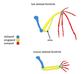
The order Chiroptera, comprising all bats, has evolved the unique mammalian adaptation of flight. Bat wings are modified tetrapod forelimbs. Because bats are mammals, the skeletal structures in their wings are morphologically homologous to the skeletal components found in other tetrapod forelimbs. Through adaptive evolution these structures in bats have undergone many morphological changes, such as webbed digits, elongation of the forelimb, and reduction in bone thickness. Recently, there have been comparative studies of mouse and bat forelimb development to understand the genetic basis of morphological evolution. Consequently, the bat wing is a valuable evo-devo model for studying the evolution of vertebrate limb diversity.

<span class="mw-page-title-main">TCF/LEF family</span> Group of genes

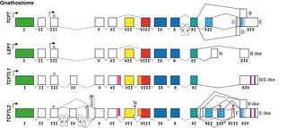
The TCF/LEF family is a group of genes that encode transcription factors which bind to DNA through a SOX-like high mobility group domain. They are involved in the Wnt signaling pathway, particularly during embryonic and stem-cell development, but also had been found to play a role in cancer and diabetes. TCF/LEF factors recruit the coactivator beta-catenin to enhancer elements of genes they target. They can also recruit members of the Groucho family of corepressors.

References

  1. 1 2 3 GRCh38: Ensembl release 89: ENSG00000121068 - Ensembl, May 2017
  2. 1 2 3 GRCm38: Ensembl release 89: ENSMUSG00000000093 - Ensembl, May 2017
  3. "Human PubMed Reference:". National Center for Biotechnology Information, U.S. National Library of Medicine.
  4. "Mouse PubMed Reference:". National Center for Biotechnology Information, U.S. National Library of Medicine.
  5. Campbell C, Goodrich K, Casey G, Beatty B (July 1995). "Cloning and mapping of a human gene (TBX2) sharing a highly conserved protein motif with the Drosophila omb gene". Genomics. 28 (2): 255–260. doi:10.1006/geno.1995.1139. PMID   8530034.
  6. EntrezGene 6909
  7. Law DJ, Gebuhr T, Garvey N, Agulnik SI, Silver LM (November 1995). "Identification, characterization, and localization to chromosome 17q21-22 of the human TBX2 homolog, member of a conserved developmental gene family". Mammalian Genome. 6 (11): 793–797. doi:10.1007/bf00539006. hdl: 2027.42/47017 . PMID   8597636. S2CID   15779406.
  8. 1 2 3 Lu J, Li XP, Dong Q, Kung HF, He ML (December 2010). "TBX2 and TBX3: the special value for anticancer drug targets". Biochimica et Biophysica Acta (BBA) - Reviews on Cancer. 1806 (2): 268–274. doi:10.1016/j.bbcan.2010.07.001. PMC   7127380 . PMID   20624445.
  9. Vance KW, Carreira S, Brosch G, Goding CR (March 2005). "Tbx2 is overexpressed and plays an important role in maintaining proliferation and suppression of senescence in melanomas". Cancer Research. 65 (6): 2260–2268. doi: 10.1158/0008-5472.CAN-04-3045 . PMID   15781639.
  10. 1 2 3 4 Farin HF, Lüdtke TH, Schmidt MK, Placzko S, Schuster-Gossler K, Petry M, et al. (2013-04-25). "Tbx2 terminates shh/fgf signaling in the developing mouse limb bud by direct repression of gremlin1". PLOS Genetics. 9 (4): e1003467. doi:10.1371/journal.pgen.1003467. PMC   3636256 . PMID   23633963.
  11. 1 2 3 4 5 Singh R, Hoogaars WM, Barnett P, Grieskamp T, Rana MS, Buermans H, et al. (April 2012). "Tbx2 and Tbx3 induce atrioventricular myocardial development and endocardial cushion formation". Cellular and Molecular Life Sciences. 69 (8): 1377–1389. doi:10.1007/s00018-011-0884-2. PMC   3314179 . PMID   22130515.
  12. Cho GS, Park DS, Choi SC, Han JK (January 2017). "Tbx2 regulates anterior neural specification by repressing FGF signaling pathway". Developmental Biology. 421 (2): 183–193. doi: 10.1016/j.ydbio.2016.11.020 . PMID   27913219.
  13. Habets PE, Moorman AF, Clout DE, van Roon MA, Lingbeek M, van Lohuizen M, et al. (May 2002). "Cooperative action of Tbx2 and Nkx2.5 inhibits ANF expression in the atrioventricular canal: implications for cardiac chamber formation". Genes & Development. 16 (10): 1234–1246. doi:10.1101/gad.222902. PMC   186286 . PMID   12023302.
  14. Aanhaanen WT, Brons JF, Domínguez JN, Rana MS, Norden J, Airik R, et al. (June 2009). "The Tbx2+ primary myocardium of the atrioventricular canal forms the atrioventricular node and the base of the left ventricle". Circulation Research. 104 (11): 1267–1274. doi: 10.1161/CIRCRESAHA.108.192450 . PMID   19423846.
  15. García-Añoveros, Jaime; Clancy, John C.; Foo, Chuan Zhi; García-Gómez, Ignacio; Zhou, Yingjie; Homma, Kazuaki; Cheatham, Mary Ann; Duggan, Anne (2022-05-04). "Tbx2 is a master regulator of inner versus outer hair cell differentiation". Nature. 605 (7909): 298–303. doi:10.1038/s41586-022-04668-3. ISSN   1476-4687. PMC   9803360 . PMID   35508658. S2CID   248527577.
  16. Pang S, Liu Y, Zhao Z, Huang W, Chen D, Yan B (September 2013). "Novel and functional sequence variants within the TBX2 gene promoter in ventricular septal defects". Biochimie. 95 (9): 1807–1809. doi:10.1016/j.biochi.2013.05.007. PMID   23727221.
  17. 1 2 Nimmakayalu M, Major H, Sheffield V, Solomon DH, Smith RJ, Patil SR, Shchelochkov OA (February 2011). "Microdeletion of 17q22q23.2 encompassing TBX2 and TBX4 in a patient with congenital microcephaly, thyroid duct cyst, sensorineural hearing loss, and pulmonary hypertension". American Journal of Medical Genetics. Part A. 155A (2): 418–423. doi:10.1002/ajmg.a.33827. PMID   21271665. S2CID   24377700.
  18. 1 2 Radio FC, Bernardini L, Loddo S, Bottillo I, Novelli A, Mingarelli R, Dallapiccola B (August 2010). "TBX2 gene duplication associated with complex heart defect and skeletal malformations". American Journal of Medical Genetics. Part A. 152A (8): 2061–2066. doi:10.1002/ajmg.a.33506. PMID   20635360. S2CID   13614834.
  19. Zhang Y, Han Q, Fan H, Li W, Xing Q, Yan B (August 2014). "Genetic analysis of the TBX2 gene promoter in indirect inguinal hernia". Hernia. 18 (4): 513–517. doi:10.1007/s10029-013-1199-z. PMID   24309999. S2CID   10028037.
  20. Puusepp H, Zilina O, Teek R, Männik K, Parkel S, Kruustük K, et al. (January 2009). "5.9 Mb microdeletion in chromosome band 17q22-q23.2 associated with tracheo-esophageal fistula and conductive hearing loss". European Journal of Medical Genetics. 52 (1): 71–74. doi:10.1016/j.ejmg.2008.09.006. PMID   18983945.
  21. Prince S, Carreira S, Vance KW, Abrahams A, Goding CR (March 2004). "Tbx2 directly represses the expression of the p21(WAF1) cyclin-dependent kinase inhibitor". Cancer Research. 64 (5): 1669–1674. doi: 10.1158/0008-5472.can-03-3286 . PMID   14996726.
  22. Rodriguez M, Aladowicz E, Lanfrancone L, Goding CR (October 2008). "Tbx3 represses E-cadherin expression and enhances melanoma invasiveness". Cancer Research. 68 (19): 7872–7881. doi: 10.1158/0008-5472.can-08-0301 . PMID   18829543.
  23. Redmond KL, Crawford NT, Farmer H, D'Costa ZC, O'Brien GJ, Buckley NE, et al. (June 2010). "T-box 2 represses NDRG1 through an EGR1-dependent mechanism to drive the proliferation of breast cancer cells". Oncogene. 29 (22): 3252–3262. doi: 10.1038/onc.2010.84 . PMID   20348948.
  24. Nandana S, Tripathi M, Duan P, Chu CY, Mishra R, Liu C, et al. (March 2017). "Bone Metastasis of Prostate Cancer Can Be Therapeutically Targeted at the TBX2-WNT Signaling Axis". Cancer Research. 77 (6): 1331–1344. doi:10.1158/0008-5472.can-16-0497. PMC   5783646 . PMID   28108510.

Further reading