The nuclear receptor 4A2 (NR4A2) (nuclear receptor subfamily 4 group A member 2) also known as nuclear receptor related 1 protein (NURR1) is a protein that in humans is encoded by the NR4A2gene.[5] NR4A2 is a member of the nuclear receptor family of intracellulartranscription factors.
NR4A2 plays a key role in the maintenance of the dopaminergic system of the brain.[6] Mutations in this gene have been associated with disorders related to dopaminergic dysfunction, including Parkinson's disease and schizophrenia. Misregulation of this gene may be associated with rheumatoid arthritis. Four transcript variants encoding four distinct isoforms have been identified for this gene. Additional alternate splice variants may exist, but their full-length nature has not been determined.[7]
This protein is thought to be critical to development of the dopaminergic phenotype in the midbrain, as mice without NR4A2 are lacking expression of this phenotype. This is further confirmed by studies showing that forced NR4A2 expression in naïve precursor cells leads to complete dopaminergic phenotype gene expression.[8]
While NR4A2 is a key protein in inducing this phenotype, there are other factors required, as expressing NR4A2 in isolation fails to produce it. One of these suggested factors is winged-helix transcription factor 2 (Foxa2). Studies have found these two factors to be within the same region of developing dopaminergic neurons, and both were required to have expression for the dopaminergic phenotype. [8]
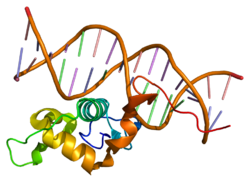
Structure
One investigation conducted research on the structure and found that NR4A2 does not contain a ligand-binding cavity but a patch filled with hydrophobic side chains. Non-polar amino acid residues of NR4A2's co-regulators, SMRT and NCoR, bind to this hydrophobic patch. Analysis of tertiary structure has shown that the binding surface of the ligand-binding domain is located on the grooves of the 11th and 12th alpha helices. This study also found essential structural components of this hydrophobic patch, to be the three amino acids residues, F574, F592, L593; mutation of any these three inhibits LBD activity.[9]
Clinical significance
Role in disease
Mutations in NR4A2 have been associated with various disorders, including Parkinson's disease, schizophrenia, manic depression, and autism. De novo gene deletions that affect NR4A2 have been identified in some individuals with intellectual disability and language impairment, some of whom meet DSM-5 criteria for an autism diagnosis.[10]
Inflammation
Research has been conducted on NR4A2's role in inflammation, and may provide important information in treating disorders caused by dopaminergic neuron disease. Inflammation in the central nervous system can result from activated microglia (macrophage analogs for the central nervous system) and other pro-inflammatory factors, such as bacterial lipopolysaccharide (LPS). LPS binds to toll-like receptors (TLR), which induces inflammatory gene expression by promoting signal-dependent transcription factors. To determine which cells are dopaminergic, experiments measured the enzyme tyrosine hydroxylase (TH), which is needed for dopamine synthesis. It has been shown that NR4A2 protects dopaminergic neurons from LPS-induced inflammation by reducing inflammatory gene expression in microglia and astrocytes. When a short hairpin RNA for NR4A2 was expressed in microglia and astrocytes, these cells produced inflammatory mediators such as TNF-alpha, nitric oxide synthase, and interleukin-1 beta (IL-1β), supporting the conclusion that reduced NR4A2 promotes inflammation and leads to cell death of dopaminergic neurons. NR4A2 interacts with the transcription factor complex NF-κB-p65 on the inflammatory gene promoters. However, NR4A2 is dependent on other factors to be able to participate in these interactions. NR4A2 needs to be sumoylated and its co-regulating factor, glycogen synthase kinase 3, needs to be phosphorylated for these interactions to occur. Sumolyated NR4A2 recruits CoREST, a complex made of several proteins that assembles chromatin remodeling enzymes. The NR4A2/CoREST complex inhibits transcription of inflammatory genes.[11]
Applications
NR4A2 induces tyrosine hydroxylase (TH) expression, which eventually leads to differentiation into dopaminergic neurons. NR4A2 has been demonstrated to induce differentiation in CNS precursor cells in vitro but they require additional factors to reach full maturity and dopaminergic differentiation.[12] Therefore, NR4A2 modulation may be promising for generation of dopaminergic neurons for Parkinson's disease research, yet implantation of these induced cells as therapy treatments, has had limited results.
Transgenic C57bl6 mice heterozygous for Nurr1 show ~50% reduced Nurr1 mRNA and ~30% reduced TH expression. Dopamine levels in the striatum were also significantly reduced and these mice show behavioral effects indicative of dopaminergic sensitivity and susceptibility to dysfunction.[13][14]
NR4A2 mRNA may be a useful biomarker for Parkinson's disease in combination with inflammatory cytokines.[15]
Knockout studies
Studies have shown that heterozygous knockout mice for the NR4A2 gene demonstrate reduced dopamine release. Initially this was compensated for by a decrease in the rate of dopamine reuptake; however, over time this reuptake could not make up for the reduced amount of dopamine being released. Coupled with the loss of dopamine receptor neurons, this can result in the onset of symptoms for Parkinson's disease.[16]
Le W, Appel SH (February 2004). "Mutant genes responsible for Parkinson's disease". Current Opinion in Pharmacology. 4 (1): 79–84. doi:10.1016/j.coph.2003.09.005. PMID15018843.
Wedler B, Wüstenberg PW, Naumann G (July 1975). "[Treatment of hypertonus in diabetes mellitus]". Zeitschrift für die Gesamte Innere Medizin und Ihre Grenzgebiete. 30 (13): 437–442. PMID4929.
Maruyama K, Sugano S (January 1994). "Oligo-capping: a simple method to replace the cap structure of eukaryotic mRNAs with oligoribonucleotides". Gene. 138 (1–2): 171–174. doi:10.1016/0378-1119(94)90802-8. PMID8125298.
Suzuki Y, Yoshitomo-Nakagawa K, Maruyama K, Suyama A, Sugano S (October 1997). "Construction and characterization of a full length-enriched and a 5'-end-enriched cDNA library". Gene. 200 (1–2): 149–156. doi:10.1016/S0378-1119(97)00411-3. PMID9373149.
Torii T, Kawarai T, Nakamura S, Kawakami H (April 1999). "Organization of the human orphan nuclear receptor Nurr1 gene". Gene. 230 (2): 225–232. doi:10.1016/S0378-1119(99)00064-5. PMID10216261.
Ichinose H, Ohye T, Suzuki T, Sumi-Ichinose C, Nomura T, Hagino Y, etal. (April 1999). "Molecular cloning of the human Nurr1 gene: characterization of the human gene and cDNAs". Gene. 230 (2): 233–239. doi:10.1016/S0378-1119(99)00065-7. PMID10216262.
Chen YH, Tsai MT, Shaw CK, Chen CH (December 2001). "Mutation analysis of the human NR4A2 gene, an essential gene for midbrain dopaminergic neurogenesis, in schizophrenic patients". American Journal of Medical Genetics. 105 (8): 753–757. doi:10.1002/ajmg.10036. PMID11803525.
Ishiguro H, Okubo Y, Ohtsuki T, Yamakawa-Kobayashi K, Arinami T (January 2002). "Mutation analysis of the retinoid X receptor beta, nuclear-related receptor 1, and peroxisome proliferator-activated receptor alpha genes in schizophrenia and alcohol dependence: possible haplotype association of nuclear-related receptor 1 gene to alcohol dependence". American Journal of Medical Genetics. 114 (1): 15–23. doi:10.1002/ajmg.1620. PMID11840500.
Le WD, Xu P, Jankovic J, Jiang H, Appel SH, Smith RG, etal. (January 2003). "Mutations in NR4A2 associated with familial Parkinson disease". Nature Genetics. 33 (1): 85–89. doi:10.1038/ng1066. PMID12496759. S2CID10699494.
Satoh J, Kuroda Y (December 2002). "The constitutive and inducible expression of Nurr1, a key regulator of dopaminergic neuronal differentiation, in human neural and non-neural cell lines". Neuropathology. 22 (4): 219–232. doi:10.1046/j.1440-1789.2002.00460.x. PMID12564761. S2CID30708166.
This page is based on this Wikipedia article Text is available under the CC BY-SA 4.0 license; additional terms may apply. Images, videos and audio are available under their respective licenses.