Pulpotomy

Last updated
Visual of tooth decay Tooth-02.png
Visual of tooth decay

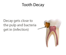
Pulpotomy is a minimally invasive procedure performed in children on a primary tooth with extensive caries but without evidence of root pathology. [1] The minimally invasive endodontic techniques of vital pulp therapy (VPT) are based on improved understanding of the capacity of pulp (nerve) tissues to heal and regenerate plus the availability of advanced endodontic materials. During caries removal, this results in a carious or mechanical pulp exposure (bleeding) from the cavity. During pulpotomy, the inflamed/diseased pulp tissue is removed from the coronal pulp chamber of the tooth leaving healthy pulp tissue which is dressed with a long-term clinically successful medicament that maintains the survival of the pulp and promotes repair. [2] There are various types of medicament placed above the vital pulp such as Buckley's Solution of formocresol, ferric sulfate, calcium hydroxide or mineral trioxide aggregate (MTA). [1] MTA is a more recent material used for pulpotomies with a high rate of success, better than formocresol or ferric sulfate. It is also recommended to be the preferred pulpotomy agent in the future. [3] [4] After the coronal pulp chamber is filled, the tooth is restored with a filling material that seals the tooth from microleakage, such as a stainless steel crown which is the most effective long-term restoration. However, if there is sufficient remaining supporting tooth structure, other filling materials such as amalgam or composite resin can provide a functional alternative when the primary tooth has a life span of two years or less. [1] The medium- to long-term treatment outcomes of pulpotomy in symptomatic permanent teeth with caries, especially in young people, indicate that pulpotomy can be a potential alternative to root canal therapy (RCT). [5]

Contents

Diagram of pulpotomy Tooth-03.png
Diagram of pulpotomy

There is another term, which is also related to vital pulp therapy, which is apexogenesis. Apexogenesis is a treatment in preserving vital pulp tissue in the apical part of a root canal to allow the completion in formation of the root apex. This clinical procedure is essentially a deep pulpotomy, aimed to preserve the pulp in immature teeth that have deep pulpal inflammation. Examples include teeth with carious exposures and trauma in which treatment of the exposed pulp is delayed and it becomes necessary to extend farther into the canal to reach healthy tissue. [6]

Objectives

In primary tooth

After the pulpotomy treatment, the radicular pulp should remain asymptomatic without any adverse clinical signs or symptoms such as sensitivity, pain, or swelling. From the radiographs, there should be absence of postoperative evidence of pathologic root resorption. There should be absence of clinical signs of infection and inflammation and no harm to the succedaneous tooth. [1] However, radiographs play a very important role, to check if pulpotomy can be done on the primary tooth. For example, the aspects we considered are the extension of caries in the primary tooth, and the development of the succedaneous permanent tooth.

The radiograph shows a primary tooth with succedaneous permanent teeth. Radiographs are needed to determine if pulpotomy can be carried out. Intraoral Periapical Radiograph (IOPA) showing Deciduous(Milky or Primary) Tooth 75 and developing crown of Permanent or Secondary Teeth 35, 36 and 37.jpg
The radiograph shows a primary tooth with succedaneous permanent teeth. Radiographs are needed to determine if pulpotomy can be carried out.

In mature permanent tooth

The tooth should be asymptomatic. There should be no clinical signs and symptoms. From the radiographs, there should be absence of postoperative evidence of pathology. [1]

In immature tooth

Pulpotomy allows the continuation of root formation, leading to tooth end closure, preservation and maintenance of pulp vitality, stronger root structure and greater structural integrity. [7]

Indications

Primary teeth

Primary/deciduous (baby) teeth in children have relatively large pulp spaces. Caries do not have to develop significantly before they reach the pulp chamber.

When the soft tissue in the pulp chamber is infected (has bacteria in it) or affected (is inflamed), it can be removed by a dentist or dental therapist under local anaesthetic. If the soft tissue in the canals is still healthy enough, a special medicated filling can be put into the chamber in an attempt to keep the remaining pulp (in the canals) alive. The process of removing the pulp from the chamber is the actual "pulpotomy", though the word is often used for the entire process including placement of the medication. There are many medicaments that can be used to fill the pulp chamber including zinc-oxide eugenol as well as mineral trioxide aggregate.

There are two types of pulpotomy techniques depending the extent of caries in a tooth and the symptoms it presents. A vital pulpotomy or a non-vital pulpotomy can be carried out. However, recent research shows that non-vital pulpotomies are rarely indicated due to their low success rates and it is therefore sometimes better to extract the tooth.

Afterwards the tooth is restored with a regular filling, either composite or amalgam, or a stainless steel crown. Due to the process of a pulpotomy causing the tooth to become slightly brittle, a stainless steel crown is normally indicated as the preferred choice of definitive restoration.

A pulpotomy can be done to both permanent and primary teeth.

Types

Primary teeth

The indication of this pulpotomy procedure is when pulp exposure occurs during caries removal in a primary tooth with a normal pulp or reversible pulpitis or after a traumatic pulp exposure. [1]   Then, the coronal tissue is amputated, and the remaining radicular tissue is judged to be vital without suppuration, pus, necrosis, or excessive bleeding which cannot be controlled by a moist cotton pellet (with saline) after several minutes, and there are no radiographic signs of infection or pathologic resorption. [1]

Pulpotomy therapy can be classified according to the following treatment objectives: devitalization (mummification, cauterization), preservation (minimal devitalization, noninductive), or regeneration (inductive, reparative). [8]

Devitalization

Multiple visits with application of formocresol in pulpotomy is used to fix the radicular pulp completely to reduce pulp infection. The radicular pulp was theoretically sterilized and devitalized, thereby reducing infection and internal resorption.

Another form of nonchemical devitalization emerged: electrosurgical pulpotomy. Electrocautery releases heat that denatures pulp and reduces bacterial contamination. Experimentally, electrosurgery has been shown to reduce pathologic root resorption and periapical pathology, and a series of pulpal effects including acute and chronic inflammation, swelling and diffuse necrosis. It is reported that this method has high success rate in pulpotomies.

However, this method may prove to be more diagnosis and technique sensitive, and it may not be suitable if apical root resorption has occurred. [8]

Preservation

Zinc oxide-eugenol (ZOE) was the first agent to be used for preservation. In recent years, glutaraldehyde has been proposed as an alternative to formocresol based on: its superior fixative properties and low toxicity. A nonaldehyde chemical, ferric sulfate, has received some attention recently as a pulpotomy agent. It minimizes the chances for inflammation and internal resorption. This category of pulp therapy is still in flux, although major changes in the future are not likely. [8]

Regeneration

The ideal pulpotomy treatment should leave the radicular pulp alive and healthy. In this case, the tooth should be filled with noxious restorative materials within, thereby diminishing the chances of internal resorption, as well as formation of reparative dentin. Calcium hydroxide was the first agent used in pulpotomies that demonstrated any capacity to induce regeneration of dentin. However, the success rate is not that high. Recent advances in the field of bone and dentin formation have opened exciting new vistas for pulp therapy, which is a factor called bone morphogenetic protein (BMP). It has bone inductive properties, that can predictably induce bone for use in the fields of orthopedic, oral, and periodontal surgery. Most importantly for dentistry, these osteogenic proteins hold promise for pulp therapy. [8]

Young permanent teeth

Partial pulpotomy for carious exposures

Partial pulpotomy is also indicated in young permanent teeth with pulp exposure due to caries, provided that the bleeding can be controlled within several minutes. It is a procedure in which the inflamed pulp tissue beneath an exposure is removed to a depth of 1 to 3mm or deeper to reach the level of healthy pulp tissue. Pulpal bleeding can be controlled by irrigation of sodium hypochlorite or chlorhexidine. The site is then covered with a pulpal medicament, calcium hydroxide or MTA, followed by a final restoration that provides a complete seal to prevent any leakage and bacterial contamination following the restoration. [1]

After the procedure, the remaining pulp should remain vital and the patient should be free of any adverse clinical signs or symptoms such as sensitivity, pain or swelling. Immature teeth should continue its normal development and apexogenesis. [1]

Partial pulpotomy for traumatic exposures

Tooth crown fractures are one of the most common dental injuries and the pulp is exposed in approximately 25% of all crown fractures. [9] Maintaining vitality of the pulp tissue in an immature tooth is important to allow continued growth of the tooth.

Partial pulpotomy due to a traumatic exposure is also known as Cvek Pulpotomy. The procedure involves removal of 1 to 3 mm (0.04 to 0.1 in) of inflamed pulp tissue beneath an exposure to reach  the level of healthy pulp tissue. The surface of the remaining pulp is then irrigated with bacteriocidal irrigants such as sodium hypochlorite or chlorhexidine until bleeding has ceased. The site is then covered with a pulpal medicament, either calcium hydroxide or MTA. [1] The remaining cavity is then restored with a material that provides a complete seal to prevent any leakage and bacterial contamination following the restoration. [10]

The remaining pulp tissue should continue to be vital after partial pulpotomy and teeth with immature roots should show continued normal development and apexogenesis. There should be no signs of pain, swelling, or sensitivity after the procedure. [1] Cvek at al reported that partial pulpotomies after a traumatic exposure had a success rate of 96%. [11]

Medicaments

Pulpotomy is a vital pulp therapy, medicaments that can promote healing and preserve the vitality of the tooth should be placed after removal of the inflamed pulp. [12]

In primary teeth medicaments such as formocresol, mineral trioxide aggregate, zinc oxide eugenol and calcium hydroxide can be used in pulpotomy. Formocresol use has been questioned due to toxicity concerns. [13] Ferric sulfate, sodium hypochlorite [14] [15] or a local anaesthetic solution containing a vasoconstrictor agent can be used to arrest any bleeding from the pulp prior to the placement of medicament. Calcium enriched mixtures have been used in permanent molar teeth with irreversible pulpitis showing positive outcomes. [16]

Ferric sulfate

Ferric sulfate is used to arrest pulpal bleeding by forming a sealing membrane through the agglutination of the blood proteins with ferric and sulfate ions. This metal-protein clot at the surface of the pulp may act as a barrier to external irritants. The physiological clot formation is thought to be able to minimize inflammation and internal resorption compared to calcium hydroxide. Most importantly, ferric sulfate causes minimal devitalization and subsequent preservation of the pulp tissue. [17] Ferric sulphate has been shown to have close to a 100% clinical success compared to formocresol (77%) with one-year follow-up. [18]

Formocresol

Formocresol has been used in pulpotomy procedures of the primary teeth since 80 years. Formocresol is both a bactericidal and devitalizing agent. It kills bacteria and converts the pulp tissue into inert compounds. This action fastens the vital pulp, maintaining them inert and conserves the primary tooth until it falls off physiologically. [19] However, formaldehyde, a primary component in formocresol has raised concerns regarding its safe of use. Formaldehyde is a hazardous substance and has perceived the need to reevaluate the use of formocresol. [19] [20]

Zinc oxide-eugenol (ZOE)

After pulpal bleeding is arrested, a suitable base such as zinc oxide-eugenol is placed to seal the tooth from microleakage. [1] ZOE is a nontoxic material for pulpal cells and possess antimicrobial as well as anti-inflammatory properties. In addition, it also has local anesthetic or soothing effect on the dental pulp. [21] [22]

Calcium hydroxide

Calcium hydroxide Ca(OH)2 is conventionally used as a pulpotomy agent of the permanent teeth but with less long-term success. [1] Calcium hydroxide is a highly alkaline (pH 12) material that has bactericidal effect and has the potential to enhance reparative dentin (dentin bridge) formation. However, it also leads to superficial necrosis of the pulp tissue in contact with the medication and has been shown to be toxic to cells in tissue culture. [12] [21] Therefore, in spite being a popular vital pulp therapy material, its use as a pulpotomy agent remains controversial.

Mineral trioxide aggregate (MTA)

MTA is a more recent material that consists of tricalcium silicate, dicalcium silicate, tricalcium aluminate, tetracalcium aluminoferrite, bismuth oxide and calcium sulfate. MTA is known to have excellent physical characteristics, biocompatibility and has the ability to stimulate cytokine release from pulpal fibroblast, which can stimulate hard tissue formation. It has better structural integrity and forms a thicker, more localized dentinal bridge. [20] [12] MTA results in a more predictable dentin bridge formation and pulpal health. [1] MTA has a high rate of success and has been shown to perform equal or better than formocresol or ferric sulfate. However, the cost, availability and difficulty in handling this material remains its current drawback. [1] [12]

Alternative pulpotomy techniques

Electro-surgery pulpotomy

Electro-surgical pulpotomy is a method of cutting and coagulating soft tissues by means of high frequency radio waves. It can control bleeding without chemical coagulation and is antibacterial. Electrosurgical pulpotomy has a success rate of 70 to 94%. However, it is considered as a sensitive technique. [19] [23]

Laser pulpotomy

The carbon dioxide laser emits an infrared beam that has an affinity for water, and is capable of producing well-localized cautery to soft tissue. Tissue is removed by ablation through conversion of the laser beam to heat. The carbon dioxide laser appears to be a promising alternative for pulpotomy therapy. [24]

Clinical significance

Conserving the vitality of pulp tissues appears to be a less painful alternative to root canal treatment (RCT) for younger patients. The high success rate reported for pulpotomy suggests that this procedure offers hope as an alternative to root canal treatment in teeth with a diagnosis of irreversible pulpitis. [25] [26]

See also

Related Research Articles

<span class="mw-page-title-main">Tooth decay</span> Deformation of teeth due to acids produced by bacteria

Tooth decay, also known as cavities or caries, is the breakdown of teeth due to acids produced by bacteria. The cavities may be a number of different colors, from yellow to black. Symptoms may include pain and difficulty eating. Complications may include inflammation of the tissue around the tooth, tooth loss and infection or abscess formation. Tooth regeneration is an on-going stem cell based field of study that is trying to reverse the effects of decay, unlike most current methods which only try to make dealing with the effects easier.

Dental products are specially fabricated materials, designed for use in dentistry. There are many different types of dental products, and their characteristics vary according to their intended purpose.

<span class="mw-page-title-main">Endodontics</span> Field of dentistry

Endodontics is the dental specialty concerned with the study and treatment of the dental pulp.

<span class="mw-page-title-main">Pulp (tooth)</span> Part in the center of a tooth made up of living connective tissue and cells called odontoblasts

The pulp is the connective tissue, nerves, blood vessels, and odontoblasts that comprise the innermost layer of a tooth. The pulp's activity and signalling processes regulate its behaviour.

<span class="mw-page-title-main">Deciduous teeth</span> First set of teeth in diphyodonts

Deciduous teeth or primary teeth, also informally known as baby teeth, tothlings, milk teeth, or temporary teeth, are the first set of teeth in the growth and development of humans and other diphyodonts, which include most mammals but not elephants, kangaroos, or manatees, which are polyphyodonts. Deciduous teeth develop during the embryonic stage of development and erupt during infancy. They are usually lost and replaced by permanent teeth, but in the absence of their permanent replacements, they can remain functional for many years into adulthood.

Pulpitis is inflammation of dental pulp tissue. The pulp contains the blood vessels, the nerves, and connective tissue inside a tooth and provides the tooth's blood and nutrients. Pulpitis is mainly caused by bacterial infection which itself is a secondary development of caries. It manifests itself in the form of a toothache.

<span class="mw-page-title-main">Dentinogenesis imperfecta</span> Medical condition

Dentinogenesis imperfecta (DI) is a genetic disorder of tooth development. It is inherited in an autosomal dominant pattern, as a result of mutations on chromosome 4q21, in the dentine sialophosphoprotein gene (DSPP). It is one of the most frequently occurring autosomal dominant features in humans. Dentinogenesis imperfecta affects an estimated 1 in 6,000-8,000 people.

A pulp polyp, also known as chronic hyperplastic pulpitis, is a "productive" inflammation of dental pulp in which the development of granulation tissue is seen in response to persistent, low-grade mechanical irritation and bacterial invasion of the pulp.

Dens evaginatus is a rare odontogenic developmental anomaly that is found in teeth where the outer surface appears to form an extra bump or cusp.

<span class="mw-page-title-main">Root canal treatment</span> Dental treatment

Root canal treatment is a treatment sequence for the infected pulp of a tooth which is intended to result in the elimination of infection and the protection of the decontaminated tooth from future microbial invasion. Root canals, and their associated pulp chamber, are the physical hollows within a tooth that are naturally inhabited by nerve tissue, blood vessels and other cellular entities. Together, these items constitute the dental pulp.

Mineral trioxide aggregate (MTA) is an alkaline, cementitious dental repair material. MTA is used for creating apical plugs during apexification, repairing root perforations during root canal therapy, and treating internal root resorption. It can be used for root-end filling material and as pulp capping material. It has better pulpotomy outcomes than calcium hydroxide or formocresol, and may be the best known material, as of 2018 data. For pulp capping, it has a success rate higher than calcium hydroxide, and indistinguishable from Biodentin.

Pulp necrosis is a clinical diagnostic category indicating the death of cells and tissues in the pulp chamber of a tooth with or without bacterial invasion. It is often the result of many cases of dental trauma, caries and irreversible pulpitis.

<span class="mw-page-title-main">Pulp stone</span>

Pulp stones are nodular, calcified masses appearing in either or both the coronal and root portion of the pulp organ in teeth. Pulp stones are not painful unless they impinge on nerves.

The Hall Technique is a minimally-invasive treatment for decayed baby back (molar) teeth. Decay is sealed under preformed crowns, avoiding injections and drilling. It is one of a number of biologically orientated strategies for managing dental decay.

<span class="mw-page-title-main">Regenerative endodontics</span> Dental specialty

Regenerative endodontic procedures is defined as biologically based procedures designed to replace damaged structures such as dentin, root structures, and cells of the pulp-dentin complex. This new treatment modality aims to promote normal function of the pulp. It has become an alternative to heal apical periodontitis. Regenerative endodontics is the extension of root canal therapy. Conventional root canal therapy cleans and fills the pulp chamber with biologically inert material after destruction of the pulp due to dental caries, congenital deformity or trauma. Regenerative endodontics instead seeks to replace live tissue in the pulp chamber. The ultimate goal of regenerative endodontic procedures is to regenerate the tissues and the normal function of the dentin-pulp complex.

<span class="mw-page-title-main">Pulp capping</span> Dental restoration technique

Pulp capping is a technique used in dental restorations to protect the dental pulp, after it has been exposed, or nearly exposed during a cavity preparation, from a traumatic injury, or by a deep cavity that reaches the center of the tooth, causing the pulp to die. Exposure of the pulp causes pulpitis. The ultimate goal of pulp capping or stepwise caries removal is to protect a healthy dental pulp, and avoid the need for root canal therapy.

Silver diammine fluoride (SDF), also known as silver diamine fluoride in most of the dental literature, is a topical medication used to treat and prevent dental caries and relieve dentinal hypersensitivity. It is a colorless or blue-tinted, odourless liquid composed of silver, ammonium and fluoride ions at a pH of 10.4 or 13. Ammonia compounds reduce the oxidative potential of SDF, increase its stability and helps to maintain a constant concentration over a period of time, rendering it safe for use in the mouth. Silver and fluoride ions possess antimicrobial properties and are used in the remineralization of enamel and dentin on teeth for preventing and arresting dental caries.

Formacresol is a mixture consisting of formalin, cresol and glycerine used in dentistry. It is used for vital pulpotomy of primary teeth and as a temporary intracanal medicament during root canal therapy.

Periapical granuloma, also sometimes referred to as a radicular granuloma or apical granuloma, is an inflammation at the tip of a dead (nonvital) tooth. It is a lesion or mass that typically starts out as an epithelial lined cyst, and undergoes an inward curvature that results in inflammation of granulation tissue at the root tips of a dead tooth. This is usually due to dental caries or a bacterial infection of the dental pulp. Periapical granuloma is an infrequent disorder that has an occurrence rate between 9.3 to 87.1 percent. Periapical granuloma is not a true granuloma due to the fact that it does not contain granulomatous inflammation; however, periapical granuloma is a common term used.

Apexification is a method of dental treatment to induce a calcific barrier in a root with incomplete formation or open apex of a tooth with necrotic pulp. Pulpal involvement usually occurs as a consequence of trauma or caries involvement of young or immature permanent teeth. As a sequelae of untreated pulp involvement, loss of pulp vitality or necrotic pulp took place for the involved teeth.

References

  1. 1 2 3 4 5 6 7 8 9 10 11 12 13 14 15 "Overview". www.aapd.org. Retrieved 2020-01-24.
  2. Cushley, Siobhan; Duncan, Henry F.; Lappin, Mark J.; Tomson, Phillip L.; Lundy, Fionnuala T.; Cooper, Paul; Clarke, Mike; El Karim, Ikhlas A. (September 2019). "Pulpotomy for mature carious teeth with symptoms of irreversible pulpitis: A systematic review" (PDF). Journal of Dentistry. 88: 103158. doi:10.1016/j.jdent.2019.06.005. ISSN   1879-176X. PMID   31229496. S2CID   195326420.
  3. Ng, F. K.; Messer, L. B. (March 2008). "Mineral trioxide aggregate as a pulpotomy medicament: a narrative review". European Archives of Paediatric Dentistry. 9 (1): 4–11. doi:10.1007/bf03321589. ISSN   1818-6300. PMID   18328232. S2CID   9601597.
  4. Seale, N. Sue; Glickman, Gerald N. (May 2008). "Contemporary perspectives on vital pulp therapy: views from the endodontists and pediatric dentists". Pediatric Dentistry. 30 (3): 261–267. ISSN   0164-1263. PMID   18615993.
  5. Barngkgei, Imad Hassan; Halboub, Esam Saleh; Alboni, Roula Safouh (2013). "Pulpotomy of Symptomatic Permanent Teeth with Carious Exposure Using Mineral Trioxide Aggregate". Iranian Endodontic Journal. 8 (2): 65–68. ISSN   1735-7497. PMC   3662039 . PMID   23717332.
  6. Cohen, Stephen (2011), "Introduction", Cohen's Pathways of the Pulp, Elsevier, pp. xvi, doi:10.1016/b978-0-323-06489-7.00037-0, ISBN   978-0-323-06489-7
  7. Nosrat, Ali; Asgary, Saeed (May 2010). "Apexogenesis treatment with a new endodontic cement: a case report". Journal of Endodontics. 36 (5): 912–914. doi:10.1016/j.joen.2009.11.025. ISSN   1878-3554. PMID   20416445.
  8. 1 2 3 4 Ranly, D. M. (November 1994). "Pulpotomy therapy in primary teeth: new modalities for old rationales". Pediatric Dentistry. 16 (6): 403–409. ISSN   0164-1263. PMID   7854945.
  9. Krastl, Gabriel; Weiger, Roland (2014-12-01). "Vital pulp therapy after trauma". ENDO - Endodontic Practice Today. 8: 293–300.
  10. Fong, Cheng D.; Davis, Martin J. (January 2002). "Partial pulpotomy for immature permanent teeth, its present and future". Pediatric Dentistry. 24 (1): 29–32. ISSN   0164-1263. PMID   11874055.
  11. Cvek, Miomir (1978-01-01). "A clinical report on partial pulpotomy and capping with calcium hydroxide in permanent incisors with complicated crown fracture". Journal of Endodontics. 4 (8): 232–237. doi:10.1016/S0099-2399(78)80153-8. ISSN   0099-2399. PMID   283188.
  12. 1 2 3 4 Taneja, Saumya; Singh, Abhinav (2019-08-01). "Evaluation of effectiveness of calcium hydroxide and MTA as pulpotomy agents in permanent teeth: A meta-analysis". Pediatric Dental Journal. 29 (2): 90–96. doi:10.1016/j.pdj.2019.04.001. ISSN   0917-2394. S2CID   155901795.
  13. Godhi B, Sood PB, Sharma A (October 2011). "Effects of mineral trioxide aggregate and formocresol on vital pulp after pulpotomy of primary molars: An in vivo study". Contemporary Clinical Dentistry. 2 (4): 296–301. doi: 10.4103/0976-237X.91792 . PMC   3276856 . PMID   22346156.
  14. Shabzendedar M, Mazhari F, Alami M, Talebi M (2013). "Sodium hypochlorite vs formocresol as pulpotomy medicaments in primary molars: 1-year follow-up". Pediatric Dentistry. 35 (4): 329–32. PMID   23930631.
  15. Al-Mutairi MA, Bawazir OA (March 2013). "Sodium hypochlorite versus Formocresol in primary molars pulpotomies: a randomized clinical trial". European Journal of Paediatric Dentistry. 14 (1): 33–6. PMID   23597217.
  16. Yazdani S, Jadidfard MP, Tahani B, Kazemian A, Dianat O, Alim Marvasti L (2014). "Health Technology Assessment of CEM Pulpotomy in Permanent Molars with Irreversible Pulpitis". Iranian Endodontic Journal. 9 (1): 23–9. PMC   3881298 . PMID   24396372.
  17. Bandi, Madhuri; Mallineni, Sreekanth Kumar; Nuvvula, Sivakumar (2017). "Clinical applications of ferric sulfate in dentistry: A narrative review". Journal of Conservative Dentistry. 20 (4): 278–281. doi: 10.4103/JCD.JCD_259_16 . ISSN   0972-0707. PMC   5721513 . PMID   29259368.
  18. Fei, A. L.; Udin, R. D.; Johnson, R. (November 1991). "A clinical study of ferric sulphate as a pulpotomy agent in primary teeth". Pediatric Dentistry. 13 (6): 327–332. ISSN   0164-1263. PMID   1843987.
  19. 1 2 3 "Formocresol, still a controversial material for pulpotomy: A critical literature review". www.jresdent.org. Retrieved 2020-01-24.
  20. 1 2 Rodd, H. D.; Waterhouse, P. J.; Fuks, A. B.; Fayle, S. A.; Moffat, M. A.; British Society of Paediatric Dentistry (September 2006). "Pulp therapy for primary molars". International Journal of Paediatric Dentistry. 16 Suppl 1 (s1): 15–23. doi:10.1111/j.1365-263X.2006.00774.x. ISSN   0960-7439. PMID   16939452.
  21. 1 2 Harandi, Azadeh; Forghani, Maryam; Ghoddusi, Jamileh (2013). "Vital Pulp Therapy with Three Different Pulpotomy Agents in Immature Molars: A Case Report". Iranian Endodontic Journal. 8 (3): 145–148. ISSN   1735-7497. PMC   3734519 . PMID   23922578.
  22. Markowitz, K.; Moynihan, M.; Liu, M.; Kim, S. (June 1992). "Biologic properties of eugenol and zinc oxide-eugenol. A clinically oriented review". Oral Surgery, Oral Medicine, and Oral Pathology. 73 (6): 729–737. doi: 10.1016/0030-4220(92)90020-q . ISSN   0030-4220. PMID   1437045.
  23. Cameron, Angus C.; Widmer, Richard P. (2013). Handbook of Pediatric Dentistry | ScienceDirect. Mosby. ISBN   9780723436959 . Retrieved 2020-01-24.
  24. Elliott, R. D.; Roberts, M. W.; Burkes, J.; Phillips, C. (September 1999). "Evaluation of the carbon dioxide laser on vital human primary pulp tissue". Pediatric Dentistry. 21 (6): 327–331. ISSN   0164-1263. PMID   10509333.
  25. "Pulpotomy for young vital pulps with carious exposure". Dental Abstracts. 64 (1): 33–34. 2019-01-01. doi:10.1016/j.denabs.2018.09.025. ISSN   0011-8486.
  26. Cushley, Siobhan; Duncan, Henry F.; Lappin, Mark J.; Tomson, Phillip L.; Lundy, Fionnuala T.; Cooper, Paul; Clarke, Mike; El Karim, Ikhlas A. (2019-09-01). "Pulpotomy for mature carious teeth with symptoms of irreversible pulpitis: A systematic review" (PDF). Journal of Dentistry. 88: 103158. doi:10.1016/j.jdent.2019.06.005. ISSN   0300-5712. PMID   31229496. S2CID   195326420.