Radiographic classification of osteoarthritis

Last updated
Radiographic classification of osteoarthritis
PurposeQuantify the degree of osteoarthritis
Based on Medical imaging

Radiographic systems to classify osteoarthritis vary by which joint is being investigated. In osteoarthritis, the choice of treatment is based on pain and decreased function, but radiography can be useful before surgery in order to prepare for the procedure.

Contents

Vertebral column

There are many grading systems for degeneration of intervertebral discs and facet joints in the cervical and lumbar vertebrae, of which the following radiographic systems can be recommended in terms of interobserver reliability: [1]

Kellgren grading of cervical disc degeneration [2]
I
  • Minimal anterior osteophytosis
II
  • Definite anterior osteophytosis
  • Possible disc space narrowing
  • Some endplate sclerosis
III
  • Moderate disc space narrowing
  • Definite endplate sclerosis
  • Osteophytosis
IV
  • Severe disc space narrowing
  • Endplate sclerosis
  • Multiple large osteophytes.
Lane grading of lumbar disc degeneration [1]
GradeJoint space narrowingOsteophytesSclerosis
0NoneNoneNone
1Definite but mild narrowingSmallPresent
2ModerateModerate
3Severe (complete joint space loss)Large

The Thomson grading system is regarded to have more academic than clinical value. [1]

Thomson grading of lumbar disc degeneration [1]
Grade Nucleus Anulus Endplate Vertebral body
IBulging gelDiscrete fibrous laminaeHyaline, uniform thicknessRounded margins
IIPeripheral white fibrous tissueMucinous material between laminaeIrregular thicknessPointed margins
IIIConsolidated fibrous tissueExtensive mucinous infiltration; loss of annular-nuclear demarcationFocal defects in cartilageSmall chondrophytes or osteophytes at margins
IVHorizontal clefts parallel to endplateFocal disruptionsFibrocartilage extending from subchondral bone; irregularity and focal sclerosis in subchondral boneOsteophytes smaller than 2 mm
VClefts extended through nucleus and annulusDiffuse sclerosisOsteophytes greater than 2 mm

Shoulder

The Samilson–Prieto classification is preferable for osteoarthritis of the glenohumeral joint. [3]

Samilson–Prieto classification [4]
GradeDescription
MildExostosis of inferior humerus and/or glenoid measuring less than 3 mm
ModerateExostosis of inferior humerus and/or glenoid measuring 3–7 mm, and slight irregularity of the joint
SevereExostosis of inferior humerus and/or glenoid measuring more than 7 mm in height as well as sclerosis and narrowing of the joint space (normal joint space is 4–5 mm). [5]

Hip

Hip joint with no signs of osteoarthritis. Hueftgelenk-gesund (crop).jpg
Hip joint with no signs of osteoarthritis.

The most commonly used radiographic classification system for osteoarthritis of the hip joint is the Kellgren–Lawrence system (or KL system). [6] It uses plain radiographs.

Kellgren–Lawrence system
GradeDescription
0No radiographic features of osteoarthritis
1Possible joint space narrowing (normal joint space is at least 2 mm at the superior acetabulum) [7] and osteophyte formation
2Definite osteophyte formation with possible joint space narrowing
3Multiple osteophytes, definite joint space narrowing, sclerosis and possible bony deformity
4Large osteophytes, marked joint space narrowing, severe sclerosis and definite bony deformity

Osteoarthritis of the hip joint may also be graded by Tönnis classification. There is no consensus whether it is more or less reliable than the Kellgren-Lawrence system. [8]

Severe (Tonnis grade 3) osteoarthritis of the hip. Severe (Tonnis grade 3) osteoarthritis of the hip.jpg
Severe (Tönnis grade 3) osteoarthritis of the hip.
Tönnis classification [9]
GradeDescription
0No osteoarthritis signs
1Mild:
  • increased osteosclerosis
  • minor joint space narrowing (normal joint space is at least 2 mm at the superior acetabulum) [7]
  • no or minor loss of head sphericity
2Moderate:
  • small bone cyst
  • moderate joint space narrowing
  • moderate loss of head sphericity
3Severe:
  • large bone cysts
  • severe joint space narrowing, or joint space obliteration
  • severe deformity of the head

Knee

For the grading of osteoarthritis in the knee, the International Knee Documentation Committee (IKDC) system is regarded to have the most favorable combination of interobserver precision and correlation to knee arthroscopy findings. [10] It was formed by a group of knee surgeons from Europe and America who met in 1987 to develop a standard form to measure results of knee ligament reconstructions. [11]

The Ahlbäck system has been found to have comparable interobserver precision and arthroscopy correlation to the IKDC system, but most of the span of the Ahlbäck system focused at various degrees of bone defect or loss, and it is therefore less useful in early osteoarthritis. [10] Systems that have been found to have lower interobserver precision and/or arthroscopy correlation are those developed by Kellgren and Lawrence, Fairbank, Brandt, and Jäger and Wirth. [10]

International Knee Documentation Committee (IKDC) system [10]
GradeFindings
ANo joint space narrowing, defined in this system as at least 4 mm joint space
BAt least 4 mm joint space, but small osteophytes, slight sclerosis, or femoral condyle flattening
C2–4 mm joint space
D< 2 mm joint space
Ahlbäck classification [12]
GradeFindings
I Joint space narrowing, with or without subchondral sclerosis. Joint space narrowing is defined by this system as a joint space less than 3 mm, or less than half of the space in the other compartment, or less than half of the space of the homologous compartment of the other knee.
IIObliteration of the joint space
IIIBone defect/loss < 5 mm
IVBone defect/loss between 5 and 10 mm
VBone defect/loss > 10 mm, often with subluxation and arthritis of the other compartment

For the patellofemoral joint, a classification by Merchant 1974 uses a 45° "skyline" view of the patella: [13]

Merchant system
StageDescription
1 (mild)Patellofemoral joint space > 3 mm
2 (moderate)Joint space < 3 mm but no bony contact
3 (severe)Bony surfaces in contact over less than one quarter of the joint surface
4 (very severe)Bony contact throughout the entire joint surface

Other joints

Ankle osteoarthritis. X-ray of ankle osteoarthritis.jpg
Ankle osteoarthritis.
  • Talus - medial malleolus: 1.70 ± 0.13 mm
  • Talus - tibial plafond: 2.04 ± 0.29 mm
  • Talus - lateral malleolus: 2.13 ± 0.20 mm

See also

Related Research Articles

<span class="mw-page-title-main">Arthroscopy</span> Medical procedure

Arthroscopy is a minimally invasive surgical procedure on a joint in which an examination and sometimes treatment of damage is performed using an arthroscope, an endoscope that is inserted into the joint through a small incision. Arthroscopic procedures can be performed during ACL reconstruction.

<span class="mw-page-title-main">Joint</span> Location at which two or more bones make contact

A joint or articulation is the connection made between bones, ossicles, or other hard structures in the body which link an animal's skeletal system into a functional whole. They are constructed to allow for different degrees and types of movement. Some joints, such as the knee, elbow, and shoulder, are self-lubricating, almost frictionless, and are able to withstand compression and maintain heavy loads while still executing smooth and precise movements. Other joints such as sutures between the bones of the skull permit very little movement in order to protect the brain and the sense organs. The connection between a tooth and the jawbone is also called a joint, and is described as a fibrous joint known as a gomphosis. Joints are classified both structurally and functionally.

<span class="mw-page-title-main">Osteoarthritis</span> Form of arthritis caused by degeneration of joints

Osteoarthritis (OA) is a type of degenerative joint disease that results from breakdown of joint cartilage and underlying bone which affects 1 in 7 adults in the United States. It is believed to be the fourth leading cause of disability in the world. The most common symptoms are joint pain and stiffness. Usually the symptoms progress slowly over years. Other symptoms may include joint swelling, decreased range of motion, and, when the back is affected, weakness or numbness of the arms and legs. The most commonly involved joints are the two near the ends of the fingers and the joint at the base of the thumbs, the knee and hip joints, and the joints of the neck and lower back. The symptoms can interfere with work and normal daily activities. Unlike some other types of arthritis, only the joints, not internal organs, are affected.

An osteotomy is a surgical operation whereby a bone is cut to shorten or lengthen it or to change its alignment. It is sometimes performed to correct a hallux valgus, or to straighten a bone that has healed crookedly following a fracture. It is also used to correct a coxa vara, genu valgum, and genu varum. The operation is done under a general anaesthetic.

<span class="mw-page-title-main">Synovial joint</span> Articulation which admits free motion in the joint; the most common type of articulation

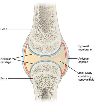
A synovial joint, also known as diarthrosis, joins bones or cartilage with a fibrous joint capsule that is continuous with the periosteum of the joined bones, constitutes the outer boundary of a synovial cavity, and surrounds the bones' articulating surfaces. This joint unites long bones and permits free bone movement and greater mobility. The synovial cavity/joint is filled with synovial fluid. The joint capsule is made up of an outer layer of fibrous membrane, which keeps the bones together structurally, and an inner layer, the synovial membrane, which seals in the synovial fluid.

<span class="mw-page-title-main">Back injury</span> Damage or wear to bones, muscles or other tissues of the back

Back injuries result from damage, wear, or trauma to the bones, muscles, or other tissues of the back. Common back injuries include sprains and strains, herniated discs, and fractured vertebrae. The lumbar spine is often the site of back pain. The area is susceptible because of its flexibility and the amount of body weight it regularly bears. It is estimated that low-back pain may affect as much as 80 to 90 percent of the general population in the United States.

<span class="mw-page-title-main">Genu valgum</span> Medical condition known as knock-knee

Genu valgum, commonly called "knock-knee", is a condition in which the knees angle in and touch each other when the legs are straightened. Individuals with severe valgus deformities are typically unable to touch their feet together while simultaneously straightening the legs. The term originates from the Latin genu, 'knee', and valgus which means "bent outwards", but is also used to describe the distal portion of the knee joint which bends outwards and thus the proximal portion seems to be bent inwards.

<span class="mw-page-title-main">Genu varum</span> Varus deformity marked by (outward) bowing at the knee

Genu varum is a varus deformity marked by (outward) bowing at the knee, which means that the lower leg is angled inward (medially) in relation to the thigh's axis, giving the limb overall the appearance of an archer's bow. Usually medial angulation of both lower limb bones is involved.

<span class="mw-page-title-main">Spondylosis</span> Degeneration of the vertebral column

Spondylosis is the degeneration of the vertebral column from any cause. In the more narrow sense it refers to spinal osteoarthritis, the age-related degeneration of the spinal column, which is the most common cause of spondylosis. The degenerative process in osteoarthritis chiefly affects the vertebral bodies, the neural foramina and the facet joints. If severe, it may cause pressure on the spinal cord or nerve roots with subsequent sensory or motor disturbances, such as pain, paresthesia, imbalance, and muscle weakness in the limbs.

<span class="mw-page-title-main">Joint dislocation</span> Medical injury

A joint dislocation, also called luxation, occurs when there is an abnormal separation in the joint, where two or more bones meet. A partial dislocation is referred to as a subluxation. Dislocations are often caused by sudden trauma on the joint like an impact or fall. A joint dislocation can cause damage to the surrounding ligaments, tendons, muscles, and nerves. Dislocations can occur in any major joint or minor joint. The most common joint dislocation is a shoulder dislocation.

<span class="mw-page-title-main">Cervical vertebrae</span> Vertebrae of the neck

In tetrapods, cervical vertebrae are the vertebrae of the neck, immediately below the skull. Truncal vertebrae lie caudal of cervical vertebrae. In sauropsid species, the cervical vertebrae bear cervical ribs. In lizards and saurischian dinosaurs, the cervical ribs are large; in birds, they are small and completely fused to the vertebrae. The vertebral transverse processes of mammals are homologous to the cervical ribs of other amniotes. Most mammals have seven cervical vertebrae, with the only three known exceptions being the manatee with six, the two-toed sloth with five or six, and the three-toed sloth with nine.

<span class="mw-page-title-main">Spondylolisthesis</span> Displacement of one spinal vertebra compared to another

Spondylolisthesis is the displacement of one spinal vertebra compared to another. While some medical dictionaries define spondylolisthesis specifically as the forward or anterior displacement of a vertebra over the vertebra inferior to it, it is often defined in medical textbooks as displacement in any direction. Spondylolisthesis is graded based upon the degree of slippage of one vertebral body relative to the subsequent adjacent vertebral body. Spondylolisthesis is classified as one of the six major etiologies: degenerative, traumatic, dysplastic, isthmic, pathologic, or post-surgical. Spondylolisthesis most commonly occurs in the lumbar spine, primarily at the L5-S1 level, with the L5 vertebral body anteriorly translating over the S1 vertebral body.

<span class="mw-page-title-main">Knee replacement</span> Surgical procedure

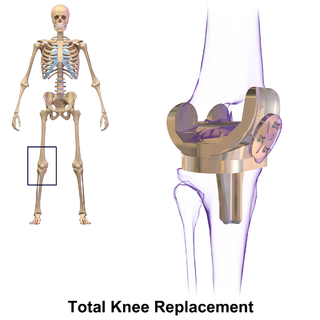
Knee replacement, also known as knee arthroplasty, is a surgical procedure to replace the weight-bearing surfaces of the knee joint to relieve pain and disability, most commonly offered when joint pain is not diminished by conservative sources. It may also be performed for other knee diseases, such as rheumatoid arthritis. In patients with severe deformity from advanced rheumatoid arthritis, trauma, or long-standing osteoarthritis, the surgery may be more complicated and carry higher risk. Osteoporosis does not typically cause knee pain, deformity, or inflammation, and is not a reason to perform knee replacement.

A meniscus transplant or meniscal transplant is a transplant of the meniscus of the knee, which separates the thigh bone (femur) from the lower leg bone (tibia). The worn or damaged meniscus is removed and is replaced with a new one from a donor. The meniscus to be transplanted is taken from a cadaver, and, as such, is known as an allograft. Meniscal transplantation is technically difficult, as it must be sized accurately for each person, positioned properly and secured to the tibial plateau. Its success also depends on donor compatibility, stability of the transplant, and long-term health of the underlying articular cartilage.

<span class="mw-page-title-main">Femoroacetabular impingement</span> Medical condition

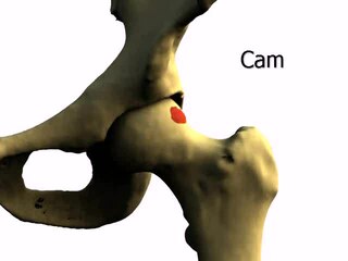
Femoroacetabular impingement (FAI) is a condition involving one or more anatomical abnormalities of the hip joint, which is a ball and socket joint. It is a common cause of hip pain and discomfort in young and middle-aged adults. It occurs when the ball shaped femoral head contacts the acetabulum abnormally or does not permit a normal range of motion in the acetabular socket. Damage can occur to the articular cartilage, or labral cartilage, or both. The condition may be symptomatic or asymptomatic. It may cause osteoarthritis of the hip. Treatment options range from conservative management to surgery.

<span class="mw-page-title-main">Facet syndrome</span> Medical condition

Facet syndrome is a syndrome in which the facet joints cause painful symptoms. In conjunction with degenerative disc disease, a distinct but functionally related condition, facet arthropathy is believed to be one of the most common causes of lower back pain.

<span class="mw-page-title-main">Diffuse idiopathic skeletal hyperostosis</span> Medical condition

Diffuse idiopathic skeletal hyperostosis (DISH) is a condition characterized by abnormal calcification/bone formation (hyperostosis) of the soft tissues surrounding the joints of the spine, and also of the peripheral or appendicular skeleton. In the spine, there is bone formation along the anterior longitudinal ligament and sometimes the posterior longitudinal ligament, which may lead to partial or complete fusion of adjacent vertebrae. The facet and sacroiliac joints tend to be uninvolved. The thoracic spine is the most common level involved. In the peripheral skeleton, DISH manifests as a calcific enthesopathy, with pathologic bone formation at sites where ligaments and tendons attach to bone.

<span class="mw-page-title-main">Spinal stenosis</span> Disease of the bony spine that results in narrowing of the spinal canal

Spinal stenosis is an abnormal narrowing of the spinal canal or neural foramen that results in pressure on the spinal cord or nerve roots. Symptoms may include pain, numbness, or weakness in the arms or legs. Symptoms are typically gradual in onset and improve with leaning forward. Severe symptoms may include loss of bladder control, loss of bowel control, or sexual dysfunction.

<span class="mw-page-title-main">Hip pain</span>

Pain in the hip is the experience of pain in the muscles or joints in the hip/ pelvic region, a condition commonly arising from any of a number of factors. Sometimes it is closely associated with lower back pain.

X-ray motion analysis is a technique used to track the movement of objects using X-rays. This is done by placing the subject to be imaged in the center of the X-ray beam and recording the motion using an image intensifier and a high-speed camera, allowing for high quality videos sampled many times per second. Depending on the settings of the X-rays, this technique can visualize specific structures in an object, such as bones or cartilage. X-ray motion analysis can be used to perform gait analysis, analyze joint movement, or record the motion of bones obscured by soft tissue. The ability to measure skeletal motions is a key aspect to one's understanding of vertebrate biomechanics, energetics, and motor control.

References

  1. 1 2 3 4 Kettler, Annette; Wilke, Hans-Joachim (2005). "Review of existing grading systems for cervical or lumbar disc and facet joint degeneration". European Spine Journal. 15 (6): 705–718. doi:10.1007/s00586-005-0954-y. ISSN   0940-6719. PMC   3489462 . PMID   16172902.
  2. Ofiram, Elisha; Garvey, Timothy A.; Schwender, James D.; Denis, Francis; Perra, Joseph H.; Transfeldt, Ensor E.; Winter, Robert B.; Wroblewski, Jill M. (2009). "Cervical degenerative index: a new quantitative radiographic scoring system for cervical spondylosis with interobserver and intraobserver reliability testing". Journal of Orthopaedics and Traumatology. 10 (1): 21–26. doi:10.1007/s10195-008-0041-3. ISSN   1590-9921. PMC   2657349 . PMID   19384631.
  3. Brox, Jens; Lereim, Paul; Merckoll, Else; Finnanger, Anne Marie (2009). "Radiographic classification of glenohumeral arthrosis". Acta Orthopaedica Scandinavica. 74 (2): 186–189. doi:10.1080/00016470310013932. ISSN   0001-6470. PMID   12807327. S2CID   36730645.
  4. Page 117 in Barbara N. W. Weissman (2009). Imaging of Arthritis and Metabolic Bone Disease. Elsevier Health Sciences. ISBN   9780323041775.
  5. "Glenohumeral joint space". radref.org., in turn citing: Petersson, Claes J.; Redlund-Johnell, Inga (2009). "Joint Space in Normal Gleno-Humeral Radiographs". Acta Orthopaedica Scandinavica. 54 (2): 274–276. doi: 10.3109/17453678308996569 . ISSN   0001-6470. PMID   6846006.
  6. Zdravko Jotanovic, Radovan Mihelic, Gordan Gulan, Branko Sestan, Zlatko Dembic (2015). "Osteoarthritis of the hip: An overview". Periodicum Biologorum. 117 (1).{{cite journal}}: CS1 maint: multiple names: authors list (link)
  7. 1 2 Lequesne, M (2004). "The normal hip joint space: variations in width, shape, and architecture on 223 pelvic radiographs". Annals of the Rheumatic Diseases. 63 (9): 1145–1151. doi:10.1136/ard.2003.018424. ISSN   0003-4967. PMC   1755132 . PMID   15308525.
  8. Terjesen, Terje; Gunderson, Ragnhild B (2012). "Radiographic evaluation of osteoarthritis of the hip". Acta Orthopaedica. 83 (2): 185–189. doi:10.3109/17453674.2012.665331. ISSN   1745-3674. PMC   3339535 . PMID   22329668.
  9. "Tönnis Classification of Osteoarthritis by Radiographic Changes". Society of Preventive Hip Surgery. Retrieved 2016-12-13.
  10. 1 2 3 4 Wright, RW (2014). "Osteoarthritis Classification Scales: Interobserver Reliability and Arthroscopic Correlation". The Journal of Bone and Joint Surgery. American Volume. 96 (14): 1145–1151. doi:10.2106/JBJS.M.00929. ISSN   0021-9355. PMC   4083772 . PMID   25031368.
  11. Hefti F, Müller W, Jakob RP, Stäubli HU (1993). "Evaluation of knee ligament injuries with the IKDC form". Knee Surg Sports Traumatol Arthrosc. 1 (3–4): 226–34. doi:10.1007/bf01560215. PMID   8536037. S2CID   22344987.
  12. Hernández-Vaquero, Daniel; Fernández-Carreira, José Manuel (2012). "Relationship between radiological grading and clinical status in knee osteoarthritis. a multicentric study". BMC Musculoskeletal Disorders. 13 (1): 194. doi: 10.1186/1471-2474-13-194 . ISSN   1471-2474. PMC   3520822 . PMID   23046837.
  13. Kim, Young-Mo; Joo, Yong-Bum (2012). "Patellofemoral Osteoarthritis". Knee Surgery & Related Research. 24 (4): 193–200. doi:10.5792/ksrr.2012.24.4.193. ISSN   2234-0726. PMC   3526755 . PMID   23269956.
  14. Page 722 in Gary S. Firestein, Ralph Budd, Sherine E Gabriel, Iain B. McInnes, James R O'Dell (2012). Kelley's Textbook of Rheumatology E-Book. Elsevier Health Sciences. ISBN   9781455737673.{{cite book}}: CS1 maint: multiple names: authors list (link)
  15. Massilla Mani, F.; Sivasubramanian, S. Satha (2016). "A study of temporomandibular joint osteoarthritis using computed tomographic imaging". Biomedical Journal. 39 (3): 201–206. doi:10.1016/j.bj.2016.06.003. ISSN   2319-4170. PMC   6138784 . PMID   27621122.
  16. Nicolas Holzer, Davide Salvo, Anne Karien Marijnissen, Aminudin Che Ahmad, Emanuele Sera, Pierre Hoffmeyer, Anne Lübbeke Wolff, Mathieu Assal (2017-09-14). "How to assess ankle osteoarthritis: comparison of the Kellgren and Lawrence scale with functional outcome and digital image analysis". Orthopaedic Proceedings. 94-B.{{cite journal}}: CS1 maint: multiple names: authors list (link)
  17. Imai, Kan; Ikoma, Kazuya; Kido, Masamitsu; Maki, Masahiro; Fujiwara, Hiroyoshi; Arai, Yuji; Oda, Ryo; Tokunaga, Daisaku; Inoue, Nozomu; Kubo, Toshikazu (2015). "Joint space width of the tibiotalar joint in the healthy foot". Journal of Foot and Ankle Research. 8 (1): 26. doi: 10.1186/s13047-015-0086-5 . ISSN   1757-1146. PMC   4490633 . PMID   26146520.
  18. Quintana, José M.; Escobar, Antonio; Arostegui, Inmaculada; Bilbao, Amaia; Azkarate, Jesús; Goenaga, J. Ignacio; Arenaza, Juan C. (2006). "Health-Related Quality of Life and Appropriateness of Knee or Hip Joint Replacement". Archives of Internal Medicine. 166 (2): 220–6. doi:10.1001/archinte.166.2.220. ISSN   0003-9926. PMID   16432092.