| Triradiate cartilage | |
|---|---|
 The acetabulum | |
| Anatomical terminology |
The triradiate cartilage (in Latin cartilago ypsiloformis) is the 'Y'-shaped epiphyseal plate between the ilium, ischium and pubis to form the acetabulum of the os coxae.
| Triradiate cartilage | |
|---|---|
| The acetabulum | |
| Anatomical terminology |
The triradiate cartilage (in Latin cartilago ypsiloformis) is the 'Y'-shaped epiphyseal plate between the ilium, ischium and pubis to form the acetabulum of the os coxae.
In children, the triradiate cartilage closes at an approximate bone age of 12 years for girls and 14 years for boys. [1]
Evaluating the position of the triradiate cartilage on an AP radiograph of the pelvis with both Perkin's line and Hilgenreiner's line can help establish a diagnosis of developmental dysplasia of the hip.
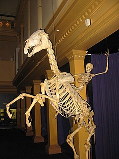
A skeleton is a structural frame that supports an animal body. There are several different skeletal types: the exoskeleton, which is the stable outer shell of an organism, the endoskeleton, which forms the support structure inside the body, and the hydroskeleton, a flexible skeleton supported by fluid pressure. The term comes from Greek σκελετός (skeletós) 'dried up'.

The larynx, commonly called the voice box, is an organ in the top of the neck involved in breathing, producing sound and protecting the trachea against food aspiration. The opening of larynx into pharynx known as the laryngeal inlet is about 4–5 centimeters in diameter. The larynx houses the vocal cords, and manipulates pitch and volume, which is essential for phonation. It is situated just below where the tract of the pharynx splits into the trachea and the esophagus. The word larynx comes from a similar Ancient Greek word.

Cartilage is a resilient and smooth elastic tissue, rubber-like padding that covers and protects the ends of long bones at the joints and nerves, and is a structural component of the rib cage, the ear, the nose, the bronchial tubes, the intervertebral discs, and many other body components. It is not as hard and rigid as bone, but it is much stiffer and much less flexible than muscle. The matrix of cartilage is made up of glycosaminoglycans, proteoglycans, collagen fibers and, sometimes, elastin.
The acetabulum is a concave surface of the pelvis. The head of the femur meets with the pelvis at the acetabulum, forming the hip joint.

In humans and other primates, the knee joins the thigh with the leg and consists of two joints: one between the femur and tibia, and one between the femur and patella. It is the largest joint in the human body. The knee is a modified hinge joint, which permits flexion and extension as well as slight internal and external rotation. The knee is vulnerable to injury and to the development of osteoarthritis.

Rhinoplasty, commonly known as a nose job, is a plastic surgery procedure for altering and reconstructing the nose. There are two types of plastic surgery used – reconstructive surgery that restores the form and functions of the nose and cosmetic surgery that changes the appearance of the nose. Reconstructive surgery seeks to resolve nasal injuries caused by various traumas including blunt, and penetrating trauma and trauma caused by blast injury. Reconstructive surgery also treats birth defects, breathing problems, and failed primary rhinoplasties. Rhinoplasty may remove a bump, narrow nostril width, change the angle between the nose and the mouth, or address injuries, birth defects, or other problems that affect breathing, such as a deviated nasal septum or a sinus condition.

Coxa vara is a deformity of the hip, whereby the angle between the head and the shaft of the femur is reduced to less than 120 degrees. This results in the leg being shortened and the development of a limp. It may be congenital and is commonly caused by injury, such as a fracture. It can also occur when the bone tissue in the neck of the femur is softer than normal, causing it to bend under the weight of the body. This may either be congenital or the result of a bone disorder. The most common cause of coxa vara is either congenital or developmental. Other common causes include metabolic bone diseases, post-Perthes deformity, osteomyelitis, and post traumatic. Shepherd's Crook deformity is a severe form of coxa vara where the proximal femur is severely deformed with a reduction in the neck shaft angle beyond 90 degrees. It is most commonly a sequela of osteogenesis imperfecta, Pagets disease, osteomyelitis, tumour and tumour-like conditions.
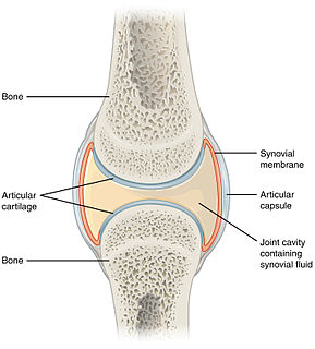
A synovial joint, also known as diarthrosis, joins bones or cartilage with a fibrous joint capsule that is continuous with the periosteum of the joined bones, constitutes the outer boundary of a synovial cavity, and surrounds the bones' articulating surfaces. This joint unites long bones and permits free bone movement and greater mobility. The synovial cavity/joint is filled with synovial fluid. The joint capsule is made up of an outer layer, the articular capsule, which keeps the bones together structurally, and an inner layer, the synovial membrane, which seals in the synovial fluid.

The nasal septum separates the left and right airways of the nasal cavity, dividing the two nostrils.
Endochondral ossification is one of the two essential processes during fetal development of the mammalian skeletal system by which bone tissue is created. Unlike intramembranous ossification, which is the other process by which bone tissue is created, cartilage is present during endochondral ossification. Endochondral ossification is also an essential process during the rudimentary formation of long bones, the growth of the length of long bones, and the natural healing of bone fractures.
The thyroid cartilage is the largest of the nine cartilages that make up the laryngeal skeleton, the cartilage structure in and around the trachea that contains the larynx. It does not completely encircle the larynx.

In vertebrate anatomy, hip refers to either an anatomical region or a joint.
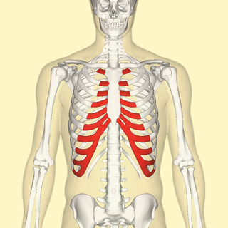
The costal cartilages are bars of hyaline cartilage that serve to prolong the ribs forward and contribute to the elasticity of the walls of the thorax. Costal cartilage is only found at the anterior ends of the ribs, providing medial extension.

Chondrogenesis is the process by which cartilage is developed.

Erythrosuchidae are a family of large basal archosauriform carnivores that lived from the later Early Triassic (Olenekian) to the early Middle Triassic (Anisian).

The epiphyseal plate is a hyaline cartilage plate in the metaphysis at each end of a long bone. It is the part of a long bone where new bone growth takes place; that is, the whole bone is alive, with maintenance remodeling throughout its existing bone tissue, but the growth plate is the place where the long bone grows longer.
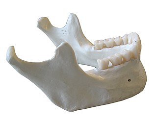
In anatomy, the mandible, lower jaw or jawbone is the largest, strongest and lowest bone in the human facial skeleton. It forms the lower jaw and holds the lower teeth in place. The mandible sits beneath the maxilla. It is the only movable bone of the skull. It is connected to the temporal bones by the temporomandibular joints.
Hilgenreiner's line is a horizontal line drawn on an AP radiograph of the pelvis running between the inferior aspects of both triradiate cartilages of the acetabulums. It is named for Heinrich Hilgenreiner.
Perkin's line is a line drawn on an AP radiograph of the pelvis perpendicular to Hilgenreiner's line at the lateral aspects of the triradiate cartilage of the acetabulum.
X-rays of hip dysplasia are one of the two main methods of medical imaging to diagnose hip dysplasia, the other one being medical ultrasonography. Ultrasound imaging yields better results defining the anatomy until the cartilage is ossified. When the infant is around 3 months old a clear roentgenographic image can be achieved. Unfortunately the time the joint gives a good x-ray image is also the point at which nonsurgical treatment methods cease to give good results.