Related Research Articles

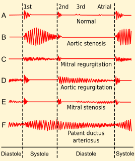
Aortic stenosis is the narrowing of the exit of the left ventricle of the heart, such that problems result. It may occur at the aortic valve as well as above and below this level. It typically gets worse over time. Symptoms often come on gradually with a decreased ability to exercise often occurring first. If heart failure, loss of consciousness, or heart related chest pain occur due to AS the outcomes are worse. Loss of consciousness typically occurs with standing or exercising. Signs of heart failure include shortness of breath especially when lying down, at night, or with exercise, and swelling of the legs. Thickening of the valve without narrowing is known as aortic sclerosis.

Mitral stenosis is a valvular heart disease characterized by the narrowing of the opening of the mitral valve of the heart. It is almost always caused by rheumatic valvular heart disease. Normally, the mitral valve is about 5 cm2 during diastole. Any decrease in area below 2 cm2 causes mitral stenosis. Early diagnosis of mitral stenosis in pregnancy is very important as the heart cannot tolerate increased cardiac output demand as in the case of exercise and pregnancy. Atrial fibrillation is a common complication of resulting left atrial enlargement, which can lead to systemic thromboembolic complications like stroke.

Interventional cardiology is a branch of cardiology that deals specifically with the catheter based treatment of structural heart diseases. Andreas Gruentzig is considered the father of interventional cardiology after the development of angioplasty by interventional radiologist Charles Dotter.

Mitral regurgitation(MR), also known as mitral insufficiency, or mitral incompetence is a form of valvular heart disease in which the mitral valve is insufficient and does not close properly when the heart pumps out blood. It is the abnormal leaking of blood backwards – regurgitation from the left ventricle, through the mitral valve, into the left atrium, when the left ventricle contracts. Mitral regurgitation is the most common form of valvular heart disease.
Aortic valve replacement is a procedure whereby the failing aortic valve of a patient's heart is replaced with an artificial heart valve. The aortic valve may need to be replaced because:
Valvular heart disease is any cardiovascular disease process involving one or more of the four valves of the heart. These conditions occur largely as a consequence of aging, but may also be the result of congenital (inborn) abnormalities or specific disease or physiologic processes including rheumatic heart disease and pregnancy.

Mitral valve repair is a cardiac surgery procedure performed by cardiac surgeons to treat stenosis (narrowing) or regurgitation (leakage) of the mitral valve. The mitral valve is the "inflow valve" for the left side of the heart. Blood flows from the lungs, where it picks up oxygen, through the pulmonary veins, to the left atrium of the heart. After the left atrium fills with blood, the mitral valve allows blood to flow from the left atrium into the heart's main pumping chamber called the left ventricle. It then closes to keep blood from leaking back into the left atrium or lungs when the ventricle contracts (squeezes) to push blood out to the body. It has two flaps, or leaflets, known as cusps.

A commissurotomy is a surgical incision of a commissure in the body, as one made in the heart at the edges of the commissure formed by cardiac valves, or one made in the brain to treat certain psychiatric disorders.
Aortic valvuloplasty also known as balloon aortic valvotomy is the widening of a stenotic aortic valve using a balloon catheter inside the valve. The balloon is placed into the aortic valve that has become stiff from calcium buildup. The balloon is then inflated in an effort to increase the opening size of the valve and improving blood flow.

Fetal surgery also known as fetal reconstructive surgery,antenatal surgery, prenatal surgery, is a growing branch of maternal-fetal medicine that covers any of a broad range of surgical techniques that are used to treat birth defects in fetuses who are still in the pregnant uterus. There are three main types: open fetal surgery, which involves completely opening the uterus to operate on the fetus; minimally invasive fetoscopic surgery, which uses small incisions and is guided by fetoscopy and sonography; and percutaneous fetal therapy, which involves placing a catheter under continuous ultrasound guidance.

Valve replacement surgery is the replacement of one or more of the heart valves with either an artificial heart valve or a bioprosthesis. It is an alternative to valve repair.
Tricuspid Valve Stenosis is a valvular heart disease that narrows the opening of the heart's tricuspid valve. It is a relatively rare condition that causes stenosis-increased restriction of blood flow through the valve.
Percutaneous aortic valve replacement (PAVR), also known as percutaneous aortic valve implantation (PAVI), transcatheter aortic valve implantation (TAVI) or transcatheter aortic valve replacement (TAVR), is the replacement of the aortic valve of the heart through the blood vessels. The replacement valve is delivered via one of several access methods: transfemoral, transapical, subclavian, direct aortic, and transcaval, among others.
Mitral valve replacement is a procedure whereby the diseased mitral valve of a patient's heart is replaced by either a mechanical or tissue (bioprosthetic) valve.
Lourdes Heart Institute and Neuro Centre (LHINC) is a new block set up in Lourdes Hospital, Cochin, Kerala, India, to cater to tertiary level care for the entire spectrum of cardiovascular and neurological disease. It was inaugurated on 16 March 2007, by Mr. A. K. Antony, the Defence Minister of India. This institute was started to meet a long-felt need to provide cardiac and neurological interventional facilities, and especially to provide interventional neurological facilities for the treatment of strokes, including selective thrombolysis and primary angioplasty for stroke which was hitherto unavailable in this part of India.
Lutembacher's syndrome is a very rare form of congenital heart disease that affects one of the chambers of the heart as well as a valve. It is commonly known as both congenital atrial septal defect (ASD) and acquired mitral stenosis (MS). Congenital atrial septal defect refers to a hole being in the septum or wall that separates the two atria; this condition is usually seen in fetuses and infants. Mitral stenosis refers to mitral valve leaflets sticking to each other making the opening for blood to pass from the atrium to the ventricles very small. With the valve being so small, blood has difficulty passing from the left atrium into the left ventricle. Septal defects that may occur with Lutembacher's syndrome include: Ostium primum atrial septal defect or ostium secundum which is more prevalent.
The following outline is provided as an overview of and topical guide to cardiology, the branch of medicine dealing with disorders of the human heart. The field includes medical diagnosis and treatment of congenital heart defects, coronary artery disease, heart failure, valvular heart disease and electrophysiology. Physicians who specialize in cardiology are called cardiologists.
Heart valve repair is a cardiac surgery procedure, carried out to repair one or more faulty heart valves. In some valvular heart diseases repair where possible is preferable to valve replacement. A mechanical heart valve is a replacement valve that is not itself subject to repair.

A hybrid cardiac surgical procedure in a narrow sense is defined as a procedure that combines a conventional, more invasive surgical part with an interventional part, using some sort of catheter-based procedure guided by fluoroscopy imaging in a hybrid operating room (OR) without interruption. The hybrid technique has a reduced risk of surgical complications and has shown decreased recovery time. It can be used to treat numerous heart diseases and conditions and with the increasing complexity of each case, the hybrid surgical technique is becoming more common.
Fetal aortic stenosis is a disorder that occurs when the fetus’ aortic valve does not fully open during development. The aortic valve is a one way valve that is located between the left ventricle and the aorta, keeping blood from leaking back into the ventricle. It has three leaflets that separate when the ventricle contracts to allow blood to move from the ventricle to the aorta. These leaflets come together when the ventricle relaxes.
References
- ↑ TheFreeDictionary > valvotomy Citing: WordNet 3.0, Farlex clipart collection. © 2003-2008 Princeton University, Farlex Inc.
Tests and procedures involving the heart | |||||||
|---|---|---|---|---|---|---|---|
| Surgery |
| ||||||
| Tests |
| ||||||
| Function tests | |||||||
| Pacing | |||||||
| | This surgery article is a stub. You can help Wikipedia by expanding it. |