AP site

Last updated

Simple representation of an AP site. Site AP.gif
Simple representation of an AP site.

In biochemistry and molecular genetics, an AP site (apurinic/apyrimidinic site), also known as an abasic site, is a location in DNA (also in RNA but much less likely) that has neither a purine nor a pyrimidine base, either spontaneously or due to DNA damage. It has been estimated that under physiological conditions 10,000 apurinic sites and 500 apyrimidinic may be generated in a cell daily. [1] [2]

Contents

AP sites can be formed by spontaneous depurination, but also occur as intermediates in base excision repair. [3] In this process, a DNA glycosylase recognizes a damaged base and cleaves the N-glycosidic bond to release the base, leaving an AP site. A variety of glycosylases that recognize different types of damage exist, including oxidized or methylated bases, or uracil in DNA. The AP site can then be cleaved by an AP endonuclease, leaving 3'-hydroxyl and deoxyribose-5-phosphate termini (see DNA structure). In alternative fashion, bifunctional glycosylase-lyases can cleave the AP site, leaving a 5' phosphate adjacent to a 3' α,β-unsaturated aldehyde. Both mechanisms form a single-strand break, which is then repaired by either short-patch or long-patch base excision repair. [4]

If left unrepaired, AP sites can lead to mutation during semiconservative replication. They can cause replication fork stalling and are bypassed by translesion synthesis. In E. coli , adenine is preferentially inserted across from AP sites, known as the "A rule". The situation is more complex in higher eukaryotes, with different nucleotides showing a preference depending on the organism and experimental conditions. [3]

Formation

AP sites form when deoxyribose is cleaved from its nitrogenous base, breaking the glycosidic linkage between the two. This can happen spontaneously, as a result of chemical activity, radiation, or due to enzyme activity. The glycosidic linkages in DNA can be broken via acid-catalyzed hydrolysis. Purine bases can be ejected under weakly acidic conditions, while pyrimidines require stronger acidity in order to be cleaved. Purines may even be removed at neutral pH, if temperature increases sufficiently. AP site formation can also be caused by various base-modifying chemicals. Alkylation, deamination, and oxidation of individual bases can all lead to the weakening of the glycosyl bond, so exposure to agents that cause those modifications can encourage AP site formation. [2]

Ionizing radiation can also lead to AP site formation. Irradiated environments contain radicals, which can contribute to AP sites in multiple ways. Hydroxyl radicals can attack the glycosidic linkages, directly creating an AP site, or make the glycosyl bond less favorable by linking to the base or the deoxyribose ring. [2]

Enzymes, namely DNA glycosylases, also commonly create AP sites, as part of the base excision repair pathway. In a given mammalian cell, 5000–10,000 apurinic sites are estimated to form per day. Apyrimidinic sites form at a rate roughly 20 times slower, with estimates at around 500 formation events per day, per cell. At rates this high, it is critical for cells to have a robust repair apparatus in place in order to prevent mutation.

Characteristics

Chemical characteristics

AP site reactivity AP lyase mechanism.png
AP site reactivity

AP sites are extremely reactive. They fluctuate between a furanose ring and an open-chain free aldehyde and free alcohol conformation. Exposure to a nucleophile can cause a β-elimination reaction, wherein the 3' phosphoester bond is broken, causing a single-stranded break. This reaction can be catalyzed by AP lyase. [2] In the presence of excess reagent, an additional elimination can occur on the 5' side. The free aldehyde can also react with nucleophilic, amine-containing aldehydes. These reactions can further promote phosphoester bond cleavage. Aldehydes containing O-HN2 groups can serve to stabilize the abasic site by reacting with the aldehyde group. This interaction does not cleave the phosphoester bond.

Biological activity

AP sites in living cells can cause various and severe consequences, including cell death. The single-stranded breaks occurring due to β-elimination require repair by DNA Ligase in order to avoid mutation. When DNA polymerase encounters an abasic site, DNA replication is usually blocked, which may itself lead to a single-stranded or double-stranded break in the DNA helix. [4] In E. coli, when the enzyme manages to bypass the abasic site, an adenine is preferentially incorporated into the new strand. [2] [3] If AP sites in DNA are not repaired, DNA replication cannot proceed normally, and significant mutations can result. [4] If mutations are merely single nucleotide polymorphisms, then the cell can potentially be unaffected. However, if more serious mutations occur, cell function can be severely impaired, growth and division may be impaired, or the cell may simply die.

Repair

AP sites are an important feature of the base excision repair pathway. DNA glycosylases first create abasic sites by recognizing and removing modified bases. Many glycosylase variants exist to deal with the multiple ways a base can be damaged. The most common circumstances are base alkylation, oxidation, and the presence of a uracil in the DNA strand. [4] Once an AP site has been successful created, an AP endonuclease catalyzes the breakage of one phosphoester bond, creating a nick in the backbone of the helix. [4] The breakage can be either 3' or 5' of the site, depending on the variant of the enzyme. End processing enzymes then prepare the site for nick ligation, which is performed by DNA polymerase. [4] The base inserted into the nick is determined by the corresponding base on the opposite strand. The nick is then sealed by DNA ligase.

The activity of AP endonuclease in the repair of AP sites in the frontal/parietal cortex, cerebellum, brain stem, midbrain and hypothalamus declines with age in rats on an ad libitum diet. [5] In calorie restricted rats, by comparison, AP endonuclease activity in these brain regions remains higher with age. [5] These findings suggest that elevated AP site repair in calorie restricted animals may delay the aging process.

Related Research Articles

Deamination is the removal of an amino group from a molecule. Enzymes that catalyse this reaction are called deaminases.

<span class="mw-page-title-main">Nuclease</span> Class of enzymes

A nuclease is an enzyme capable of cleaving the phosphodiester bonds between nucleotides of nucleic acids. Nucleases variously effect single and double stranded breaks in their target molecules. In living organisms, they are essential machinery for many aspects of DNA repair. Defects in certain nucleases can cause genetic instability or immunodeficiency. Nucleases are also extensively used in molecular cloning.

In molecular biology, endonucleases are enzymes that cleave the phosphodiester bond within a polynucleotide chain. Some, such as deoxyribonuclease I, cut DNA relatively nonspecifically, while many, typically called restriction endonucleases or restriction enzymes, cleave only at very specific nucleotide sequences. Endonucleases differ from exonucleases, which cleave the ends of recognition sequences instead of the middle (endo) portion. Some enzymes known as "exo-endonucleases", however, are not limited to either nuclease function, displaying qualities that are both endo- and exo-like. Evidence suggests that endonuclease activity experiences a lag compared to exonuclease activity.

DNA glycosylases are a family of enzymes involved in base excision repair, classified under EC number EC 3.2.2. Base excision repair is the mechanism by which damaged bases in DNA are removed and replaced. DNA glycosylases catalyze the first step of this process. They remove the damaged nitrogenous base while leaving the sugar-phosphate backbone intact, creating an apurinic/apyrimidinic site, commonly referred to as an AP site. This is accomplished by flipping the damaged base out of the double helix followed by cleavage of the N-glycosidic bond.

<span class="mw-page-title-main">Base excision repair</span> DNA repair process

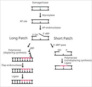
Base excision repair (BER) is a cellular mechanism, studied in the fields of biochemistry and genetics, that repairs damaged DNA throughout the cell cycle. It is responsible primarily for removing small, non-helix-distorting base lesions from the genome. The related nucleotide excision repair pathway repairs bulky helix-distorting lesions. BER is important for removing damaged bases that could otherwise cause mutations by mispairing or lead to breaks in DNA during replication. BER is initiated by DNA glycosylases, which recognize and remove specific damaged or inappropriate bases, forming AP sites. These are then cleaved by an AP endonuclease. The resulting single-strand break can then be processed by either short-patch or long-patch BER.

<span class="mw-page-title-main">Depurination</span>

Depurination is a chemical reaction of purine deoxyribonucleosides, deoxyadenosine and deoxyguanosine, and ribonucleosides, adenosine or guanosine, in which the β-N-glycosidic bond is hydrolytically cleaved releasing a nucleic base, adenine or guanine, respectively. The second product of depurination of deoxyribonucleosides and ribonucleosides is sugar, 2'-deoxyribose and ribose, respectively. More complex compounds containing nucleoside residues, nucleotides and nucleic acids, also suffer from depurination. Deoxyribonucleosides and their derivatives are substantially more prone to depurination than their corresponding ribonucleoside counterparts. Loss of pyrimidine bases occurs by a similar mechanism, but at a substantially lower rate.

A nick is a discontinuity in a double stranded DNA molecule where there is no phosphodiester bond between adjacent nucleotides of one strand typically through damage or enzyme action. Nicks allow DNA strands to untwist during replication, and are also thought to play a role in the DNA mismatch repair mechanisms that fix errors on both the leading and lagging daughter strands.

<span class="mw-page-title-main">AP endonuclease</span> Enzyme involved in DNA repair

Apurinic/apyrimidinic (AP) endonuclease is an enzyme that is involved in the DNA base excision repair pathway (BER). Its main role in the repair of damaged or mismatched nucleotides in DNA is to create a nick in the phosphodiester backbone of the AP site created when DNA glycosylase removes the damaged base.

<span class="mw-page-title-main">Crosslinking of DNA</span> Phenomenon in genetics

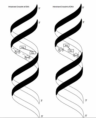
In genetics, crosslinking of DNA occurs when various exogenous or endogenous agents react with two nucleotides of DNA, forming a covalent linkage between them. This crosslink can occur within the same strand (intrastrand) or between opposite strands of double-stranded DNA (interstrand). These adducts interfere with cellular metabolism, such as DNA replication and transcription, triggering cell death. These crosslinks can, however, be repaired through excision or recombination pathways.

<span class="mw-page-title-main">Nucleotidyltransferase</span>

Nucleotidyltransferases are transferase enzymes of phosphorus-containing groups, e.g., substituents of nucleotidylic acids or simply nucleoside monophosphates. The general reaction of transferring a nucleoside monophosphate moiety from A to B, can be written as:

Deoxyribonuclease IV (phage-T4-induced) is catalyzes the degradation nucleotides in DsDNA by attacking the 5'-terminal end.

<span class="mw-page-title-main">APEX1</span> Protein-coding gene in the species Homo sapiens

DNA-(apurinic or apyrimidinic site) lyase is an enzyme that in humans is encoded by the APEX1 gene.

<span class="mw-page-title-main">LIG1</span> Protein-coding gene in the species Homo sapiens

DNA ligase 1 is an enzyme that in humans is encoded by the LIG1 gene. DNA ligase I is the only known eukaryotic DNA ligase involved in both DNA replication and repair, making it the most studied of the ligases.

The enzyme DNA-(apurinic or apyrimidinic site) lyase, also referred to as DNA-(apurinic or apyrimidinic site) 5'-phosphomonoester-lyase or DNA AP lyase catalyzes the cleavage of the C-O-P bond 3' from the apurinic or apyrimidinic site in DNA via β-elimination reaction, leaving a 3'-terminal unsaturated sugar and a product with a terminal 5'-phosphate. In the 1970s, this class of enzyme was found to repair at apurinic or apyrimidinic DNA sites in E. coli and in mammalian cells. The major active enzyme of this class in bacteria, and specifically, E. coli is endonuclease type III. This enzyme is part of a family of lyases that cleave carbon-oxygen bonds.

<span class="mw-page-title-main">Uracil-DNA glycosylase</span> Enzyme that repairs DNA damage

Uracil-DNA glycosylase is also known as UNG or UDG. Its most important function is to prevent mutagenesis by eliminating uracil from DNA molecules by cleaving the N-glycosidic bond and initiating the base-excision repair (BER) pathway.

<span class="mw-page-title-main">DNA-3-methyladenine glycosylase</span> Protein-coding gene in the species Homo sapiens

DNA-3-methyladenine glycosylase also known as 3-alkyladenine DNA glycosylase (AAG) or N-methylpurine DNA glycosylase (MPG) is an enzyme that in humans is encoded by the MPG gene.

<span class="mw-page-title-main">NEIL1</span> Protein-coding gene in the species Homo sapiens

Endonuclease VIII-like 1 is an enzyme that in humans is encoded by the NEIL1 gene.

<span class="mw-page-title-main">FPG IleRS zinc finger</span>

The FPG IleRS zinc finger domain represents a zinc finger domain found at the C-terminal in both DNA glycosylase/AP lyase enzymes and in isoleucyl tRNA synthetase. In these two types of enzymes, the C-terminal domain forms a zinc finger.

<span class="mw-page-title-main">H2TH domain</span>

In molecular biology, the H2TH domain is a DNA-binding domain found in DNA glycosylase/AP lyase enzymes, which are involved in base excision repair of DNA damaged by oxidation or by mutagenic agents. Most damage to bases in DNA is repaired by the base excision repair pathway. These enzymes are primarily from bacteria, and have both DNA glycosylase activity EC 3.2.2.- and AP lyase activity EC 4.2.99.18. Examples include formamidopyrimidine-DNA glycosylases and endonuclease VIII (Nei).

DNA-deoxyinosine glycosylase is an enzyme with systematic name DNA-deoxyinosine deoxyribohydrolase. This enzyme is involved in DNA damage repair and targets hypoxanthine bases.

References

  1. Tropp, Burton (2012). Molecular Biology. Sudbury, MA: Jones & Bartlett Learning. p. 455. ISBN   978-1-4496-0091-4.
  2. 1 2 3 4 5 Borlé, Myriam (1987). "Formation, detection, and repair of AP sites". Mutation Research. 181 (1): 45–56. doi:10.1016/0027-5107(87)90286-7. PMID   2444877.
  3. 1 2 3 Abasic sites in DNA: repair and biological consequences in Saccharomyces cerevisiae. DNA Repair (Amst). 2004 Jan 5;3(1):1-12.
  4. 1 2 3 4 5 6 Lindhal, Tomas (1993). "Instability and decay of the primary structure of DNA". Nature. 362 (6422): 709–715. Bibcode:1993Natur.362..709L. doi:10.1038/362709a0. PMID   8469282. S2CID   4283694.
  5. 1 2 Kisby GE, Kohama SG, Olivas A, Churchwell M, Doerge D, Spangler E, de Cabo R, Ingram DK, Imhof B, Bao G, Kow YW. Effect of caloric restriction on base-excision repair (BER) in the aging rat brain. Exp Gerontol. 2010 Mar;45(3):208-16. doi: 10.1016/j.exger.2009.12.003. Epub 2009 Dec 11. PMID 20005284; PMCID: PMC2826610