
The ventricular system is a set of four interconnected cavities (ventricles) in the brain, where the cerebrospinal fluid (CSF) is produced. Within each ventricle is a region of choroid plexus, a network of ependymal cells involved in the production of CSF. The ventricular system is continuous with the central canal of the spinal cord, allowing for the flow of CSF to circulate. All of the ventricular system and the central canal of the spinal cord are lined with ependyma, a specialised form of epithelium.
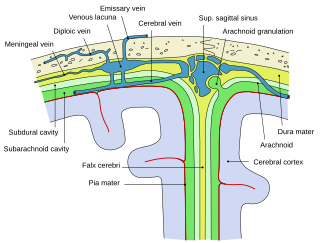
Arachnoid granulations are small protrusions of the arachnoid mater into the outer membrane of the dura mater. They protrude into the dural venous sinuses of the brain, and allow cerebrospinal fluid (CSF) to exit the subarachnoid space and enter the blood stream.

Dura mater is a thick membrane made of dense irregular connective tissue that surrounds the brain and spinal cord. It is the outermost of the three layers of membrane called the meninges that protect the central nervous system. The other two meningeal layers are the arachnoid mater and the pia mater. The dura surrounds the brain and the spinal cord and is responsible for keeping in the cerebrospinal fluid. It is derived from neural crest cells.

A hematoma or haematoma is a localized bleeding outside of blood vessels, due to either disease or trauma including injury or surgery and may involve blood continuing to seep from broken capillaries. A hematoma is benign and is initially in liquid form spread among the tissues including in sacs between tissues where it may coagulate and solidify before blood is reabsorbed into blood vessels. An ecchymosis is a hematoma of the skin larger than 10mm.

The spinal canal is the space in the vertebral column formed by the vertebrae through which the spinal cord passes. It is a process of the dorsal body cavity. This canal is enclosed within the vertebral foramen of the vertebrae. In the intervertebral spaces, the canal is protected by the ligamentum flavum posteriorly and the posterior longitudinal ligament anteriorly.

Intracranial hemorrhage (ICH), also known as intracranial bleed, is bleeding within the skull. Subtypes are intracerebral bleeds, subarachnoid bleeds, epidural bleeds, and subdural bleeds.

In the spine, the epidural space is an anatomic space that is the outermost part of the spinal canal. It is the space within the canal lying outside the dura mater. In humans the epidural space contains lymphatics, spinal nerve roots, loose connective tissue, fatty tissue, small arteries, and a network of internal vertebral venous plexuses.

Arachnoiditis is an inflammatory condition of the arachnoid mater or 'arachnoid', one of the membranes known as meninges that surround and protect the nerves of the central nervous system, including the brain and spinal cord. The arachnoid can become inflamed because of adverse reactions to chemicals, infection from bacteria or viruses, as the result of direct injury to the spine, chronic compression of spinal nerves, complications from spinal surgery or other invasive spinal procedures, or the accidental intrathecal injection of steroids intended for the epidural space. Inflammation can sometimes lead to the formation of scar tissue and adhesion that can make the spinal nerves "stick" together, a condition where such tissue develops in and between the leptomeninges. The condition is extremely painful, especially when progressing to adhesive arachnoiditis. Another form of the condition is arachnoiditis ossificans, in which the arachnoid becomes ossified, or turns to bone, and is thought to be a late-stage complication of the adhesive form of arachnoiditis.

The subarachnoid cisterns are spaces formed by openings in the subarachnoid space, an anatomic space in the meninges of the brain. The space separates two of the meninges, the arachnoid mater and the pia mater. These cisterns are filled with cerebrospinal fluid.
The cisterna magna is one of three principal openings in the subarachnoid space between the arachnoid and pia mater layers of the meninges surrounding the brain. The openings are collectively referred to as the subarachnoid cisterns. The cisterna magna is located between the cerebellum and the dorsal surface of the medulla oblongata. Cerebrospinal fluid produced in the fourth ventricle drains into the cisterna magna via the lateral apertures and median aperture.
The subdural space is a potential space that can be opened by the separation of the arachnoid mater from the dura mater as the result of trauma, pathologic process, or the absence of cerebrospinal fluid as seen in a cadaver. In the cadaver, due to the absence of cerebrospinal fluid in the subarachnoid space, the arachnoid mater falls away from the dura mater. It may also be the site of trauma, such as a subdural hematoma, causing abnormal separation of dura and arachnoid mater. Hence, the subdural space is referred to as "potential" or "artificial" space.

The cerebellopontine angle (CPA) is located between the cerebellum and the pons.
The cerebellopontine angle is the site of the cerebellopontine angle cistern one of the subarachnoid cisterns that contains cerebrospinal fluid, arachnoid tissue, cranial nerves, and associated vessels. The cerebellopontine angle is also the site of a set of neurological disorders known as the cerebellopontine angle syndrome.

The dural venous sinuses are venous channels found between the endosteal and meningeal layers of dura mater in the brain. They receive blood from internal and external veins of the brain, receive cerebrospinal fluid (CSF) from the subarachnoid space via arachnoid granulations, and mainly empty into the internal jugular vein.
Neoplastic or malignant meningitis, also called carcinomatous meningitis, leptomeningeal carcinoma, leptomeningeal carcinomatosis, leptomeningeal metastasis, meningeal carcinomatosis, meningeal metastasis, and meningitis carcinomatosa, is the development of meningitis due to infiltration of the subarachnoid space by cancerous cells. Malignant cells come from primary cancer such as breast cancer or from a primary brain tumor like medulloblastoma. Neoplastic Meningitis (NM) was first reported in the 1870s with the most common cause being breast cancer, lung cancer, and malignant melanoma.
A Rich focus is a tuberculous granuloma occurring within the cortex or meninges of the brain that ruptures into the subarachnoid space, causing tuberculous meningitis. The Rich focus is named for Arnold Rice Rich, a pathologist at Johns Hopkins Hospital, who along with his colleague Howard McCordock first described the post-mortem finding of caseous foci within the cerebral cortex or meninges which appeared to predate the development of meningitis. Prior to their research the prevailing view had been that meningitis occurred as a result of the dissemination of tuberculous bacilli associated with miliary tuberculosis and that these processes occurred at the same time.

A laminar organization describes the way certain tissues, such as bone membrane, skin, or brain tissues, are arranged in layers.

