Cerebrospinal fluid (CSF) is a clear, colorless body fluid found within the tissue that surrounds the brain and spinal cord of all vertebrates.
The ventricular system is a set of four interconnected cavities known as cerebral ventricles in the brain. Within each ventricle is a region of choroid plexus which produces the circulating cerebrospinal fluid (CSF). The ventricular system is continuous with the central canal of the spinal cord from the fourth ventricle, allowing for the flow of CSF to circulate.

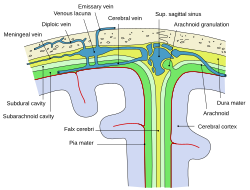
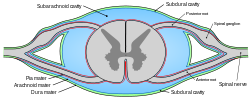
In anatomy, the meninges are the three membranes that envelop the brain and spinal cord. In mammals, the meninges are the dura mater, the arachnoid mater, and the pia mater. Cerebrospinal fluid is located in the subarachnoid space between the arachnoid mater and the pia mater. The primary function of the meninges is to protect the central nervous system.

Pia mater, often referred to as simply the pia, is the delicate innermost layer of the meninges, the membranes surrounding the brain and spinal cord. Pia mater is medieval Latin meaning "tender mother". The other two meningeal membranes are the dura mater and the arachnoid mater. Both the pia and arachnoid mater are derivatives of the neural crest while the dura is derived from embryonic mesoderm. The pia mater is a thin fibrous tissue that is permeable to water and small solutes. The pia mater allows blood vessels to pass through and nourish the brain. The perivascular space between blood vessels and pia mater is proposed to be part of a pseudolymphatic system for the brain. When the pia mater becomes irritated and inflamed the result is meningitis.

Arachnoid granulations are small protrusions of the arachnoid mater into the outer membrane of the dura mater. They protrude into the dural venous sinuses of the brain, and allow cerebrospinal fluid (CSF) to exit the subarachnoid space and enter the blood stream.

Dura mater is a thick membrane made of dense irregular connective tissue that surrounds the brain and spinal cord. It is the outermost of the three layers of membrane called the meninges that protect the central nervous system. The other two meningeal layers are the arachnoid mater and the pia mater. The dura surrounds the brain and the spinal cord. It envelops the arachnoid mater, which is responsible for keeping in the cerebrospinal fluid. It is derived primarily from the neural crest cell population, with postnatal contributions of the paraxial mesoderm.

A hematoma, also spelled haematoma, or blood suffusion is a localized bleeding outside of blood vessels, due to either disease or trauma including injury or surgery and may involve blood continuing to seep from broken capillaries. A hematoma is benign and is initially in liquid form spread among the tissues including in sacs between tissues where it may coagulate and solidify before blood is reabsorbed into blood vessels. An ecchymosis is a hematoma of the skin larger than 10 mm.

A subdural hematoma (SDH) is a type of bleeding in which a collection of blood—usually but not always associated with a traumatic brain injury—gathers between the inner layer of the dura mater and the arachnoid mater of the meninges surrounding the brain. It usually results from tears in bridging veins that cross the subdural space.

The spinal canal is the canal that contains the spinal cord within the vertebral column. The spinal canal is formed by the vertebrae through which the spinal cord passes. It is a process of the dorsal body cavity. This canal is enclosed within the foramen of the vertebrae. In the intervertebral spaces, the canal is protected by the ligamentum flavum posteriorly and the posterior longitudinal ligament anteriorly.

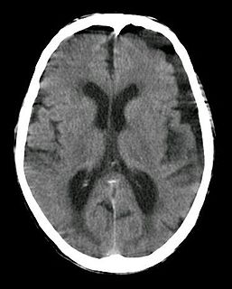
Intracranial hemorrhage (ICH), also known as intracranial bleed, is bleeding within the skull. Subtypes are intracerebral bleeds, subarachnoid bleeds, epidural bleeds, and subdural bleeds. More often than not it ends in a lethal outcome.

In anatomy, the epidural space is the potential space between the dura mater and vertebrae (spine)

The cisterna magna is one of three principal openings in the subarachnoid space between the arachnoid and pia mater layers of the meninges surrounding the brain. The openings are collectively referred to as the subarachnoid cisterns. The cisterna magna is located between the cerebellum and the dorsal surface of the medulla oblongata. Cerebrospinal fluid produced in the fourth ventricle drains into the cisterna magna via the lateral apertures and median aperture.
The arachnoid mater is one of the three meninges, the protective membranes that cover the brain and spinal cord. The arachnoid mater is a derivative of the neural crest mesectoderm in the embryo.
Arachnoid cysts are cerebrospinal fluid covered by arachnoidal cells and collagen that may develop between the surface of the brain and the cranial base or on the arachnoid membrane, one of the three meningeal layers that cover the brain and the spinal cord. Primary arachnoid cysts are a congenital disorder whereas secondary arachnoid cysts are the result of head injury or trauma. Most cases of primary cysts begin during infancy; however, onset may be delayed until adolescence.
The cranial cavity, also known as intracranial space, is the space within the skull that accommodates the brain. The skull minus the mandible is called the cranium. The cavity is formed by eight cranial bones known as the neurocranium that in humans includes the skull cap and forms the protective case around the brain. The remainder of the skull is called the facial skeleton. Meninges are protective membranes that surround the brain to minimize damage of the brain when there is head trauma. Meningitis is the inflammation of meninges caused by bacterial or viral infections.

The dural venous sinuses are venous channels found between the endosteal and meningeal layers of dura mater in the brain. They receive blood from the cerebral veins, receive cerebrospinal fluid (CSF) from the subarachnoid space via arachnoid granulations, and mainly empty into the internal jugular vein.

The superior sagittal sinus, within the human head, is an unpaired area along the attached margin of the falx cerebri. It allows blood to drain from the lateral aspects of anterior cerebral hemispheres to the confluence of sinuses. Cerebrospinal fluid drains through arachnoid granulations into the superior sagittal sinus and is returned to venous circulation.
A subdural hygroma (SDG) is a collection of cerebrospinal fluid (CSF), without blood, located under the dural membrane of the brain. Most subdural hygromas are believed to be derived from chronic subdural hematomas. They are commonly seen in elderly people after minor trauma but can also be seen in children following infection or trauma. One of the common causes of subdural hygroma is a sudden decrease in pressure as a result of placing a ventricular shunt. This can lead to leakage of CSF into the subdural space especially in cases with moderate to severe brain atrophy. In these cases the symptoms such as mild fever, headache, drowsiness and confusion can be seen, which are relieved by draining this subdural fluid.
Bridging veins are veins in the subarachnoid space that puncture the dura mater and empty into the dural venous sinuses. A rupture of a bridging vein causes a subdural hematoma.

The thecal sac or dural sac is the membranous sheath (theca) or tube of dura mater that surrounds the spinal cord and the cauda equina. The thecal sac contains the cerebrospinal fluid which provides nutrients and buoyancy to the spinal cord. From the skull the tube adheres to bone at the foramen magnum and extends down to the second sacral vertebra where it tapers to cover over the filum terminale. Along most of the spinal canal it is separated from the inner surface by the epidural space. The sac has projections that follow the spinal nerves along their paths out of the vertebral canal which become the dural root sheaths.