Related Research Articles

The third ventricle is one of the four connected ventricles of the ventricular system within the mammalian brain. It is a slit-like cavity formed in the diencephalon between the two thalami, in the midline between the right and left lateral ventricles, and is filled with cerebrospinal fluid (CSF).

The subarachnoid cisterns are spaces formed by openings in the subarachnoid space, an anatomic space in the meninges of the brain. The space is situated between the two meninges, the arachnoid mater and the pia mater. These cisterns are filled with cerebrospinal fluid (CSF).
The cisterna magna is the largest of the subarachnoid cisterns. It occupies the space created by the angle between the caudal/inferior surface of the cerebellum, and the dorsal/posterior surface of the medulla oblongata. The fourth ventricle communicates with the cistern via the unpaired midline median aperture. It is continuous inferiorly with the subarachnoid space of the spinal canal.

In human anatomy, the left gastric artery arises from the celiac artery and runs along the superior portion of the lesser curvature of the stomach before anastomosing with the right gastric artery. It also issues esophageal branches that supply lower esophagus and ascend through the esophageal hiatus to form anastomoses with the esophageal branches of thoracic part of aorta.

The ethmoid sinuses or ethmoid air cells of the ethmoid bone are one of the four paired paranasal sinuses. Unlike the other three pairs of paranasal sinuses which consist of one or two large cavities, the ethmoidal sinuses entail a number of small air-filled cavities. The cells are located within the lateral mass (labyrinth) of each ethmoid bone and are variable in both size and number. The cells are grouped into anterior, middle, and posterior groups; the groups differ in their drainage modalities, though all ultimately drain into either the superior or the middle nasal meatus of the lateral wall of the nasal cavity.

The stylopharyngeus muscle is a muscle in the head. It originates from the temporal styloid process. Some of its fibres insert onto the thyroid cartilage, while others end by intermingling with proximal structures. It is innervated by the glossopharyngeal nerve. It acts to elevate the larynx and pharynx, and dilate the pharynx, thus facilitating swallowing.

The cerebellopontine angle (CPA) is located between the cerebellum and the pons. The cerebellopontine angle is the site of the cerebellopontine angle cistern.

The median portion of the wall of the forebrain consists of a thin lamina, the lamina terminalis, which stretches from the interventricular foramen to the recess at the base of the optic stalk and contains the vascular organ of the lamina terminalis, which regulates the osmotic concentration of the blood. The lamina terminalis is immediately anterior to the tuber cinereum; together they form the pituitary stalk.
The tuber cinereum is the portion of hypothalamus forming the floor of the third ventricle situated between the optic chiasm, and the mammillary bodies. The tuberal region is one of the three regions of the hypothalamus, the other two being the chiasmatic region and the mamillary region.

The arcuate line of rectus sheath is a line of demarcation corresponding to the free inferior margin of the posterior layer of the rectus sheath inferior to which only the anterior layer of the rectus sheath is present and the rectus abdominis muscle is therefore in direct contact with the transversalis fascia. The arcuate line is concave inferior-wards.

The deep artery of the penis is a small collateral branch of the internal pudendal artery that supplies the corpus spongiosum. The artery enters the crus of penis at the crus' anterior extremity.

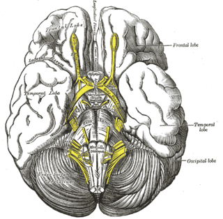
The terminal nerve, also known as cranial nerve 0 or simply as CN 0, is a nerve that was not included in the seminal classification of the cranial nerves as CN I through CN XII but is now generally classified as a cranial nerve. It was discovered by German scientist Gustav Fritsch in 1878 in the brains of sharks. It was first found in humans in 1913. A 1990 study has indicated that the terminal nerve is a common finding in the adult human brain. The nerve has been called unofficially by other names, including cranial nerve XIII, zero nerve, nerve N, and NT.
The pontine cistern is a subarachnoid cistern situated ventrally/anteriorly to the pons. It contains the basilar artery. Each lateral aperture opens into the pontine cistern just posterior to the cranial nerve VIII.
The interpeduncular cistern is the subarachnoid cistern situated between the dorsum sellae (anteriorly) and the two cerebral peduncles of the mesencephalon (midbrain). Its roof is represented by the floor of the third ventricle. Its floor is formed by the arachnoid membrane extending between the temporal lobes of either side. Anteriorly, it extends to the optic chiasm.
The chiasmatic cistern or suprasellar cistern is a small subarachnoid cistern related to the optic chiasm.
The cistern of lateral cerebral fossa is an elongated subarachnoid cistern formed by arachnoid mater bridging the lateral sulcus between the frontal, temporal, and parietal opercula. The cistern contains the middle cerebral artery (MCA) and its branches, and the two middle cerebral veins (MCVs).
The quadrigeminal cistern is a subarachnoid cistern situated between splenium of corpus callosum, and the superior surface of the cerebellum. It contains a part of the great cerebral vein, the posterior cerebral artery, quadrigeminal artery, glossopharyngeal nerve, and the pineal gland.
The ambient cistern is a bilaterally paired subarachnoid cistern situated at either lateral aspect of the mesencephalon (midbrain). Each ambient cistern has a supratentorial compartment and an infratentorial compartment. Each is continuous anteriorly with the interpeduncular cistern, and posteriorly with the quadrigeminal cistern.
The pericallosal cistern is one of the subarachnoid cisterns. It is situated atop the corpus callosum, extending from its splenium (rostrally/anteriorly) to its genu (caudally/posteriorly).
References
- ↑ "cystern of lamina terminalis - Dictionnaire médical de l'Académie de Médecine". www.academie-medecine.fr. Retrieved 2024-05-24.
- 1 2 3 4 J. Randy Jinkins (2000). "The Subarachnoid Cisterns, Fissures, and Spaces". Atlas of neuroradiologic embryology, anatomy, and variants. Lippincott Williams & Wilkins. p. 261. ISBN 0-7817-1652-7.
- 1 2 3 4 Shafique, Shiza; Rayi, Appaji (2023), "Anatomy, Head and Neck, Subarachnoid Space", StatPearls, Treasure Island (FL): StatPearls Publishing, PMID 32491453 , retrieved 2023-08-01
- 1 2 Standring, Susan (2020). Gray's Anatomy: The Anatomical Basis of Clinical Practice (42th ed.). New York. p. 413. ISBN 978-0-7020-7707-4. OCLC 1201341621.
{{cite book}}: CS1 maint: location missing publisher (link)
| | This neuroanatomy article is a stub. You can help Wikipedia by expanding it. |