Falx cerebelli

Last updated
Falx cerebelli
Sobo 1909 589.png
Falx cerebelli seen in back portion of skull.
Gray130.png
Occipital bone. Inner surface. (Portions "for faulx cerebelli" identified at center left.)
Details
Part of Meninges
Identifiers
Latin falx cerebelli
NeuroNames 1238
TA98 A14.1.01.106
TA2 5377
FMA 83974
Anatomical terms of neuroanatomy

The falx cerebelli is a small sickle-shaped fold of dura mater projecting forwards into the posterior cerebellar notch as well as projecting into the vallecula of the cerebellum between the two cerebellar hemispheres. [1]

Contents

The name comes from two Latin words: falx, meaning "curved blade or scythe", and cerebellum, meaning "little brain". [2]

Anatomy

The falx cerebelli is a small midline fold of dura mater projecting anterior-ward from the skull and into the space between the cerebellar hemispheres. [3] It generally measures between 2.8 and 4.5 cm in length, and approximately 1–2 mm in thickness. [4]

Attachments

Superiorly, it (with its upwardly directed base) attaches at the midline to the posterior portion of the inferior surface of the tentorium cerebelli. [3]

Posteriorly, it attaches to the internal occipital crest; the inferior-most extremity of its posterior attachment frequently divides into two small folds that terminate at either side of the foramen magnum. [3]

Anatomical relations

The occipital sinus is contained within the posterior extremity of the falx cerebelli where it attaches to the internal occipital crest. [3]

Anatomical variation

In its lower portion the falx cerebelli diminishes very rapidly in height and as it descends, it can divide into two smaller folds or diverging limbs, [5] which are lost on the sides of the foramen magnum. Other variations such as duplication, [6] triplication, [7] absence, [8] and fenestration are much less common. As dural venous sinuses are concurrent with the development of dural folds, duplication of the falx cerebelli is usually associated with duplicated occipital sinus. [9] Knowledge of these variations is important in preventing iatrogenic injuries in this region.[ citation needed ]

See also

Related Research Articles

Articles related to anatomy include:

<span class="mw-page-title-main">Dura mater</span> Outermost layer of the protective tissues around the central nervous system (meninges)

In neuroanatomy, dura mater is a thick membrane made of dense irregular connective tissue that surrounds the brain and spinal cord. It is the outermost of the three layers of membrane called the meninges that protect the central nervous system. The other two meningeal layers are the arachnoid mater and the pia mater. It envelops the arachnoid mater, which is responsible for keeping in the cerebrospinal fluid. It is derived primarily from the neural crest cell population, with postnatal contributions of the paraxial mesoderm.

<span class="mw-page-title-main">Great cerebral vein</span>

The great cerebral vein is one of the large blood vessels in the skull draining the cerebrum of the brain. It is also known as the vein of Galen, named for its discoverer, the Greek physician Galen.

<span class="mw-page-title-main">Epidural space</span> Space between the dura mater and vertebrae

In anatomy, the epidural space is the potential space between the dura mater and vertebrae (spine).

<span class="mw-page-title-main">Posterior cranial fossa</span> Area of the cranium containing the brainstem and cerebellum

The posterior cranial fossa is the part of the cranial cavity located between the foramen magnum, and tentorium cerebelli. It is formed by the sphenoid bones, temporal bones, and occipital bone. It lodges the cerebellum, and parts of the brainstem.

<span class="mw-page-title-main">Falx cerebri</span> Anatomical structure of the brain

The falx cerebri is a large, crescent-shaped fold of dura mater that descends vertically into the longitudinal fissure between the cerebral hemispheres of the human brain, separating the two hemispheres and supporting dural sinuses that provide venous and CSF drainage to the brain. It is attached to the crista galli anteriorly, and blends with the tentorium cerebelli posteriorly.

<span class="mw-page-title-main">Cerebellar tentorium</span> Vertebrate brain structure separating the cerebellum from the occipital lobes

The cerebellar tentorium or tentorium cerebelli is an extension of the dura mater between the inferior aspect of the occipital lobes and the superior aspect of the cerebellum. The free border of the tentorium gives passage to the midbrain.

<span class="mw-page-title-main">Confluence of sinuses</span> Venous sinus in the skull

The confluence of sinuses, torcular Herophili, or torcula is the connecting point of the superior sagittal sinus, straight sinus, and occipital sinus. It is below the internal occipital protuberance of the skull. It drains venous blood from the brain into the transverse sinuses. It may be affected by arteriovenous fistulas, a thrombus, major trauma, or surgical damage, and may be imaged with many radiology techniques.

<span class="mw-page-title-main">Straight sinus</span> Venous sinus draining the brain

The straight sinus, also known as tentorial sinus or the sinus rectus, is an area within the skull beneath the brain. It receives blood from the inferior sagittal sinus and the great cerebral vein, and drains into the confluence of sinuses.

<span class="mw-page-title-main">Occipital sinus</span> Organ In the brain

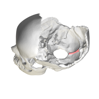
The occipital sinus is the smallest of the dural venous sinuses. It is usually unpaired, and is sometimes altogether absent. It is situated in the attached margin of the falx cerebelli. It commences near the foramen magnum, and ends by draining into the confluence of sinuses.

<span class="mw-page-title-main">Sigmoid sinus</span>

The sigmoid sinuses, also known as the pars sigmoid, are paired dural venous sinuses within the skull that receive blood from posterior transverse sinuses.

<span class="mw-page-title-main">Transverse sinuses</span>

The transverse sinuses, within the human head, are two areas beneath the brain which allow blood to drain from the back of the head. They run laterally in a groove along the interior surface of the occipital bone. They drain from the confluence of sinuses to the sigmoid sinuses, which ultimately connect to the internal jugular vein. See diagram : labeled under the brain as "SIN. TRANS.".

<span class="mw-page-title-main">Articularis cubiti muscle</span> Elbow muscle

The Articularis cubiti muscle is a muscle of the elbow.

<span class="mw-page-title-main">Squamous part of occipital bone</span>

The squamous part of occipital bone is situated above and behind the foramen magnum, and is curved from above downward and from side to side.

<span class="mw-page-title-main">Mastoid foramen</span> Hole in the back of the temporal bone

The mastoid foramen is a hole in the posterior border of the temporal bone. It transmits an emissary vein between the sigmoid sinus and the suboccipital venous plexus, and a small branch of the occipital artery, the posterior meningeal artery to the dura mater.

<span class="mw-page-title-main">Internal occipital crest</span>

In the occipital bone, the lower division of the cruciate eminence is prominent, and is named the internal occipital crest; it bifurcates near the foramen magnum and gives attachment to the falx cerebelli; in the attached margin of this falx is the occipital sinus, which is sometimes duplicated.

<span class="mw-page-title-main">Mastoid cells</span> Air-filled cavities in the temporal bone

The mastoid cells are air-filled cavities within the mastoid process of the temporal bone of the cranium. The mastoid cells are a form of skeletal pneumaticity. Infection in these cells is called mastoiditis.

<span class="mw-page-title-main">Cerebellar veins</span> Veins that drain the cerebellum

The cerebellar veins are veins which drain the cerebellum. They consist of the superior cerebellar veins and the inferior cerebellar veins. The superior cerebellar veins drain to the straight sinus and the internal cerebral veins. The inferior cerebellar veins drain to the transverse sinus, the superior petrosal sinus, and the occipital sinus.

<span class="mw-page-title-main">Calvaria (skull)</span> Top part of the skull

The calvaria is the top part of the skull. It is the superior part of the neurocranium and covers the cranial cavity containing the brain. It forms the main component of the skull roof.

<span class="mw-page-title-main">Outline of human anatomy</span> Overview of and topical guide to human anatomy

The following outline is provided as an overview of and topical guide to human anatomy:

References

  1. Atlas and textbook of human anatomy. Atlas der deskriptiven Anatomie des Menschen.English. Saunders. 1909.
  2. Sihler, Andrew L. (1995). New Comparative Grammar of Greek and Latin. Oxford University Press. ISBN   978-0-19-508345-3. Retrieved 12 March 2013.
  3. 1 2 3 4 Standring, Susan (2021). Gray's Anatomy: The Anatomical Basis of Clinical Practice (42nd ed.). [New York]: Elsavier. ISBN   978-0-7020-7707-4. OCLC   1201341621.
  4. Shoja MM, Tubbs RS, Khaki AA, Shokouhi G. A rare variation of the posterior cranial fossa: duplicated falx cerebelli, occipital venous sinus, and internal occipital crest. Folia Morphol (Warsz) 2006;65(2):171–173.
  5. Atlas and textbook of human anatomy. Atlas der deskriptiven Anatomie des Menschen.English. Saunders. 1909.
  6. Shoja MM, Tubbs RS, Khaki AA, Shokouhi G. A rare variation of the posterior cranial fossa: duplicated falx cerebelli, occipital venous sinus, and internal occipital crest. Folia Morphol (Warsz) 2006;65(2):171–173.
  7. Shoja MM, Tubbs RS, Loukas M, Shokouhi G, Oakes WJ. A complex dural-venous variation in the posterior cranial fossa: a triplicate falx cerebelli and an aberrant venous sinus. Folia Morphol (Warsz) 2007;66(2):148–51.
  8. Tubbs Rs, Dockery SE, Salter G, Elton S, Blount JP, Grabb PA, Oakes WJ. Absence of the falx cerebelli in a Chiari II malformation. Clin Anat. 2002;15(3):193–195.
  9. Shoja MM, Tubbs RS, Shokouhi GH, Ashrafian A, Oakes WJ. Abstract presented at the 23rd Annual Meeting of the American Association of Clinical Anatomists. Milwaukee, Wisconsin: 2006. A triple dural-venous variation in the posterior cranial fossa: A duplicated plus accessory falx cerebelli and an aberrant venous sinus.