Related Research Articles

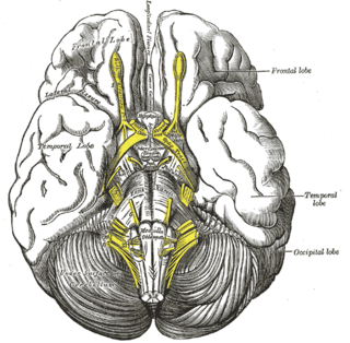
In neuroanatomy, the optic nerve, also known as the second cranial nerve, cranial nerve II, or simply CN II, is a paired cranial nerve that transmits visual information from the retina to the brain. In humans, the optic nerve is derived from optic stalks during the seventh week of development and is composed of retinal ganglion cell axons and glial cells; it extends from the optic disc to the optic chiasma and continues as the optic tract to the lateral geniculate nucleus, pretectal nuclei, and superior colliculus.

The oculomotor nerve, also known as the third cranial nerve, cranial nerve III, or simply CN III, is a cranial nerve that enters the orbit through the superior orbital fissure and innervates extraocular muscles that enable most movements of the eye and that raise the eyelid. The nerve also contains fibers that innervate the intrinsic eye muscles that enable pupillary constriction and accommodation. The oculomotor nerve is derived from the basal plate of the embryonic midbrain. Cranial nerves IV and VI also participate in control of eye movement.

The sella turcica is a saddle-shaped depression in the body of the sphenoid bone of the human skull and of the skulls of other hominids including chimpanzees, gorillas and orangutans. It serves as a cephalometric landmark. The pituitary gland or hypophysis is located within the most inferior aspect of the sella turcica, the hypophyseal fossa.

The subarachnoid cisterns are spaces formed by openings in the subarachnoid space, an anatomic space in the meninges of the brain. The space is situated between the two meninges, the arachnoid mater and the pia mater. These cisterns are filled with cerebrospinal fluid (CSF).
The cisterna magna is the largest of the subarachnoid cisterns. It occupies the space created by the angle between the caudal/inferior surface of the cerebellum, and the dorsal/posterior surface of the medulla oblongata. The fourth ventricle communicates with the cistern via the unpaired midline median aperture. It is continuous inferiorly with the subarachnoid space of the spinal canal.

The anterior choroidal artery is a bilaterally paired artery of the brain. It is typically a branch of the internal carotid artery which supplies the choroid plexus of lateral ventricle and third ventricle as well as numerous structures of the brain.
In human anatomy, the left and right posterior communicating arteries are small arteries at the base of the brain that form part of the circle of Willis.
In human anatomy, the anterior communicating artery is a blood vessel of the brain that connects the left and right anterior cerebral arteries.

The cerebellopontine angle (CPA) is located between the cerebellum and the pons. The cerebellopontine angle is the site of the cerebellopontine angle cistern.
The tuber cinereum is the portion of hypothalamus forming the of the floor of the third ventricle situated between the situated between the optic chiasm, and the mammillary bodies. The tuberal region one of the three regions of the hypothalamus.

The middle cranial fossa is formed by the sphenoid bones, and the temporal bones. It lodges the temporal lobes, and the pituitary gland. It is deeper than the anterior cranial fossa, is narrow medially and widens laterally to the sides of the skull. It is separated from the posterior cranial fossa by the clivus and the petrous crest.

The long posterior ciliary arteries are arteries of the orbit. There are long posterior ciliary arteries two on each side of the body. They are branches of the ophthalmic artery. They pass forward within the eye to reach the ciliary body where they ramify and anastomose with the anterior ciliary arteries, thus forming the major arterial circle of the iris.The long posterior ciliary arteries contribute arterial supply to the choroid, ciliary body, and iris.
The short posterior ciliary arteries are a number of branches of the ophthalmic artery. They pass forward with the optic nerve to reach the eyeball, piercing the sclera around the entry of the optic nerve into the eyeball.
The pontine cistern is a subarachnoid cistern situated ventrally/anteriorly to the pons. It contains the basilar artery. Each lateral aperture opens into the pontine cistern just posterior to the cranial nerve VIII.
The interpeduncular cistern is the subarachnoid cistern situated between the dorsum sellae (anteriorly) and the two cerebral peduncles of the mesencephalon (midbrain). Its roof is represented by the floor of the third ventricle. Its floor is formed by the arachnoid membrane extending between the temporal lobes of either side. Anteriorly, it extends to the optic chiasm.
The cistern of lateral cerebral fossa is an elongated subarachnoid cistern formed by arachnoid mater bridging the lateral sulcus between the frontal, temporal, and parietal opercula. The cistern contains the middle cerebral artery (MCA) and its branches, and the two middle cerebral veins (MCVs).
The quadrigeminal cistern is a subarachnoid cistern situated between splenium of corpus callosum, and the superior surface of the cerebellum. It contains a part of the great cerebral vein, the posterior cerebral artery, quadrigeminal artery, glossopharyngeal nerve, and the pineal gland.
The cistern of lamina terminalis is one of the a subarachnoid cisterns. It is situated rostral/anterior to the lamina terminalis and anterior commissure between the two frontal lobes of the cerebrum. It is situated rostral/anterior to the third ventricle. The cistern is an extension of interpeduncular cistern. The cistern of lamina terminalis interconnects the chiasmatic cistern and pericallosal cistern.
The cerebellopontine cistern is a paired subarachnoid cistern at the cerebellopontine angle, an angle created between the cerebellum and the pons on either side. Each cerebellopontine cistern is continuous anteromedially with the pontine cistern.
References
- 1 2 Standring, Susan (2020). Gray's Anatomy: The Anatomical Basis of Clinical Practice (42th ed.). New York. p. 413. ISBN 978-0-7020-7707-4. OCLC 1201341621.
{{cite book}}: CS1 maint: location missing publisher (link) - 1 2 3 Shafique, Shiza; Rayi, Appaji (2023), "Anatomy, Head and Neck, Subarachnoid Space", StatPearls, Treasure Island (FL): StatPearls Publishing, PMID 32491453 , retrieved 2023-08-01
- 1 2 Sinnatamby, Chummy S. (2011). Last's Anatomy (12th ed.). p. 440. ISBN 978-0-7295-3752-0.
- ↑ J. Randy Jinkins (2000). "The Subarachnoid Cisterns, Fissures, and Spaces". Atlas of neuroradiologic embryology, anatomy, and variants. Lippincott Williams & Wilkins. p. 261. ISBN 0-7817-1652-7.