Anterior cerebral artery

Last updated
Anterior cerebral artery
Gray's Anatomy plate 517 brain.png
Outer surface of cerebral hemisphere, showing areas supplied by cerebral arteries. Areas supplied by the anterior cerebral artery are shown in blue.
Gray518.png
Medial surface of cerebral hemisphere. Areas supplied by the anterior cerebral artery are shown in blue.
Details
Source Internal carotid artery
Vein Cerebral veins
Supplies Cerebrum
Identifiers
Latin arteria cerebri anterior
MeSH D020771
TA98 A12.2.07.022
TA2 4502
FMA 50028
Anatomical terminology

The anterior cerebral artery (ACA) is one of a pair of cerebral arteries that supplies oxygenated blood to most midline portions of the frontal lobes and superior medial parietal lobes of the brain. The two anterior cerebral arteries arise from the internal carotid artery and are part of the circle of Willis. The left and right anterior cerebral arteries are connected by the anterior communicating artery.

Contents

Anterior cerebral artery syndrome refers to symptoms that follow a stroke occurring in the area normally supplied by one of the arteries. It is characterized by weakness and sensory loss in the lower leg and foot opposite to the lesion and behavioral changes.

Structure

Arteries of the brain. Anterior cerebral artery labeled at the left (twice) Sobotta.3.1909.549.png
Arteries of the brain. Anterior cerebral artery labeled at the left (twice)

The anterior cerebral artery is divided into 5 segments. Its smaller branches: the callosal (supracallosal) arteries are considered to be the A4 and A5 segments. [1] [2]

Development

The anterior cerebral artery develops from a primitive anterior division of the internal carotid artery that initially supplies the optic and olfactory regions. This anterior division, which appears at the twenty-eighth day of development, also forms the middle cerebral artery and the anterior choroidal artery. The anterior cerebral arteries grow toward each other and form the anterior communicating artery at the 21–24 mm stage of the embryo. [5]

Variation

The anterior cerebral artery shows considerable variation. In a study made using MRA, the most common variation was an underdeveloped A1 segment (5.6%), followed by the presence of an extra A2 segment (3%). In 2% of cases there was only one A2 segment. [6]

Function

The anterior cerebral artery supplies a part of the frontal lobe, specifically its medial surface and the upper border. It also supplies the front four–fifths of the corpus callosum, and provides blood to deep structures such as the anterior limb of the internal capsule, part of the caudate nucleus, and the anterior part of the globus pallidus. [7]

Clinical significance

Areas of the brain supplied by left anterior cerebral artery (represented in green color)

Occlusion

Strokes that occur in a part of the artery prior to the anterior communicating usually do not produce many symptoms because of collateral circulation. If a blockage occurs in the A2 segment or later, the following signs and symptoms may be noted: [8]

Additional images

See also

Related Research Articles

Articles related to anatomy include:

<span class="mw-page-title-main">Circle of Willis</span> Circulatory anastomosis that supplies blood to the brain and surrounding structures

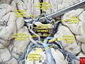
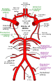
The circle of Willis is a circulatory anastomosis that supplies blood to the brain and surrounding structures in reptiles, birds and mammals, including humans. It is named after Thomas Willis (1621–1675), an English physician.

<span class="mw-page-title-main">External carotid artery</span> Major artery of the head and neck

The external carotid artery is a major artery of the head and neck. It arises from the common carotid artery when it splits into the external and internal carotid artery. The external carotid artery supplies blood to the face, brain and neck.

<span class="mw-page-title-main">Internal carotid artery</span> Artery of the human brain

The internal carotid artery is an artery in the neck which supplies the anterior circulation of the brain.

<span class="mw-page-title-main">Internal capsule</span> White matter structure situated in the inferomedial part of each cerebral hemisphere of the brain

The internal capsule is a white matter structure situated in the inferomedial part of each cerebral hemisphere of the brain. It carries information past the basal ganglia, separating the caudate nucleus and the thalamus from the putamen and the globus pallidus. The internal capsule contains both ascending and descending axons, going to and coming from the cerebral cortex. It also separates the caudate nucleus and the putamen in the dorsal striatum, a brain region involved in motor and reward pathways.

<span class="mw-page-title-main">Lateral ventricles</span> Two largest ventricles in each cerebral hemisphere

The lateral ventricles are the two largest ventricles of the brain and contain cerebrospinal fluid. Each cerebral hemisphere contains a lateral ventricle, known as the left or right lateral ventricle, respectively.

<span class="mw-page-title-main">Basilar artery</span> Artery that supplies the brain with blood

The basilar artery is one of the arteries that supplies the brain with oxygen-rich blood.

<span class="mw-page-title-main">Vertebral artery</span> Major arteries of the neck

The vertebral arteries are major arteries of the neck. Typically, the vertebral arteries originate from the subclavian arteries. Each vessel courses superiorly along each side of the neck, merging within the skull to form the single, midline basilar artery. As the supplying component of the vertebrobasilar vascular system, the vertebral arteries supply blood to the upper spinal cord, brainstem, cerebellum, and posterior part of brain.

<span class="mw-page-title-main">Subarachnoid cisterns</span> Spaces around the brain filled with cerebrospinal fluid

The subarachnoid cisterns are spaces formed by openings in the subarachnoid space, an anatomic space in the meninges of the brain. The space is situated between the two meninges, the arachnoid mater and the pia mater. These cisterns are filled with cerebrospinal fluid (CSF).

<span class="mw-page-title-main">Middle cerebral artery</span> Paired artery that supplies blood to the cerebrum

The middle cerebral artery (MCA) is one of the three major paired cerebral arteries that supply blood to the cerebrum. The MCA arises from the internal carotid artery and continues into the lateral sulcus where it then branches and projects to many parts of the lateral cerebral cortex. It also supplies blood to the anterior temporal lobes and the insular cortices.

<span class="mw-page-title-main">Anterior choroidal artery</span>

The anterior choroidal artery is a bilaterally paired artery of the brain. It is typically a branch of the internal carotid artery which supplies the choroid plexus of lateral ventricle and third ventricle as well as numerous structures of the brain.

<span class="mw-page-title-main">Posterior cerebral artery</span> Artery which supplies blood to the occipital lobe of the brain

The posterior cerebral artery (PCA) is one of a pair of cerebral arteries that supply oxygenated blood to the occipital lobe, part of the back of the human brain. The two arteries originate from the distal end of the basilar artery, where it bifurcates into the left and right posterior cerebral arteries. These anastomose with the middle cerebral arteries and internal carotid arteries via the posterior communicating arteries.

<span class="mw-page-title-main">Posterior communicating artery</span> Arteries at the base of the brain that form part of the circle of Willis

In human anatomy, the left and right posterior communicating arteries are small arteries at the base of the brain that form part of the circle of Willis.

<span class="mw-page-title-main">Anterior communicating artery</span>

In human anatomy, the anterior communicating artery is a blood vessel of the brain that connects the left and right anterior cerebral arteries.

<span class="mw-page-title-main">Cerebral arteries</span> 3 main pairs of arteries which supply blood to the cerebrum of the brain

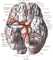
The cerebral arteries describe three main pairs of arteries and their branches, which perfuse the cerebrum of the brain. The three main arteries are the:

<span class="mw-page-title-main">Anterior cerebral artery syndrome</span> Medical condition

Anterior cerebral artery syndrome is a condition whereby the blood supply from the anterior cerebral artery (ACA) is restricted, leading to a reduction of the function of the portions of the brain supplied by that vessel: the medial aspects of the frontal and parietal lobes, basal ganglia, anterior fornix and anterior corpus callosum.

<span class="mw-page-title-main">Leptomeningeal collateral circulation</span>

The leptomeningeal collateral circulation is a network of small blood vessels in the brain that connects branches of the middle, anterior and posterior cerebral arteries, with variation in its precise anatomy between individuals. During a stroke, leptomeningeal collateral vessels allow limited blood flow when other, larger blood vessels provide inadequate blood supply to a part of the brain.

The recurrent artery of Heubner, Heubner's artery or distal medial striate artery is an artery in the head. It is named after the German paediatrician Otto Heubner. It is a branch of the anterior cerebral artery. Its vascular territory is the anteromedial section of the caudate nucleus and the anterioinferior section of the internal capsule, as well as parts of the putamen and septal nuclei.

<span class="mw-page-title-main">Orbitofrontal artery</span>

The orbitofrontal artery is one of the branches of the anterior cerebral artery, that supplies blood to the cerebrum. The orbitofrontal artery is usually the first cortical branch of the A2 segment, arising from the subcallosal segment to supply the inferior and inferomedial surfaces of the frontal lobe including the gyri recti.

References

  1. 1 2 Krayenbühl, Hugo; Yaşargil, Mahmut Gazi; Huber, Peter; Bosse, George (1982), Cerebral Angiography, Thieme, pp. 79–91, ISBN   978-0-86577-067-6
  2. Biller, J (2007). "Neurovascular System". Textbook of Clinical Neurology (Third Edition). Chapter 22: 405–434.
  3. SCHNEIDER, MARCO A. STEFANI; MARRONE, ANTONIO C. H.; SEVERINO, ANTONIO G.; JACKOWSKI, ANDREA P.; WALLACE, M. CHRISTOPHER (2000), "Anatomic Variations of Anterior Cerebral Artery Cortical Branches", Clinical Anatomy, 13 (4): 231–236, doi: 10.1002/1098-2353(2000)13:4<231::aid-ca1>3.0.co;2-t , PMID   10873213, S2CID   6349855
  4. Osborn, Anne G.; Jacobs, John M. (1999), Diagnostic Cerebral Angiography, Lippincott Williams & Wilkins, pp. 143–144, ISBN   978-0-397-58404-8
  5. Menshawi, K; Mohr, JP; Gutierrez, J (May 2015). "A Functional Perspective on the Embryology and Anatomy of the Cerebral Blood Supply". Journal of Stroke. 17 (2): 144–58. doi:10.5853/jos.2015.17.2.144. PMC   4460334 . PMID   26060802.
  6. Uchino, A; Nomiyama, K; Takase, Y; Kudo, S (September 2006). "Anterior cerebral artery variations detected by MR angiography". Neuroradiology. 48 (9): 647–52. doi:10.1007/s00234-006-0110-3. PMID   16786350. S2CID   8019565.
  7. Ropper, A.; Samuels, M.; Klein, J. (2014). Adams and Victor's Principles of Neurology (10th ed.). McGraw-Hill. p. 798. ISBN   978-0071794794.
  8. Longo, D; Fauci, A; Kasper, D; Hauser, S; Jameson, J; Loscalzo, J (2012). Harrison's Principles of Internal Medicine (18th ed.). New York: McGraw-Hill. p. 3286. ISBN   978-0071748896.