
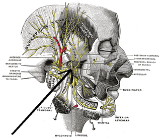
The mandibular nerve (V3) is the largest of the three divisions of the trigeminal nerve, the fifth cranial nerve (CN V).

The sphenoid bone is an unpaired bone of the neurocranium. It is situated in the middle of the skull towards the front, in front of the temporal bone and the basilar part of the occipital bone. The sphenoid bone is one of the seven bones that articulate to form the orbit. Its shape somewhat resembles that of a butterfly or bat with its wings extended.
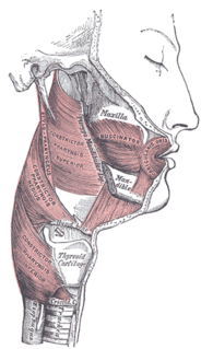
There are four classical muscles of mastication. During mastication, three muscles of mastication are responsible for adduction of the jaw, and one helps to abduct it. All four move the jaw laterally. Other muscles, usually associated with the hyoid, such as the mylohyoid muscle, are responsible for opening the jaw in addition to the lateral pterygoid.

In human anatomy, the masseter is one of the muscles of mastication. Found only in mammals, it is particularly powerful in herbivores to facilitate chewing of plant matter. The most obvious muscle of mastication is the masseter muscle, since it is the most superficial and one of the strongest.

The buccal nerve is a nerve in the face. It is a branch of the mandibular nerve and transmits sensory information from skin over the buccal membrane and from the second and third molar teeth. Not to be confused with the buccal branch of the facial nerve which transmits motor information to the buccinator muscle.
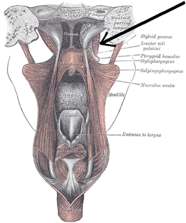
The tensor veli palatini muscle is a broad, thin, ribbon-like muscle in the head that tenses the soft palate.

The pterygoid processes of the sphenoid, one on either side, descend perpendicularly from the regions where the body and the greater wings of the sphenoid bone unite.
The greater wing of the sphenoid bone, or alisphenoid, is a bony process of the sphenoid bone; there is one on each side, extending from the side of the body of the sphenoid and curving upward, laterally, and backward.

The pterygoid plexus is a venous plexus of considerable size, and is situated between the temporalis muscle and lateral pterygoid muscle, and partly between the two pterygoid muscles.

The maxillary artery supplies deep structures of the face. It branches from the external carotid artery just deep to the neck of the mandible.
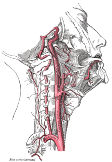
The ascending palatine artery is an artery in the head that branches off the facial artery and runs up the superior pharyngeal constrictor muscle.
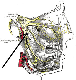
External Pterygoid Nerve : The nerve to the Pterygoideus externus frequently arises in conjunction with the buccinator nerve, but it may be given off separately from the anterior division of the mandibular nerve.

The infratemporal fossa is an irregularly shaped cavity, situated below and medial to the zygomatic arch. It is not fully enclosed by bone in all directions, and it contains superficial muscles that are visible during dissection after removing skin and fascia: namely, the lower part of the temporalis muscle, the lateral pterygoid, and the medial pterygoid.
The medial pterygoid nerve is a branch of the mandibular nerve that innervates the medial pterygoid muscle, tensor veli palatini and tensor tympani.
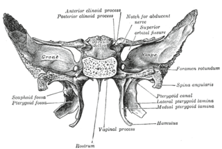
The pterygoid fossa is an anatomical term for the fossa formed by the divergence of the lateral pterygoid plate and the medial pterygoid plate of the sphenoid bone.
The pterygomandibular raphe is a ligamentous band of the buccopharyngeal fascia, attached superiorly to the pterygoid hamulus of the medial pterygoid plate, and inferiorly to the posterior end of the mylohyoid line of the mandible.

At the lower part of the infratemporal surface of the maxilla is a rounded eminence, the maxillary tuberosity, especially prominent after the growth of the wisdom tooth; it is rough on its lateral side for articulation with the pyramidal process of the palatine bone and in some cases articulates with the lateral pterygoid plate of the sphenoid.
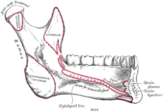
The pterygoid fovea is a concave surface on the uppermost medial side of the ramus of the mandible.
The public domain consists of all the creative works to which no exclusive intellectual property rights apply. Those rights may have expired, been forfeited, expressly waived, or may be inapplicable.

Gray's Anatomy is an English language textbook of human anatomy originally written by Henry Gray and illustrated by Henry Vandyke Carter. Earlier editions were called Anatomy: Descriptive and Surgical, Anatomy of the Human Body and Gray's Anatomy: Descriptive and Applied, but the book's name is commonly shortened to, and later editions are titled, Gray's Anatomy. The book is widely regarded as an extremely influential work on the subject, and has continued to be revised and republished from its initial publication in 1858 to the present day. The latest edition of the book, the 41st, was published in September 2015.


