Middle meningeal artery

Last updated
Middle meningeal artery
Middle meningeal artery.png
Plan of branches of the maxillary artery.
Gray1198.png
Relations of the brain and middle meningeal artery to the surface of the skull.
Details
Source Maxillary artery
BranchesAnterior: posterior: superior tympanic artery
Vein Middle meningeal vein
Supplies Meninges
Identifiers
Latin arteria meningea media
TA98 A12.2.05.061
TA2 4431
FMA 49711
Anatomical terminology

The middle meningeal artery ( Latin : arteria meningea media) is typically the third branch of the first portion of the maxillary artery. After branching off the maxillary artery in the infratemporal fossa, it runs through the foramen spinosum to supply the dura mater (the outer meningeal layer) and the calvaria. The middle meningeal artery is the largest of the three (paired) arteries that supply the meninges, the others being the anterior meningeal artery and the posterior meningeal artery.

Contents

The anterior branch of the middle meningeal artery runs beneath the pterion. It is vulnerable to injury at this point, where the skull is thin. Rupture of the artery may give rise to an epidural hematoma. In the dry cranium, the middle meningeal, which runs within the dura mater surrounding the brain, makes a deep groove in the calvarium.

The middle meningeal artery is intimately associated with the auriculotemporal nerve, which wraps around the artery making the two easily identifiable in the dissection of human cadavers and also easily damaged in surgery.

Structure

It ascends between the sphenomandibular ligament and the lateral pterygoid muscle, and between the two roots of the auriculotemporal nerve to the foramen spinosum of the sphenoid bone, through which it enters the cranium; it then runs forward in a groove on the great wing of the sphenoid bone, and divides into two branches, anterior and posterior.

The anterior branch, the larger, crosses the great wing of the sphenoid, reaches the groove, or canal, in the sphenoidal angle of the parietal bone, and then divides into branches that spread out between the dura mater and internal surface of the cranium, some passing upward as far as the vertex, and others backward to the occipital region.

The posterior branch curves backward on the squamous part of the temporal bone, and, reaching the parietal bone some distance in front of its mastoid angle, divides into branches that supply the posterior part of the dura mater and cranium.

The branches of the middle meningeal artery are distributed partly to the dura mater, but chiefly to the bones; they anastomose with the arteries of the opposite side, and with the anterior and posterior meningeal arteries. The very smallest distal branches anastomose through the skull with small arterioles from the scalp.

On entering the cranium, the middle meningeal artery gives off the following branches:

  1. Numerous small vessels supply the trigeminal ganglion and the dura mater
  2. A superficial petrosal branch enters the hiatus of the facial canal, supplies the facial nerve, and anastomoses with the stylomastoid branch of the posterior auricular artery.
  3. A superior tympanic artery runs in the canal of the tensor tympani muscle, and supplies this muscle and the lining of the canal.
  4. Orbital branches pass through the superior orbital fissure or through separate canals in the great wing of the sphenoid, to anastomose with the lacrimal or other branches of the ophthalmic artery.
  5. Temporal branches pass through foramina in the great wing of the sphenoid, and anastomose in the temporal fossa with the deep temporal arteries.

Variation

In approximately half of subjects, it branches into an accessory meningeal artery.

Very rarely, the ophthalmic artery may arise as a branch of the middle meningeal artery.

The middle meningeal artery may arise not only from the maxillary artery but also from the ophthalmic artery, or lacrimal artery. [1]

Clinical relevance

An injured middle meningeal artery is the most common cause of an epidural hematoma. A head injury (e.g., from a road traffic accident or sports injury) is required to rupture the artery. Emergency treatment requires decompression of the haematoma, usually by craniotomy. Subdural bleeding is usually venous in nature, rather than arterial.

The middle meningeal artery runs in a groove on the inside of the cranium. This can clearly be seen on a lateral skull X-ray, where it may be mistaken for a fracture of the skull. On a dry specimen, the groove is easy to see. This means that the artery is easy to study, even in specimens centuries old, and several classifications of the branches have been proposed, e.g. Adachi's classification of 1928.

Additional images

See also

Related Research Articles

Articles related to anatomy include:

<span class="mw-page-title-main">Sphenoid bone</span> Bone of the neurocranium

The sphenoid bone is an unpaired bone of the neurocranium. It is situated in the middle of the skull towards the front, in front of the basilar part of the occipital bone. The sphenoid bone is one of the seven bones that articulate to form the orbit. Its shape somewhat resembles that of a butterfly or bat with its wings extended.

<span class="mw-page-title-main">Internal carotid artery</span> Artery of the human brain

The internal carotid artery is an artery in the neck which supplies the anterior circulation of the brain.

<span class="mw-page-title-main">Orbit (anatomy)</span> Cavity or socket of the skull in which the eye and its appendages are situated

In anatomy, the orbit is the cavity or socket/hole of the skull in which the eye and its appendages are situated. "Orbit" can refer to the bony socket, or it can also be used to imply the contents. In the adult human, the volume of the orbit is 30 millilitres, of which the eye occupies 6.5 ml. The orbital contents comprise the eye, the orbital and retrobulbar fascia, extraocular muscles, cranial nerves II, III, IV, V, and VI, blood vessels, fat, the lacrimal gland with its sac and duct, the eyelids, medial and lateral palpebral ligaments, cheek ligaments, the suspensory ligament, septum, ciliary ganglion and short ciliary nerves.

<span class="mw-page-title-main">Dura mater</span> Outermost layer of the protective tissues around the central nervous system (meninges)

In neuroanatomy, dura mater is a thick membrane made of dense irregular connective tissue that surrounds the brain and spinal cord. It is the outermost of the three layers of membrane called the meninges that protect the central nervous system. The other two meningeal layers are the arachnoid mater and the pia mater. It envelops the arachnoid mater, which is responsible for keeping in the cerebrospinal fluid. It is derived primarily from the neural crest cell population, with postnatal contributions of the paraxial mesoderm.

<span class="mw-page-title-main">Greater petrosal nerve</span> Nerve in the skull

The greater petrosal nerve is a nerve of the head mainly containing pre-ganglionic parasympathetic fibres which ultimately synapse in the pterygopalatine ganglion. It branches from the facial nerve and is derived from the parasympathetic part of the nervus intermedius component of CN VII, with its cell bodies located in the superior salivary nucleus. In the connective tissue substance of the foramen lacerum, the greater petrosal nerve unites with the (sympathetic) deep petrosal nerve to form the nerve of the pterygoid canal which proceeds to the pterygopalatine ganglion.

<span class="mw-page-title-main">Foramen spinosum</span> Hole in the sphenoid bone of the skull

The foramen spinosum is a small open hole in the greater wing of the sphenoid bone that gives passage to the middle meningeal artery and vein, and the meningeal branch of the mandibular nerve.

<span class="mw-page-title-main">Maxillary nerve</span> Branch of the trigeminal nerve responsible for the mid-face

In neuroanatomy, the maxillary nerve (V2) is one of the three branches or divisions of the trigeminal nerve, the fifth (CN V) cranial nerve. It comprises the principal functions of sensation from the maxilla, nasal cavity, sinuses, the palate and subsequently that of the mid-face, and is intermediate, both in position and size, between the ophthalmic nerve and the mandibular nerve.

<span class="mw-page-title-main">Pterygopalatine fossa</span> Fossa in the skull

In human anatomy, the pterygopalatine fossa is a fossa in the skull. A human skull contains two pterygopalatine fossae—one on the left side, and another on the right side. Each fossa is a cone-shaped paired depression deep to the infratemporal fossa and posterior to the maxilla on each side of the skull, located between the pterygoid process and the maxillary tuberosity close to the apex of the orbit. It is the indented area medial to the pterygomaxillary fissure leading into the sphenopalatine foramen. It communicates with the nasal and oral cavities, infratemporal fossa, orbit, pharynx, and middle cranial fossa through eight foramina.

<span class="mw-page-title-main">Greater wing of sphenoid bone</span> Large part of the skull front behind the eye socket

The greater wing of the sphenoid bone, or alisphenoid, is a bony process of the sphenoid bone, positioned in the skull behind each eye. There is one on each side, extending from the side of the body of the sphenoid and curving upward, laterally, and backward.

<span class="mw-page-title-main">Lesser wing of sphenoid bone</span>

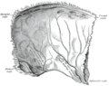
The lesser wings of the sphenoid or orbito-sphenoids are two thin triangular plates, which arise from the upper and anterior parts of the body, and, projecting lateralward, end in sharp points [Fig. 1].

<span class="mw-page-title-main">Anterior ethmoidal artery</span>

The anterior ethmoidal artery is a branch of the ophthalmic artery in the orbit. It exits the orbit through the anterior ethmoidal foramen alongside the anterior ethmoidal nerve. It contributes blood supply to the ethmoid sinuses, frontal sinuses, the dura mater, lateral nasal wall, and nasal septum. It issues a meningeal branch, and nasal branches.

<span class="mw-page-title-main">Meningeal branch of the mandibular nerve</span>

The meningeal branch of the mandibular nerve is a sensory branch of the mandibular nerve that enters the middle cranial fossa through either the foramen spinosum or foramen ovale to innervate the meninges of this fossa as well as the mastoid air cells.

<span class="mw-page-title-main">Maxillary artery</span> Artery supplying face structures in humans

The maxillary artery supplies deep structures of the face. It branches from the external carotid artery just deep to the neck of the mandible.

<span class="mw-page-title-main">Petrous part of the temporal bone</span> Feature at the base of the human skull

The petrous part of the temporal bone is pyramid-shaped and is wedged in at the base of the skull between the sphenoid and occipital bones. Directed medially, forward, and a little upward, it presents a base, an apex, three surfaces, and three angles, and houses in its interior, the components of the inner ear. The petrous portion is among the most basal elements of the skull and forms part of the endocranium. Petrous comes from the Latin word petrosus, meaning "stone-like, hard". It is one of the densest bones in the body. In other mammals, it is a separate bone, the petrosal bone.

<span class="mw-page-title-main">Middle cranial fossa</span>

The middle cranial fossa is formed by the sphenoid bones, and the temporal bones. It lodges the temporal lobes, and the pituitary gland. It is deeper than the anterior cranial fossa, is narrow medially and widens laterally to the sides of the skull. It is separated from the posterior cranial fossa by the clivus and the petrous crest.

<span class="mw-page-title-main">Infratemporal fossa</span> Cavity that is part of the skull

The infratemporal fossa is an irregularly shaped cavity that is a part of the skull. It is situated below and medial to the zygomatic arch. It is not fully enclosed by bone in all directions. It contains superficial muscles, including the lower part of the temporalis muscle, the lateral pterygoid muscle, and the medial pterygoid muscle. It also contains important blood vessels such as the middle meningeal artery, the pterygoid plexus, and the retromandibular vein, and nerves such as the mandibular nerve (CN V3) and its branches.

<span class="mw-page-title-main">Middle meningeal nerve</span>

The middle meningeal nerve (meningeal or dural branch) is given off from the maxillary nerve (CN V2) directly after its origin from the trigeminal ganglion, before CN V2 enters the foramen rotundum.

<span class="mw-page-title-main">Base of skull</span> Inferior area of the skull, composed of the endocranium and lower parts of the skull roof

The base of skull, also known as the cranial base or the cranial floor, is the most inferior area of the skull. It is composed of the endocranium and the lower parts of the calvaria.

<span class="mw-page-title-main">Outline of human anatomy</span> Overview of and topical guide to human anatomy

The following outline is provided as an overview of and topical guide to human anatomy:

References

PD-icon.svgThis article incorporates text in the public domain from page 560 of the 20th edition of Gray's Anatomy (1918)

  1. Krayenbühl, Niklaus; Isolan, Gustavo Rassier; Al-Mefty, Ossama (2 August 2008). "The foramen spinosum: a landmark in middle fossa surgery" (PDF). Neurosurgical Review. 31 (4): 397–402. doi:10.1007/s10143-008-0152-6. PMID   18677523. S2CID   37126697.