
In anatomy, the orbit is the cavity or socket/hole of the skull in which the eye and its appendages are situated. "Orbit" can refer to the bony socket, or it can also be used to imply the contents. In the adult human, the volume of the orbit is 30 millilitres, of which the eye occupies 6.5 ml. The orbital contents comprise the eye, the orbital and retrobulbar fascia, extraocular muscles, cranial nerves II, III, IV, V, and VI, blood vessels, fat, the lacrimal gland with its sac and duct, the eyelids, medial and lateral palpebral ligaments, cheek ligaments, the suspensory ligament, septum, ciliary ganglion and short ciliary nerves.

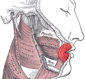
The buccinator is a thin quadrilateral muscle occupying the interval between the maxilla and the mandible at the side of the face. It forms the anterior part of the cheek or the lateral wall of the oral cavity.
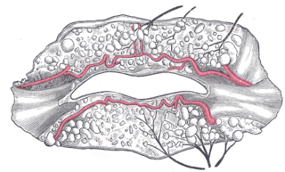
In human anatomy, the orbicularis oris muscle is a complex of muscles in the lips that encircles the mouth. It is not a true sphincter, as was once thought, as it is actually composed of four independent quadrants that interlace and give only an appearance of circularity.

The levator labii superioris is a muscle of the human body used in facial expression. It is a broad sheet, the origin of which extends from the side of the nose to the zygomatic bone.

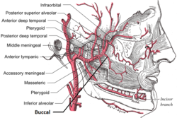
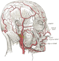
The facial artery is a branch of the external carotid artery that supplies structures of the superficial face.
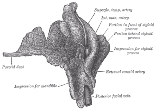
The parotid duct or Stensen duct is a salivary duct. It is the route that saliva takes from the major salivary gland, the parotid gland, into the mouth. It opens into the mouth opposite the second upper molar tooth.

The buccal nerve is a sensory nerve of the face arising from the mandibular nerve. It conveys sensory information from the skin of the cheek, and parts of the oral mucosa, periodontium, and gingiva.

In neuroanatomy, the maxillary nerve (V2) is one of the three branches or divisions of the trigeminal nerve, the fifth (CN V) cranial nerve. It comprises the principal functions of sensation from the maxilla, nasal cavity, sinuses, the palate and subsequently that of the mid-face, and is intermediate, both in position and size, between the ophthalmic nerve and the mandibular nerve.

The pterygoid plexus is a fine venous plexus upon and within the lateral pterygoid muscle. It drains by a short maxillary vein.

The transverse facial artery is an artery that branches from the superficial temporal artery and runs across the face.
The inferior labial artery arises near the angle of the mouth as a branch of the facial artery; it passes upward and forward beneath the triangularis and, penetrating the orbicularis oris, runs in a tortuous course along the edge of the lower lip between this muscle and the mucous membrane.

The maxillary artery supplies deep structures of the face. It branches from the external carotid artery just deep to the neck of the mandible.

The infraorbital artery is a small artery in the head that arises from the maxillary artery and passes through the inferior orbital fissure to enter the orbit, then passes forward along the floor of the orbit, finally exiting the orbit through the infraorbital foramen to reach the face.
The angular artery is an artery of the face. It is the terminal part of the facial artery. It ascends to the medial angle of the eye's orbit. It is accompanied by the angular vein. It ends by anastomosing with the dorsal nasal branch of the ophthalmic artery. It supplies the lacrimal sac, the orbicularis oculi muscle, and the outer side of the nose.

The buccal branches of the facial nerve, are of larger size than the rest of the branches, pass horizontally forward to be distributed below the orbit and around the mouth.
The zygomatic branches of the facial nerve (malar branches) are nerves of the face. They run across the zygomatic bone to the lateral angle of the orbit. Here, they supply the orbicularis oculi muscle, and join with filaments from the lacrimal nerve and the zygomaticofacial branch of the maxillary nerve (CN V2).

The buccal space is a fascial space of the head and neck. It is a potential space in the cheek, and is paired on each side. The buccal space is superficial to the buccinator muscle and deep to the platysma muscle and the skin. The buccal space is part of the subcutaneous space, which is continuous from head to toe.
The lateral nasal branch of facial artery is derived from the facial artery as that vessel ascends along the side of the nose.

The human nose is the first organ of the respiratory system. It is also the principal organ in the olfactory system. The shape of the nose is determined by the nasal bones and the nasal cartilages, including the nasal septum which separates the nostrils and divides the nasal cavity into two.

The facial lymph nodes comprise three groups:
