Occipital artery

Last updated
Occipital artery
Gray508.png
The arteries of the face and scalp. (Occipital visible at center left.)
Occipital artery.PNG
Superficial dissection of the right side of the neck, showing the carotid and subclavian arteries.
Details
Source External carotid artery
Vein Occipital vein
Supplies Occiput, scalp
Identifiers
Latin arteria occipitalis
TA98 A12.2.05.030
TA2 4398
FMA 49586
Anatomical terminology

The occipital artery is a branch of the external carotid artery that provides arterial supply to the back of the scalp, sternocleidomastoid muscles, and deep muscles of the back and neck.

Contents

Structure

Origin

The occipital artery arises from (the posterior aspect of) the external carotid artery (some 2 cm distal to the origin of the external carotid artery). [1]

Course and relations

At its origin, the hypoglossal nerve (CN XII) crosses artery superficially as the nerve passes posteroanteriorly. [1]

The artery passes superoposteriorly deep to the posterior belly of the digastricus muscle. It crosses the internal carotid artery and vein, the vagus nerve (CN X), accessory nerve (CN XI), and hypoglossal nerve (CN XII). [1]

It next ascends to the interval between the transverse process of the atlas and the mastoid process of the temporal bone, and passes horizontally backward, grooving the surface of the latter bone, being covered by the sternocleidomastoideus, splenius capitis, longissimus capitis, and digastricus, and resting upon the rectus capitis lateralis, the obliquus superior, and semispinalis capitis.

It then changes its course and runs vertically upward, pierces the fascia connecting the cranial attachment of the trapezius with the sternocleidomastoideus, and ascends in a tortuous course in the superficial fascia of the scalp, where it divides into numerous branches, which reach as high as the vertex of the skull and anastomose with the posterior auricular and superficial temporal arteries.

Distribution

  1. Muscular branches: supply the digastric, stylohyoid, splenius, and longus capitis muscles.
  2. Sternocleidomastoid branch: This branch divides into upper and lower branches in the carotid triangle. The upper branch accompanies the accessory nerve to the sternocleidomastoid, and the lower branch arises near the origin of the occipital artery before entering the sternocleidomastoid muscle. Occasionally, this branch arises directly from the external carotid artery.
  3. Auricular branch: supplies the back of the ear. In many specimens, this branch gives rise to the mastoid branch, which supplies the dura mater, diploe, and mastoid air cells. In other specimens, the mastoid artery is a branch of the occipital artery, rather than the auricular branch.
  4. Meningeal branch: supplies the dura mater in the posterior cranial fossa
  5. Descending branches: This is the largest branch. It descends on the posterior aspect of the neck, and divides into a superficial and deep portion. The superficial portion supplies the trapezius muscle and anastomoses with the ascending branch of the transverse cervical. The deep portion anastomoses with the vertebral artery and with the a. profunda cervicalis, a branch of the costocervical trunk. Thus, branches of the occipital artery participate in anastomosis between the external carotid and the subclavian artery, thereby providing collateral circulation. [2]

Its terminal portion is accompanied by the greater occipital nerve.

Additional images

Related Research Articles

<span class="mw-page-title-main">Lesser occipital nerve</span> Nerve of the head and neck

The lesser occipital nerve is a cutaneous spinal nerve of the cervical plexus. It arises from second cervical (spinal) nerve (C2). It innervates the skin of the back of the upper neck and of the scalp posterior to the ear.

<span class="mw-page-title-main">Great auricular nerve</span> Cutaneous nerve of the head

The great auricular nerve is a cutaneous (sensory) nerve of the head. It originates from the second and third cervical (spinal) nerves (C2-C3) of the cervical plexus. It provides sensory innervation to the skin over the parotid gland and the mastoid process, parts of the outer ear, and to the parotid gland and its fascia.

<span class="mw-page-title-main">External carotid artery</span> Major artery of the head and neck

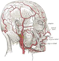
The external carotid artery is a major artery of the head and neck. It arises from the common carotid artery when it splits into the external and internal carotid artery. The external carotid artery supplies blood to the face, brain and neck.

<span class="mw-page-title-main">Internal carotid artery</span> Artery of the human brain

The internal carotid artery is an artery in the neck which supplies the anterior circulation of the brain.

<span class="mw-page-title-main">Scalp</span> Anatomical area bordered by the face at the front, and by the neck at the sides and back

The scalp is the area of the head where head hair grows. It is made up of skin, layers of connective and fibrous tissues, and the membrane of the skull. Anatomically, the scalp is part of the epicranium, a collection of structures covering the cranium. The scalp is bordered by the face at the front, and by the neck at the sides and back. The scientific study of hair and scalp is called trichology.

<span class="mw-page-title-main">Levator scapulae muscle</span> Slender skeletal muscle at the back and side of the neck

The levator scapulae is a slender skeletal muscle situated at the back and side of the neck. It originates from the transverse processes of the four uppermost cervical vertebrae; it inserts onto the upper portion of the medial border of the scapula. It is innervated by the cervical nerves C3-C4, and frequently also by the dorsal scapular nerve. As the Latin name suggests, its main function is to lift the scapula.

<span class="mw-page-title-main">Splenius capitis muscle</span>

The splenius capitis is a broad, straplike muscle in the back of the neck. It pulls on the base of the skull from the vertebrae in the neck and upper thorax. It is involved in movements such as shaking the head.

<span class="mw-page-title-main">Superficial temporal artery</span> Major artery of the head

In human anatomy, the superficial temporal artery is a major artery of the head. It arises from the external carotid artery when it splits into the superficial temporal artery and maxillary artery.

<span class="mw-page-title-main">Posterior triangle of the neck</span> Region of the neck

The posterior triangle is a region of the neck.

<span class="mw-page-title-main">Superior thyroid artery</span>

The superior thyroid artery arises from the external carotid artery just below the level of the greater cornu of the hyoid bone and ends in the thyroid gland.

<span class="mw-page-title-main">Transverse cervical artery</span> Artery of the neck

The transverse cervical artery is an artery in the neck and a branch of the thyrocervical trunk, running at a higher level than the suprascapular artery.

<span class="mw-page-title-main">Inferior thyroid artery</span> Artery of the neck

The inferior thyroid artery is an artery in the neck. It arises from the thyrocervical trunk and passes upward, in front of the vertebral artery and longus colli muscle. It then turns medially behind the carotid sheath and its contents, and also behind the sympathetic trunk, the middle cervical ganglion resting upon the vessel.

<span class="mw-page-title-main">Posterior auricular artery</span> Small artery

The posterior auricular artery is a small artery that arises from the external carotid artery. It ascends along the side of the head. It supplies several muscles of the neck and several structures of the head.

<span class="mw-page-title-main">Ascending pharyngeal artery</span>

The ascending pharyngeal artery is an artery of the neck that supplies the pharynx.

<span class="mw-page-title-main">Posterior auricular nerve</span> Nerve of the head

The posterior auricular nerve is a nerve of the head. It is a branch of the facial nerve. It communicates with branches from the vagus nerve, the great auricular nerve, and the lesser occipital nerve. Its auricular branch supplies the posterior auricular muscle, the intrinsic muscles of the auricle, and gives sensation to the auricle. Its occipital branch supplies the occipitalis muscle.

<span class="mw-page-title-main">Submandibular triangle</span>

The submandibular triangle corresponds to the region of the neck immediately beneath the body of the mandible.

<span class="mw-page-title-main">Carotid triangle</span>

The carotid triangle is a portion of the anterior triangle of the neck.

<span class="mw-page-title-main">Posterior branches of cervical nerves</span>

The posterior branches of cervical nerves branch from the dorsal rami of the cervical nerves.

<span class="mw-page-title-main">Outline of human anatomy</span> Overview of and topical guide to human anatomy

The following outline is provided as an overview of and topical guide to human anatomy:

<span class="mw-page-title-main">Descending branch of occipital artery</span>

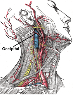
The descending branch of occipital artery, the largest branch of the occipital, descends on the back of the neck, and divides into a superficial and deep portion.

References

PD-icon.svgThis article incorporates text in the public domain from page 556 of the 20th edition of Gray's Anatomy (1918)

  1. 1 2 3 Standring, Susan (2020). Gray's Anatomy: The Anatomical Basis of Clinical Practice (42th ed.). New York. p. 586. ISBN   978-0-7020-7707-4. OCLC   1201341621.{{cite book}}: CS1 maint: location missing publisher (link)
  2. Gray, Henry (1918). Anatomy of the Human Body.