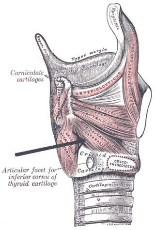
The lateral cricoarytenoid is an intrinsic muscle of the larynx. It attaches at the cricoid cartilage anteriorly, and at the arytenoid cartilage of the same side posteriorly. It is innervated by the recurrent laryngeal nerve. It acts to close the rima glottidis, thus closing the airway.

The falx cerebri is a large, crescent-shaped fold of dura mater that descends vertically into the longitudinal fissure between the cerebral hemispheres of the human brain, separating the two hemispheres and supporting dural sinuses that provide venous and CSF drainage to the brain. It is attached to the crista galli anteriorly, and blends with the tentorium cerebelli posteriorly.

The rectus capitis posterior major is a muscle in the upper back part of the neck. It is one of the suboccipital muscles. Its inferior attachment is at the spinous process of the axis ; its superior attachment is onto the outer surface of the occipital bone on and around the side part of the inferior nuchal line. The muscle is innervated by the suboccipital nerve. The muscle acts to extend the head and rorate the head to its side.
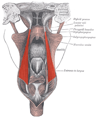
The salpingopharyngeus muscle is a muscle of the pharynx. It arises from the lower part of the cartilage of the Eustachian tube, and inserts into the palatopharyngeus muscle by blending with its posterior fasciculus. It is innervated by vagus nerve via the pharyngeal plexus. It raises the pharynx and larynx during deglutition (swallowing) and laterally draws the pharyngeal walls up. It opens the pharyngeal orifice of the Eustachian tube during swallowing to allow for the equalization of pressure between it and the pharynx.

The nasociliary nerve is a branch of the ophthalmic nerve (CN V1) (which is in turn a branch of the trigeminal nerve (CN V)). It is intermediate in size between the other two branches of the ophthalmic nerve, the frontal nerve and lacrimal nerve.

The occipital artery is a branch of the external carotid artery that provides arterial supply to the back of the scalp, sternocleidomastoid muscles, and deep muscles of the back and neck.

The ascending pharyngeal artery is an artery of the neck that supplies the pharynx.

The anterior ethmoidal artery is a branch of the ophthalmic artery in the orbit. It exits the orbit through the anterior ethmoidal foramen alongside the anterior ethmoidal nerve. It contributes blood supply to the ethmoid sinuses, frontal sinuses, the dura mater, lateral nasal wall, and nasal septum. It issues a meningeal branch, and nasal branches.

The infratrochlear nerve is a branch of the nasociliary nerve (itself a branch of the ophthalmic nerve (CN V1)) in the orbit. It exits the orbit inferior to the trochlea of superior oblique. It provides sensory innervation to structures of the orbit and skin of adjacent structures.
The anterior ethmoidal nerve is a nerve of the head. It is a branch of the nasociliary nerve (itself a branch of the ophthalmic nerve (V1)). It arises in the orbit, and enters first the cranial cavity and then the nasal cavity. It provides sensory innervation to part of the meninges, parts of the nasal cavity, and part of the skin of the nose.
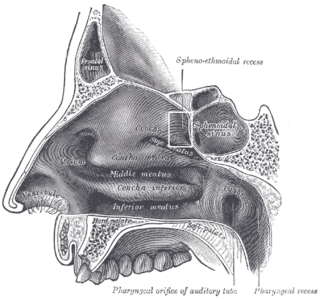
The sphenoethmoidal recess is a small triangular space of the superior nasal meatus of the nasal cavity into which the sphenoidal sinus and the posterior ethmoidal air cells open. The sphenoethmoidal recess is situated superoposterior to the superior nasal concha, between the superior nasal concha and the anterior aspect of the body of the sphenoid bone.

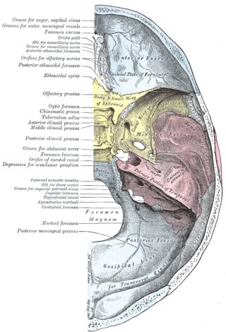
The middle cranial fossa is formed by the sphenoid bones, and the temporal bones. It lodges the temporal lobes, and the pituitary gland. It is deeper than the anterior cranial fossa, is narrow medially and widens laterally to the sides of the skull. It is separated from the posterior cranial fossa by the clivus and the petrous crest.
The posterior meningeal artery is one of the meningeal branches of the ascending pharyngeal artery. It passes through the jugular foramen to enter the posterior cranial fossa. It is the largest vessel supplying the dura of the posterior cranial fossa.
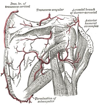
The anterior humeral circumflex artery is an artery in the arm. It is one of two circumflexing arteries that branch from the axillary artery, the other being the posterior humeral circumflex artery. The anterior humeral circumflex artery is considerably smaller than the posterior and arises nearly opposite to it, from the lateral side of the axillary artery.

The dorsal nasal artery is an artery of the face. It is one of the two terminal branches of the ophthalmic artery. It contributes arterial supply to the lacrimal sac, and outer surface of the nose.

The supratrochlear artery is one of the terminal branches of the ophthalmic artery. It arises within the orbit. It exits the orbit alongside the supratrochlear nerve. It contributes arterial supply to the skin, muscles and pericranium of the forehead.

The short posterior ciliary arteries are a number of branches of the ophthalmic artery. They pass forward with the optic nerve to reach the eyeball, piercing the sclera around the entry of the optic nerve into the eyeball.

The ethmoidal infundibulum is a funnel-shaped/slit-like/curved opening/passage/space/cleft upon the anterosuperior portion of the middle nasal meatus at the hiatus semilunaris. The anterior ethmoidal air cells, and (usually) the frontonasal duct open into the ethmoidal infundibulum. The ethmoidal infundibulum extends anterosuperiorly from its opening into the nasal cavity.

The sphenoparietal sinus is a paired dural venous sinus situated along the posterior edge of the lesser wing of either sphenoid bone. It drains into the cavernous sinus.
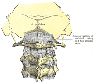
The posterior atlantooccipital membrane is a broad but thin membrane extending between the to the posterior margin of the foramen magnum above, and posterior arch of atlas below. It forms the floor of the suboccipital triangle.
