Articles related to anatomy include:

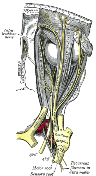
The oculomotor nerve, also known as the third cranial nerve, cranial nerve III, or simply CN III, is a cranial nerve that enters the orbit through the superior orbital fissure and innervates extraocular muscles that enable most movements of the eye and that raise the eyelid. The nerve also contains fibers that innervate the intrinsic eye muscles that enable pupillary constriction and accommodation. The oculomotor nerve is derived from the basal plate of the embryonic midbrain. Cranial nerves IV and VI also participate in control of eye movement.
The supraorbital nerve is one of two branches of the frontal nerve, itself a branch of the ophthalmic nerve. The other branch of the frontal nerve is the supratrochlear nerve.

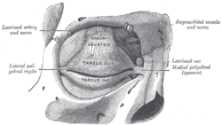
In anatomy, the orbit is the cavity or socket/hole of the skull in which the eye and its appendages are situated. "Orbit" can refer to the bony socket, or it can also be used to imply the contents. In the adult human, the volume of the orbit is 30 millilitres, of which the eye occupies 6.5 ml. The orbital contents comprise the eye, the orbital and retrobulbar fascia, extraocular muscles, cranial nerves II, III, IV, V, and VI, blood vessels, fat, the lacrimal gland with its sac and duct, the eyelids, medial and lateral palpebral ligaments, cheek ligaments, the suspensory ligament, septum, ciliary ganglion and short ciliary nerves.

The middle meningeal artery is typically the third branch of the first portion of the maxillary artery. After branching off the maxillary artery in the infratemporal fossa, it runs through the foramen spinosum to supply the dura mater and the calvaria. The middle meningeal artery is the largest of the three (paired) arteries that supply the meninges, the others being the anterior meningeal artery and the posterior meningeal artery.

The levator palpebrae superioris is the muscle in the orbit that elevates the upper eyelid.

The extraocular muscles, or extrinsic ocular muscles, are the seven extrinsic muscles of the eye in humans and other animals. Six of the extraocular muscles, the four recti muscles, and the superior and inferior oblique muscles, control movement of the eye. The other muscle, the levator palpebrae superioris, controls eyelid elevation. The actions of the six muscles responsible for eye movement depend on the position of the eye at the time of muscle contraction.

The ophthalmic artery (OA) is an artery of the head. It is the first branch of the internal carotid artery distal to the cavernous sinus. Branches of the ophthalmic artery supply all the structures in the orbit around the eye, as well as some structures in the nose, face, and meninges. Occlusion of the ophthalmic artery or its branches can produce sight-threatening conditions.

The ophthalmic nerve (CN V1) is a sensory nerve of the head. It is one of three divisions of the trigeminal nerve (CN V), a cranial nerve. It has three major branches which provide sensory innervation to the eye, and the skin of the upper face and anterior scalp, as well as other structures of the head.

The superior ophthalmic vein is a vein of the orbit that drains venous blood from structures of the upper orbit. It is formed by the union of the angular vein, and supraorbital vein. It passes backwards within the orbit alongside the ophthalmic artery, then exits the orbit through the superior orbital fissure to drain into the cavernous sinus.
The frontal nerve is the largest branch of the ophthalmic nerve (V1), itself a branch of the trigeminal nerve (CN V). It supplies sensation to the skin of the forehead, the mucosa of the frontal sinus, and the skin of the upper eyelid. It may be affected by schwannoma.

The lacrimal nerve is the smallest of the three main branches of the ophthalmic nerve (CN V1) (itself a branch of the trigeminal nerve (CN V)).

The supratrochlear nerve is a branch of the frontal nerve, itself a branch of the ophthalmic nerve (CN V1) from the trigeminal nerve (CN V). It provides sensory innervation to the skin of the forehead and the upper eyelid.

The supraorbital vein is a vein of the forehead. It communicates with the frontal branch of the superficial temporal vein. It passes through the supraorbital notch, and merges with the angular vein to form the superior ophthalmic vein. The supraorbital vein helps to drain blood from the forehead, eyebrow, and upper eyelid.
The lacrimal artery is an artery of the orbit. It is a branch of the ophthalmic artery. It accompanies the lacrimal nerve along the upper border of the lateral rectus muscle, travelling forward to reach the lacrimal gland. It supplies the lacrimal gland, two rectus muscles of the eye, the eyelids, and the conjunctiva.

The infraorbital artery is a small artery in the head that arises from the maxillary artery and passes through the inferior orbital fissure to enter the orbit, then passes forward along the floor of the orbit, finally exiting the orbit through the infraorbital foramen to reach the face.

The angular artery is an artery of the face. It is the terminal part of the facial artery. It ascends to the medial angle of the eye's orbit. It is accompanied by the angular vein. It ends by anastomosing with the dorsal nasal branch of the ophthalmic artery. It supplies the lacrimal sac, the orbicularis oculi muscle, and the outer side of the nose.

The supratrochlear artery is one of the terminal branches of the ophthalmic artery. It arises within the orbit. It exits the orbit alongside the supratrochlear nerve. It contributes arterial supply to the skin, muscles and pericranium of the forehead.

The anterior ciliary arteries are seven arteries in each eye-socket that arise from muscular branches of the ophthalmic artery and supply the conjunctiva, sclera, rectus muscles, and the ciliary body. The arteries end by anastomosing with branches of the long posterior ciliary arteries to form the circulus arteriosus major.

The following outline is provided as an overview of and topical guide to human anatomy: