Inferior thyroid artery

Last updated

Inferior thyroid artery
Thyrocervical trunk.png
Thyrocervical trunk and its branches, including inferior thyroid artery. Superficial dissection of the right side of the neck.
Gray1177.png
Details
Source Thyrocervical trunk
Vein Inferior thyroid veins
Supplies Thyroid gland
Identifiers
Latin arteria thyreoidea inferior
TA98 A12.2.08.043
TA2 4591
FMA 10662
Anatomical terminology

The inferior thyroid artery is an artery in the neck. It arises from the thyrocervical trunk and passes upward, in front of the vertebral artery and longus colli muscle. It then turns medially behind the carotid sheath and its contents, and also behind the sympathetic trunk, the middle cervical ganglion resting upon the vessel.

Contents

Reaching the lower border of the thyroid gland it divides into two branches, which supply the postero-inferior parts of the gland, and anastomose with the superior thyroid artery, and with the corresponding artery of the opposite side.

Structure

The branches of the inferior thyroid artery are the inferior laryngeal, the oesophageal, the tracheal, the ascending cervical and the pharyngeal arteries.

Branches

Inferior laryngeal artery

The inferior laryngeal artery - accompanied by the recurrent laryngeal nerve - passes superior-ward upon the trachea [1] deep to the inferior pharyngeal constrictor muscle to reach the posterior surface of the larynx.[ citation needed ] At the inferior border of the inferior pharyngeal constrictor muscle, the artery enters the larynx. [1]

The artery supplies the muscles and mucosa of the larynx. [1]

It forms anastomoses with its contralateral partner, and the superior laryngeal branch of the superior thyroid artery. [1]

Tracheal branches

The tracheal branches are distributed on the trachea, and anastomose inferiorly with the bronchial arteries. [1]

Esophageal branches

The esophageal branches supply the esophagus, and anastomose with the esophageal branches of the thoracic aorta. [1]

Ascending cervical artery

The ascending cervical artery is a small branch which arises from the inferior thyroid artery as it turns medial-ward posterior to the carotid sheath. [1]

The artery ascends upon the anterior tubercles of the transverse processes of the cervical vertebrae between the anterior scalene muscle and longus capitis muscle. [1]

The ascending cervical artery gives twigs to the neck muscles and these anastomose with branches of the vertebral arteries. One or two spinal branches are sent into the spinal canal, through the intervertebral foramina to be distributed to the spinal cord and its membranes, and to the bodies of the vertebrae. It then anastomoses with the ascending pharyngeal and occipital arteries.

Pharyngeal branches

The pharyngeal branches are distributed to the inferior portion of the pharynx. [1]

Glandular branches

The glandular branches are supply the inferior and posterior portions of the thyroid gland. They anastomose with the ipsilateral superior thyroid artery, and the contralateral inferior thyroid artery. The ascending branch supplies the parathyroid gland as well. [1]

Clinical significance

The relationship between the recurrent laryngeal nerve and inferior thyroid artery is highly variable. [2] The recurrent laryngeal nerve passes upward generally behind, but occasionally in front of, the inferior thyroid artery. This makes it vulnerable to injury during surgery that involves ligating the inferior thyroid artery, such as excision of the lower pole of the thyroid gland. Also as the parathyroid is mainly supplied by inferior thyroid artery accidental ligation during thyroidectomy can cause hypoparathyroidism.

The injection of dye into the inferior thyroid artery can be used as an alternate method in identification the recurrent laryngeal nerve. [3]

Additional images

See also

Related Research Articles

<span class="mw-page-title-main">Vagus nerve</span> Cranial nerve X, for visceral innervation

The vagus nerve, also known as the tenth cranial nerve, cranial nerve X, or simply CN X, is a cranial nerve that carries sensory fibers that create a pathway that interfaces with the parasympathetic control of the heart, lungs, and digestive tract. It comprises two nerves—the left and right vagus nerves—but they are typically referred to collectively as a single subsystem. The vagus is the longest nerve of the autonomic nervous system in the human body and comprises both sensory and motor fibers. The sensory fibers originate from neurons of the nodose ganglion, whereas the motor fibers come from neurons of the dorsal motor nucleus of the vagus and the nucleus ambiguus. The vagus was also historically called the pneumogastric nerve.

Articles related to anatomy include:

<span class="mw-page-title-main">External carotid artery</span> Major artery of the head and neck

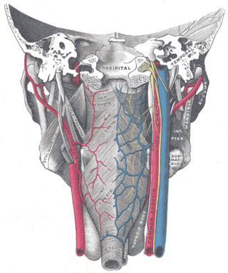
The external carotid artery is a major artery of the head and neck. It arises from the common carotid artery when it splits into the external and internal carotid artery. The external carotid artery supplies blood to the face, brain and neck.

<span class="mw-page-title-main">Recurrent laryngeal nerve</span> Nerve in the human body

The recurrent laryngeal nerve (RLN) is a branch of the vagus nerve that supplies all the intrinsic muscles of the larynx, with the exception of the cricothyroid muscles. There are two recurrent laryngeal nerves, right and left. The right and left nerves are not symmetrical, with the left nerve looping under the aortic arch, and the right nerve looping under the right subclavian artery then traveling upwards. They both travel alongside the trachea. Additionally, the nerves are among the few nerves that follow a recurrent course, moving in the opposite direction to the nerve they branch from, a fact from which they gain their name.

<span class="mw-page-title-main">Posterior cricoarytenoid muscle</span> Muscle of the larynx

The posterior cricoarytenoid muscle is a intrinsic muscle of the larynx. It arises from the cricoid cartilage; it inserts onto the arytenoid cartilage of the same side. It is innervated by the recurrent laryngeal nerve. Each acts to open the vocal folds by pulling the vocal fold of the same side laterally. It participates in the production of sounds.

<span class="mw-page-title-main">Facial artery</span>

The facial artery is a branch of the external carotid artery that supplies structures of the superficial face.

<span class="mw-page-title-main">Common carotid artery</span> One of the two arteries that supply the head and neck with blood

In anatomy, the left and right common carotid arteries (carotids) are arteries that supply the head and neck with oxygenated blood; they divide in the neck to form the external and internal carotid arteries.

<span class="mw-page-title-main">Sternothyroid muscle</span>

The sternothyroid muscle is an infrahyoid muscle of the neck. It acts to depress the hyoid bone.

<span class="mw-page-title-main">Thyrohyoid muscle</span> Neck muscle that depresses the hyoid bone and elevates the larynx

The thyrohyoid muscle is a small skeletal muscle of the neck. Above, it attaches onto the greater cornu of the hyoid bone; below, it attaches onto the oblique line of the thyroid cartilage. It is innervated by fibres derived from the cervical spinal nerve 1 that run with the hypoglossal nerve to reach this muscle. The thyrohyoid muscle depresses the hyoid bone and elevates the larynx during swallowing. By controlling the position and shape of the larynx, it aids in making sound.

<span class="mw-page-title-main">Inferior pharyngeal constrictor muscle</span> Skeletal muscle of the pharynx

The inferior pharyngeal constrictor muscle is a skeletal muscle of the neck. It is the thickest of the three outer pharyngeal muscles. It arises from the sides of the cricoid cartilage and the thyroid cartilage. It is supplied by the vagus nerve. It is active during swallowing, and partially during breathing and speech. It may be affected by Zenker's diverticulum.

<span class="mw-page-title-main">Stylopharyngeus muscle</span> Muscle

The stylopharyngeus muscle is a muscle in the head. It originates from the temporal styloid process. Some of its fibres insert onto the thyroid cartilage, while others end by intermingling with proximal structures. It is innervated by the glossopharyngeal nerve. It acts to elevate the larynx and pharynx, and dilate the pharynx, thus facilitating swallowing.

<span class="mw-page-title-main">Superior thyroid artery</span>

The superior thyroid artery arises from the external carotid artery just below the level of the greater cornu of the hyoid bone and ends in the thyroid gland.

<span class="mw-page-title-main">Ascending pharyngeal artery</span>

The ascending pharyngeal artery is an artery of the neck that supplies the pharynx.

<span class="mw-page-title-main">Superior laryngeal nerve</span> Branch of the vagus nerve

The superior laryngeal nerve is a branch of the vagus nerve. It arises from the middle of the inferior ganglion of vagus nerve and additionally also receives a sympathetic branch from the superior cervical ganglion.

In anatomy, arterial tree is used to refer to all arteries and/or the branching pattern of the arteries. This article regards the human arterial tree. Starting from the aorta:

<span class="mw-page-title-main">Carotid triangle</span>

The carotid triangle is a portion of the anterior triangle of the neck.

<span class="mw-page-title-main">Inferior cervical ganglion</span>

The inferior cervical ganglion is one of the three cervical sympathetic ganglia. It situated between the base of the transverse process of the last cervical vertebra and the neck of the first rib, on the medial side of the costocervical artery.

<span class="mw-page-title-main">Pharyngeal plexus of vagus nerve</span> Nerve fibers innervating most of the palate and pharynx

The pharyngeal plexus is a nerve plexus located upon the outer surface of the pharynx. It contains a motor component, a sensory component, and sympathetic component.

<span class="mw-page-title-main">Outline of human anatomy</span> Overview of and topical guide to human anatomy

The following outline is provided as an overview of and topical guide to human anatomy:

References

PD-icon.svgThis article incorporates text in the public domain from page 581 of the 20th edition of Gray's Anatomy (1918)

  1. 1 2 3 4 5 6 7 8 9 10 Standring, Susan (2020). Gray's Anatomy: The Anatomical Basis of Clinical Practice (42th ed.). New York. p. 590. ISBN   978-0-7020-7707-4. OCLC   1201341621.{{cite book}}: CS1 maint: location missing publisher (link)
  2. Yalçin B (February 2006). "Anatomic configurations of the recurrent laryngeal nerve and inferior thyroid artery". Surgery. 139 (2): 181–7. doi:10.1016/j.surg.2005.06.035. PMID   16455326.
  3. Hepgul G, Kucukyilmaz M, Koc O, Duzkoylu Y, Sari YS, Erbil Y (2013). "The identification of recurrent laryngeal nerve by injection of blue dye into the inferior thyroid artery in elusive locations". Journal of Thyroid Research. 2013: 539274. doi: 10.1155/2013/539274 . PMC   3563180 . PMID   23401846.