Related Research Articles

The soft palate is, in mammals, the soft tissue constituting the back of the roof of the mouth. The soft palate is part of the palate of the mouth; the other part is the hard palate. The soft palate is distinguished from the hard palate at the front of the mouth in that it does not contain bone.

In anatomy, the palatine bones are two irregular bones of the facial skeleton in many animal species, located above the uvula in the throat. Together with the maxilla, they comprise the hard palate.

Palatine tonsils, commonly called the tonsils and occasionally called the faucial tonsils, are tonsils located on the left and right sides at the back of the throat, which can often be seen as flesh-colored, pinkish lumps. Tonsils only present as "white lumps" if they are inflamed or infected with symptoms of exudates and severe swelling.

The renal arteries are paired arteries that supply the kidneys with blood. Each is directed across the crus of the diaphragm, so as to form nearly a right angle.
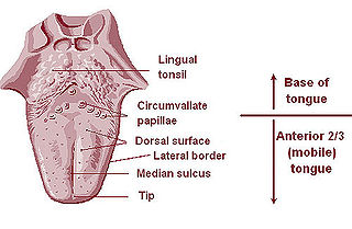
The lingual tonsils are a collection of lymphatic tissue located in the lamina propria of the root of the tongue. This lymphatic tissue consists of the lymphatic nodules rich in cells of the immune system (immunocytes). The immunocytes initiate the immune response when the lingual tonsils get in contact with invading microorganisms.

The pharyngeal arches, also known as visceral arches, are structures seen in the embryonic development of vertebrates that are recognisable precursors for many structures. In fish, the arches are known as the branchial arches, or gill arches.

The lingual artery arises from the external carotid artery between the superior thyroid artery and facial artery. It can be located easily in the tongue.

The anterior interosseous artery is an artery in the forearm. It is a branch of the common interosseous artery.
The nasopalatine nerve (also long sphenopalatine nerve) is a nerve of the head. It is a sensory branch of the maxillary nerve (CN V2) that passes through the pterygopalatine ganglion (without synapsing) and then through the sphenopalatine foramen to enter the nasal cavity, and finally out of the nasal cavity through the incisive canal and then the incisive fossa to enter the hard palate. It provides sensory innervation to the posteroinferior part of the nasal septum, and gingiva just posterior to the upper incisor teeth.

The descending palatine artery is a branch of the third part of the maxillary artery supplying the hard and soft palate.
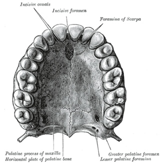
The incisive canals are two bony canals of the anterior hard palate connecting the nasal cavity and the oral cavity. An incisive canal courses through each maxilla. Below, the two incisive canals typically converge medially.
The greater palatine artery is a branch of the descending palatine artery and contributes to the blood supply of the hard palate and nasal septum.

The ascending palatine artery is an artery is a branch of the facial artery which ascends along the neck before splitting into two terminal branches; one branch supplies the soft palate, and the other supplies the palatine tonsil and pharyngotympanic tube.

The palatine nerves are distributed to the roof of the mouth, soft palate, tonsil, and lining membrane of the nasal cavity.
The greater palatine nerve is a branch of the pterygopalatine ganglion. This nerve is also referred to as the anterior palatine nerve, due to its location anterior to the lesser palatine nerve. It carries both general sensory fibres from the maxillary nerve, and parasympathetic fibers from the nerve of the pterygoid canal. It may be anaesthetised for procedures of the mouth and maxillary (upper) teeth.

The greater palatine canal is a passage in the skull that transmits the descending palatine artery, vein, and greater and lesser palatine nerves between the pterygopalatine fossa and the oral cavity.
The lesser palatine nerves (posterior palatine nerve) are branches of the maxillary nerve (CN V2). They descends through the greater palatine canal alongside the greater palatine nerve, and emerge (separately) through the lesser palatine foramen to pass posteriorward. They supply the soft palate, tonsil, and uvula.

The human nose is the first organ of the respiratory system. It is also the principal organ in the olfactory system. The shape of the nose is determined by the nasal bones and the nasal cartilages, including the nasal septum, which separates the nostrils and divides the nasal cavity into two.
The tonsillar branches of glossopharyngeal nerve supply the palatine tonsil, forming around it a plexus from which filaments are distributed to the soft palate and fauces, where they communicate with the palatine nerves.

The pharynx is the part of the throat behind the mouth and nasal cavity, and above the esophagus and trachea. It is found in vertebrates and invertebrates, though its structure varies across species. The pharynx carries food to the esophagus and air to the larynx. The flap of cartilage called the epiglottis stops food from entering the larynx.
References
- ↑ Choi, Jinho; Park, Hyung-Sik (1 January 2003). "The clinical anatomy of the maxillary artery in the pterygopalatine fossa". Journal of Oral and Maxillofacial Surgery . 61 (1): 72–78. doi:10.1053/joms.2003.50012. ISSN 0278-2391.
- 1 2 3 Miwa, Yoko; Asaumi, Rieko; Kawai, Taisuke; Maeda, Yuuki; Sato, Iwao (1 February 2018). "Morphological observation and CBCT of the bony canal structure of the groove and the location of blood vessels and nerves in the palatine of elderly human cadavers". Surgical and Radiologic Anatomy . 40 (2): 199–206. doi:10.1007/s00276-017-1952-6. ISSN 1279-8517.
- ↑ "Lesser palatine arteries" at Dorland's Medical Dictionary
- ↑ Maistry, T.; Lazarus, L.; Partab, P.; Satyapal, K. S. (2012). "An Anatomical Study of the Arterial Supply to the Soft Palate". International Journal of Morphology. 30 (3): 847–857. doi: 10.4067/S0717-95022012000300014 . S2CID 39720355.
- ↑ Zhang, K. Q. (1 January 1994). "Artery supply of the lip and palate in normal and cleft patients". Chinese Journal of Stomatology. 29 (1): 30–3, 63. ISSN 1002-0098. PMID 7995132.
| | This cardiovascular system article is a stub. You can help Wikipedia by expanding it. |