Aurora kinase A

Last updated
AURKA
Protein AURKA PDB 1mq4.png
Available structures
PDB Ortholog search: PDBe RCSB
Identifiers
Aliases AURKA , AIK, ARK1, AURA, AURORA2, BTAK, PPP1R47, STK15, STK6, STK7, aurora kinase A
External IDs OMIM: 603072 MGI: 894678 HomoloGene: 2670 GeneCards: AURKA
Orthologs
SpeciesHumanMouse
Entrez
Ensembl
UniProt
RefSeq (mRNA)

NM_011497
NM_001291185

RefSeq (protein)

NP_001278114
NP_035627

Location (UCSC) Chr 20: 56.37 – 56.39 Mb Chr 2: 172.2 – 172.21 Mb
PubMed search [3] [4]
Wikidata
View/Edit Human View/Edit Mouse

Aurora kinase A also known as serine/threonine-protein kinase 6 is an enzyme that in humans is encoded by the AURKA gene. [5] [6]

Aurora A is a member of a family of mitotic serine/threonine kinases. It is implicated with important processes during mitosis and meiosis whose proper function is integral for healthy cell proliferation. Aurora A is activated by one or more phosphorylations [7] and its activity peaks during the G2 phase to M phase transition in the cell cycle. [8]

Discovery

The aurora kinases were first identified in 1990 during a cDNA screen of Xenopus eggs. [7] The kinase discovered, Eg2, is now referred to as Aurora A. [9] However, Aurora A's meiotic and mitotic significance was not recognized until 1998. [7]

Aurora kinase family

The human genome contains three members of the aurora kinase family: Aurora kinase A, Aurora kinase B and Aurora C kinase. The Xenopus, Drosophila , and Caenorhabditis elegans genomes, on the other hand, contain orthologues only to Aurora A and Aurora B. [7]

In all studied species, the three Aurora mitotic kinases localize to the centrosome [9] during different phases of mitosis. [7] The family members have highly conserved C-terminal catalytic domains. Their N-terminal domains, however, exhibit a large degree of variance in the size and sequence. [9]

Aurora A and Aurora B kinases play important roles in mitosis. The Aurora kinase A is associated with centrosome maturation and separation and thereby regulates spindle assembly and stability. The Aurora kinase B is a chromosome passenger protein and regulates chromosome segregation and cytokinesis.

Although there is evidence to suggest that Aurora C might be a chromosomal passenger protein, the cellular function of it is less clear.

Localization

Aurora A localizes next to the centrosome late in the G1 phase and early in the S phase. As the cell cycle progresses, concentrations of Aurora A increase and the kinase associates with the mitotic poles and the adjacent spindle microtubules. Aurora A remains associated with the spindles through telophase. [7] Right before mitotic exit, Aurora A relocalizes to the mid-zone of the spindle. [10]

Mitosis

During mitosis, a mitotic spindle is assembled by using microtubules to tether together the mother centrosome to its daughter. The resulting mitotic spindle is then used to propel apart the sister chromosomes into what will become the two new daughter cells. Aurora A is critical for proper formation of mitotic spindle. It is required for the recruitment of several different proteins important to the spindle formation. Among these target proteins are TACC, a microtubule-associated protein that stabilizes centrosomal microtubules and Kinesin 5, a motor protein involved in the formation of the bipolar mitotic spindle. [7] γ-tubulins, the base structure from which centrosomal microtubules polymerize, are also recruited by Aurora A. Without Aurora A the centrosome does not accumulate the quantity of γ-tubulin that normal centrosomes recruit prior to entering anaphase. Though the cell cycle continues even in the absence of sufficient γ-tubulin, the centrosome never fully matures; it organizes fewer aster microtubules than normal. [8]

Furthermore, Aurora A is necessary for the proper separation of the centrosomes after the mitotic spindle has been formed. Without Aurora A, the mitotic spindle, depending on the organism, will either never separate or will begin to separate only to collapse back onto itself. [8] In the case of the former, it has been suggested that Aurora A cooperates with the kinase Nek2 in Xenopus to dissolve the structure tethering the cell's centrosomes together. Therefore, without proper expression of Aurora A, the cell's centrosomes are never able to separate. [10]

Aurora A also assures proper organization and alignment of the chromosomes during prometaphase. It is directly involved in the interaction of the kinetochore, the part of the chromosome at which the mitotic spindle attaches and pulls, and the mitotic spindle's extended microtubules. It is speculated that Aurora B cooperates with Aurora A to complete this task. In the absence of Aurora A mad2, a protein that normally dissipates once a proper kinetochore-microtubule connection is made, remains present even into metaphase. [10]

Finally, Aurora A helps orchestrate an exit from mitosis by contributing to the completion of cytokinesis- the process by which the cytoplasm of the parent cell is split into two daughter cells. During cytokinesis the mother centriole returns to the mid-body of the mitotic cell at the end of mitosis and causes the central microtubules to release from the mid-body. The release allows mitosis to run to completion. Though the exact mechanism by which Aurora A aids cytokinesis is unknown, it is well documented that it relocalizes to the mid-body immediately before the completion of mitosis. [10]

Intriguingly, abolishment of Aurora A through RNAi interference results in different mutant phenotypes in different organisms and cell types. [10] For example, deletion of Aurora A in C. elegans results in an initial separation of the cell's centrosomes followed by an immediate collapse of the asters. In Xenopus, deletion disallows the mitotic spindle from ever even forming. [8] And in Drosophila, flies without Aurora A will effectively form spindles and separate but the aster microtubules will be dwarfed. These observations suggests that while Aurora-A has orthologues in many different organisms, it may play a similar but slightly different role in each. [10]

Meiosis

Aurora A phosphorylation directs the cytoplasmic polyadenylation translation of mRNA's, like the MAP kinase kinase kinase protein MOS, that are vital to the completion of meiosis in Xenopus Oocytes. [9] Prior to the first meiotic metaphase, Aurora A induces the synthesis of MOS. The MOS protein accumulates until it exceeds a threshold and then transduces the phosphorylation cascade in the map kinase pathway. This signal subsequently activates the kinase RSK which in turn binds to the protein Myt1. Myt1, in complex with RSK, is now unable to inhibit cdc2. As a consequence, cdc2 permits entry into meiosis. [7] A similar Aurora A dependent process regulates the transition from meiosis I-meiosis II.

Furthermore, Aurora A has been observed to have a biphasic pattern of activation during progression through meiosis. It has been suggested that the fluctuations, or phases, of Aurora A activation are dependent on a positive-feedback mechanism with a p13SUC1-associated protein kinase [10]

Protein translation

Aurora A is not only implicated with the translation of MOS during meiosis but also in the polyadenylation and subsequent translation of neural mRNAs whose protein products are associated with synaptic plasticity. [10]

Clinical significance

Aurora A dysregulation has been associated with high occurrence of cancer. For example, one study showed over-expression of Aurora A in 94 percent of the invasive tissue growth in breast cancer, while surrounding, healthy tissues had normal levels of Aurora A expression. [7] Aurora A has also been shown to be involved in the Epithelial–mesenchymal transition and Neuroendocrine Transdifferentiation of Prostate Cancer cells in aggressive disease. [11]

Dysregulation of Aurora A may lead to cancer because Aurora A is required for the completion of cytokinesis. If the cell begins mitosis, duplicates its DNA, but is then not able to divide into two separate cells it becomes an aneuploid- containing more chromosomes than normal. Aneuploidy is a trait of many cancerous tumors. [10] Ordinarily, Aurora A expression levels are kept in check by the tumor suppressor protein p53. [7]

Mutations of the chromosome region that contains Aurora A, 20q13, are generally considered to have a poor prognosis. [7]

Osimertinib and rociletinib, two anti cancer drugs for lung cancer, work by shutting off mutant EGFR, which initially kills cancerous tumors, but the tumors rewire and activate Aurora kinase A, becoming cancerous growths again. According to a 2018 study, targeting both EGFR and Aurora prevents return of drug resistant tumors. [12]

Interactions

Aurora A kinase has been shown to interact with:

Related Research Articles

<span class="mw-page-title-main">Mitosis</span> Process in which chromosomes are replicated and separated into two new identical nuclei

Mitosis is a part of the cell cycle in which replicated chromosomes are separated into two new nuclei. Cell division by mitosis is an equational division which gives rise to genetically identical cells in which the total number of chromosomes is maintained. Mitosis is preceded by the S phase of interphase and is followed by telophase and cytokinesis, which divide the cytoplasm, organelles, and cell membrane of one cell into two new cells containing roughly equal shares of these cellular components. The different stages of mitosis altogether define the mitotic phase of a cell cycle—the division of the mother cell into two daughter cells genetically identical to each other.

<span class="mw-page-title-main">Cell division</span> Process by which living cells divide

Cell division is the process by which a parent cell divides into two daughter cells. Cell division usually occurs as part of a larger cell cycle in which the cell grows and replicates its chromosome(s) before dividing. In eukaryotes, there are two distinct types of cell division: a vegetative division (mitosis), producing daughter cells genetically identical to the parent cell, and a cell division that produces haploid gametes for sexual reproduction (meiosis), reducing the number of chromosomes from two of each type in the diploid parent cell to one of each type in the daughter cells. Mitosis is a part of the cell cycle, in which, replicated chromosomes are separated into two new nuclei. Cell division gives rise to genetically identical cells in which the total number of chromosomes is maintained. In general, mitosis is preceded by the S stage of interphase and is followed by telophase and cytokinesis; which divides the cytoplasm, organelles, and cell membrane of one cell into two new cells containing roughly equal shares of these cellular components. The different stages of mitosis all together define the M phase of an animal cell cycle—the division of the mother cell into two genetically identical daughter cells. To ensure proper progression through the cell cycle, DNA damage is detected and repaired at various checkpoints throughout the cycle. These checkpoints can halt progression through the cell cycle by inhibiting certain cyclin-CDK complexes. Meiosis undergoes two divisions resulting in four haploid daughter cells. Homologous chromosomes are separated in the first division of meiosis, such that each daughter cell has one copy of each chromosome. These chromosomes have already been replicated and have two sister chromatids which are then separated during the second division of meiosis. Both of these cell division cycles are used in the process of sexual reproduction at some point in their life cycle. Both are believed to be present in the last eukaryotic common ancestor.

<span class="mw-page-title-main">Centrosome</span> Cell organelle in animal cell helping in cell division

In cell biology, the centrosome is an organelle that serves as the main microtubule organizing center (MTOC) of the animal cell, as well as a regulator of cell-cycle progression. The centrosome provides structure for the cell. The centrosome is thought to have evolved only in the metazoan lineage of eukaryotic cells. Fungi and plants lack centrosomes and therefore use other structures to organize their microtubules. Although the centrosome has a key role in efficient mitosis in animal cells, it is not essential in certain fly and flatworm species.

<span class="mw-page-title-main">Spindle apparatus</span> Feature of biological cell structure

In cell biology, the spindle apparatus is the cytoskeletal structure of eukaryotic cells that forms during cell division to separate sister chromatids between daughter cells. It is referred to as the mitotic spindle during mitosis, a process that produces genetically identical daughter cells, or the meiotic spindle during meiosis, a process that produces gametes with half the number of chromosomes of the parent cell.

<span class="mw-page-title-main">Telophase</span> Final stage of a cell division for eukaryotic cells both in mitosis and meiosis

Telophase is the final stage in both meiosis and mitosis in a eukaryotic cell. During telophase, the effects of prophase and prometaphase are reversed. As chromosomes reach the cell poles, a nuclear envelope is re-assembled around each set of chromatids, the nucleoli reappear, and chromosomes begin to decondense back into the expanded chromatin that is present during interphase. The mitotic spindle is disassembled and remaining spindle microtubules are depolymerized. Telophase accounts for approximately 2% of the cell cycle's duration.

<span class="mw-page-title-main">Spindle checkpoint</span> Cell cycle checkpoint

The spindle checkpoint, also known as the metaphase-to-anaphase transition, the spindle assembly checkpoint (SAC), the metaphase checkpoint, or the mitotic checkpoint, is a cell cycle checkpoint during metaphase of mitosis or meiosis that prevents the separation of the duplicated chromosomes (anaphase) until each chromosome is properly attached to the spindle. To achieve proper segregation, the two kinetochores on the sister chromatids must be attached to opposite spindle poles. Only this pattern of attachment will ensure that each daughter cell receives one copy of the chromosome. The defining biochemical feature of this checkpoint is the stimulation of the anaphase-promoting complex by M-phase cyclin-CDK complexes, which in turn causes the proteolytic destruction of cyclins and proteins that hold the sister chromatids together.

<span class="mw-page-title-main">Kinetochore</span> Protein complex that allows microtubules to attach to chromosomes during cell division

A kinetochore is a disc-shaped protein structure associated with duplicated chromatids in eukaryotic cells where the spindle fibers attach during cell division to pull sister chromatids apart. The kinetochore assembles on the centromere and links the chromosome to microtubule polymers from the mitotic spindle during mitosis and meiosis. The term kinetochore was first used in a footnote in a 1934 Cytology book by Lester W. Sharp and commonly accepted in 1936. Sharp's footnote reads: "The convenient term kinetochore has been suggested to the author by J. A. Moore", likely referring to John Alexander Moore who had joined Columbia University as a freshman in 1932.

<span class="mw-page-title-main">Aurora kinase B</span> Protein

Aurora kinase B is a protein that functions in the attachment of the mitotic spindle to the centromere.

<span class="mw-page-title-main">Aurora inhibitor</span>

Aurora kinase inhibitors are a putative drug class for treating cancer. The Aurora kinase enzymes could be potential targets for novel small-molecule enzyme inhibitors.

Polo-like kinases (Plks) are regulatory serine/threonine kinases of the cell cycle involved in mitotic entry, mitotic exit, spindle formation, cytokinesis, and meiosis. Only one Plk is found in the genomes of the fly Drosophila melanogaster (Polo), budding yeast (Cdc5) and fission yeast (Plo1). Vertebrates and other animals, however, have many Plk family members including Plk1, Plk2/Snk, Plk3/Prk/FnK, Plk4/Sak and Plk5. Of the vertebrate Plk family members, the mammalian Plk1 has been most extensively studied. During mitosis and cytokinesis, Plks associate with several structures including the centrosome, kinetochores, and the central spindle.

<span class="mw-page-title-main">PLK1</span> Mammalian protein found in Homo sapiens

Serine/threonine-protein kinase PLK1, also known as polo-like kinase 1 (PLK-1) or serine/threonine-protein kinase 13 (STPK13), is an enzyme that in humans is encoded by the PLK1 gene.

<span class="mw-page-title-main">NEK2</span> Protein-coding gene in the species Homo sapiens

Serine/threonine-protein kinase Nek2 is an enzyme that in humans is encoded by the NEK2 gene.

<span class="mw-page-title-main">KIF23</span> Protein-coding gene in the species Homo sapiens

Kinesin-like protein KIF23 is a protein that in humans is encoded by the KIF23 gene.

<span class="mw-page-title-main">INCENP</span> Protein-coding gene in the species Homo sapiens

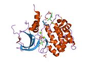
Inner centromere protein is a protein that in humans is encoded by the INCENP gene. It is a regulatory protein in the chromosome passenger complex (CPC). It is involved in regulation of the catalytic proteins Aurora B and Aurora C. It acts in association with two other proteins - Survivin and Borealin. These proteins form a tight three-helical bundle. The N-terminal domain of INCENP is the domain involved in formation of this three-helical bundle while its C-terminal domain is responsible for the interaction with Aurora B.

<span class="mw-page-title-main">TPX2</span> Protein-coding gene in the species Homo sapiens

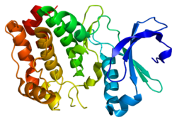
Targeting protein for Xklp2 is a protein that in humans is encoded by the TPX2 gene. It is one of the many spindle assembly factors that play a key role in inducing microtubule assembly and growth during M phase.

<span class="mw-page-title-main">Aurora kinase C</span> Protein-coding gene in the species Homo sapiens

Aurora kinase C, also Serine/threonine-protein kinase 13 is an enzyme that in humans is encoded by the AURKC gene.

<span class="mw-page-title-main">CKAP5</span> Protein-coding gene in the species Homo sapiens

Cytoskeleton-associated protein 5 is a microtubule-associated protein that in humans is encoded by the CKAP5 gene. It is the homolog of the Xenopus protein XMAP215 and is also known as ch-Tog.

Cdc14 and Cdc14 are a gene and its protein product respectively. Cdc14 is found in most of the eukaryotes. Cdc14 was defined by Hartwell in his famous screen for loci that control the cell cycle of Saccharomyces cerevisiae. Cdc14 was later shown to encode a protein phosphatase. Cdc14 is dual-specificity, which means it has serine/threonine and tyrosine-directed activity. A preference for serines next to proline is reported. Many early studies, especially in the budding yeast Saccharomyces cerevisiae, demonstrated that the protein plays a key role in regulating late mitotic processes. However, more recent work in a range of systems suggests that its cellular function is more complex.

<span class="mw-page-title-main">Centrosome cycle</span> Centrioles are nine triplets microtubules

Centrosomes are the major microtubule organizing centers (MTOC) in mammalian cells. Failure of centrosome regulation can cause mistakes in chromosome segregation and is associated with aneuploidy. A centrosome is composed of two orthogonal cylindrical protein assemblies, called centrioles, which are surrounded by a protein dense amorphous cloud of pericentriolar material (PCM). The PCM is essential for nucleation and organization of microtubules. The centrosome cycle is important to ensure that daughter cells receive a centrosome after cell division. As the cell cycle progresses, the centrosome undergoes a series of morphological and functional changes. Initiation of the centrosome cycle occurs early in the cell cycle in order to have two centrosomes by the time mitosis occurs.

<span class="mw-page-title-main">Kinesin-like protein KIF11</span> Protein-coding gene in the species Homo sapiens

Kinesin-like protein KIF11 is a molecular motor protein that is essential in mitosis. In humans it is coded for by the gene KIF11. Kinesin-like protein KIF11 is a member of the kinesin superfamily, which are nanomotors that move along microtubule tracks in the cell. Named from studies in the early days of discovery, it is also known as Kinesin-5, or as BimC, Eg5 or N-2, based on the founding members of this kinesin family.

References

  1. 1 2 3 GRCh38: Ensembl release 89: ENSG00000087586 Ensembl, May 2017
  2. 1 2 3 GRCm38: Ensembl release 89: ENSMUSG00000027496 Ensembl, May 2017
  3. "Human PubMed Reference:". National Center for Biotechnology Information, U.S. National Library of Medicine.
  4. "Mouse PubMed Reference:". National Center for Biotechnology Information, U.S. National Library of Medicine.
  5. Sen S, Zhou H, White RA (May 1997). "A putative serine/threonine kinase encoding gene BTAK on chromosome 20q13 is amplified and overexpressed in human breast cancer cell lines". Oncogene. 14 (18): 2195–200. doi: 10.1038/sj.onc.1201065 . PMID   9174055.
  6. Zhou H, Kuang J, Zhong L, Kuo WL, Gray JW, Sahin A, Brinkley BR, Sen S (October 1998). "Tumour amplified kinase STK15/BTAK induces centrosome amplification, aneuploidy and transformation". Nat. Genet. 20 (2): 189–93. doi:10.1038/2496. PMID   9771714. S2CID   40012197.
  7. 1 2 3 4 5 6 7 8 9 10 11 Crane R, Gadea B, Littlepage L, Wu H, Ruderman JV (2004). "Aurora A, meiosis and mitosis". Biol. Cell. 96 (3): 215–29. doi: 10.1016/j.biolcel.2003.09.008 . PMID   15182704. S2CID   29416056.
  8. 1 2 3 4 Hannak E, Kirkham M, Hyman AA, Oegema K (December 2001). "Aurora-A kinase is required for centrosome maturation in Caenorhabditis elegans". J. Cell Biol. 155 (7): 1109–16. doi:10.1083/jcb.200108051. PMC   2199344 . PMID   11748251.
  9. 1 2 3 4 Ma C, Cummings C, Liu XJ (March 2003). "Biphasic activation of Aurora-A kinase during the meiosis I- meiosis II transition in Xenopus oocytes". Mol. Cell. Biol. 23 (5): 1703–16. doi:10.1128/MCB.23.5.1703-1716.2003. PMC   151708 . PMID   12588989.
  10. 1 2 3 4 5 6 7 8 9 Marumoto T, Honda S, Hara T, Nitta M, Hirota T, Kohmura E, Saya H (December 2003). "Aurora-A kinase maintains the fidelity of early and late mitotic events in HeLa cells". J. Biol. Chem. 278 (51): 51786–95. doi: 10.1074/jbc.M306275200 . PMID   14523000.
  11. Nouri M, Ratther E, Stylianou N, Nelson CC, Hollier BG, Williams ED (2014). "Androgen-targeted therapy-induced epithelial mesenchymal plasticity and neuroendocrine transdifferentiation in prostate cancer: an opportunity for intervention". Front Oncol. 4: 370. doi: 10.3389/fonc.2014.00370 . PMC   4274903 . PMID   25566507.
  12. "Cancer researchers identify 'Achilles heel' of drug-resistant tumors".
  13. Sakai H, Urano T, Ookata K, Kim MH, Hirai Y, Saito M, Nojima Y, Ishikawa F (December 2002). "MBD3 and HDAC1, two components of the NuRD complex, are localized at Aurora-A-positive centrosomes in M phase". J. Biol. Chem. 277 (50): 48714–23. doi: 10.1074/jbc.M208461200 . PMID   12354758.
  14. Otto, Tobias; Horn, Sebastian; Brockmann, Markus; Eilers, Ursula; Schüttrumpf, Lars; Popov, Nikita; Kenney, Anna Marie; Schulte, Johannes H.; Beijersbergen, Roderick; Christiansen, Holger; Berwanger, Bernd; Eilers, Martin (2009). "Stabilization of N-Myc Is a Critical Function of Aurora A in Human Neuroblastoma". Cancer Cell. 15 (1). Elsevier BV: 67–78. doi:10.1016/j.ccr.2008.12.005. ISSN   1535-6108.
  15. Du J, Hannon GJ (December 2002). "The centrosomal kinase Aurora-A/STK15 interacts with a putative tumor suppressor NM23-H1". Nucleic Acids Res. 30 (24): 5465–75. doi:10.1093/nar/gkf678. PMC   140054 . PMID   12490715.
  16. Chen SS, Chang PC, Cheng YW, Tang FM, Lin YS (September 2002). "Suppression of the STK15 oncogenic activity requires a transactivation-independent p53 function". EMBO J. 21 (17): 4491–9. doi:10.1093/emboj/cdf409. PMC   126178 . PMID   12198151.
  17. Delaval B, Ferrand A, Conte N, Larroque C, Hernandez-Verdun D, Prigent C, Birnbaum D (June 2004). "Aurora B -TACC1 protein complex in cytokinesis". Oncogene. 23 (26): 4516–22. doi: 10.1038/sj.onc.1207593 . PMID   15064709.
  18. Conte N, Delaval B, Ginestier C, Ferrand A, Isnardon D, Larroque C, Prigent C, Séraphin B, Jacquemier J, Birnbaum D (November 2003). "TACC1-chTOG-Aurora A protein complex in breast cancer". Oncogene. 22 (50): 8102–16. doi: 10.1038/sj.onc.1206972 . PMID   14603251.
  19. Kufer TA, Silljé HH, Körner R, Gruss OJ, Meraldi P, Nigg EA (August 2002). "Human TPX2 is required for targeting Aurora-A kinase to the spindle". J. Cell Biol. 158 (4): 617–23. doi:10.1083/jcb.200204155. PMC   2174010 . PMID   12177045.
  20. Ewart-Toland A, Briassouli P, de Koning JP, Mao JH, Yuan J, Chan F, MacCarthy-Morrogh L, Ponder BA, Nagase H, Burn J, Ball S, Almeida M, Linardopoulos S, Balmain A (August 2003). "Identification of Stk6/STK15 as a candidate low-penetrance tumor-susceptibility gene in mouse and human". Nat. Genet. 34 (4): 403–12. doi:10.1038/ng1220. PMID   12881723. S2CID   29442841.

Further reading