Rhodopsin kinase

Last updated
G protein-coupled receptor kinase 1
Identifiers
SymbolGRK1
Alt. symbolsRHOK
NCBI gene 6011
HGNC 10013
OMIM 180381
RefSeq NM_002929
UniProt Q15835
Other data
EC number 2.7.11.14
Locus Chr. 13 q34

Rhodopsin kinase (EC 2.7.11.14, rod opsin kinase, G-protein-coupled receptor kinase 1, GPCR kinase 1, GRK1, opsin kinase, opsin kinase (phosphorylating), rhodopsin kinase (phosphorylating), RK, STK14) is a serine/threonine-specific protein kinase involved in phototransduction. [1] [2] [3] [4] [5] [6] [7] [8] This enzyme catalyses the following chemical reaction:

Contents

ATP + rhodopsin ADP + phospho-rhodopsin

Mutations in rhodopsin kinase are associated with a form of night blindness called Oguchi disease. [9]

Function and mechanism of action

Rhodopsin kinase is a member of the family of G protein-coupled receptor kinases, and is officially named G protein-coupled receptor kinase 1, or GRK1. Rhodopsin kinase is found primarily in mammalian retinal rod cells, where it phosphorylates light-activated rhodopsin, a member of the family of G protein-coupled receptors that recognizes light. Phosphorylated, light-activated rhodopsin binds to the protein arrestin to terminate the light-activated signaling cascade. The related GRK7, also known as cone opsin kinase, serves a similar function in retinal cone cells subserving high-acuity color vision in the fovea. [10] The post-translational modification of GRK1 by farnesylation and α-carboxyl methylation is important for regulating the ability of the enzyme to recognize rhodopsin in rod outer segment disk membranes. [11] [12]

Arrestin-1 bound to rhodopsin prevents rhodopsin activation of the transducin protein to turn off photo-transduction completely. [13] [14]

Rhodopsin kinase is inhibited by the calcium-binding protein recoverin in a graded manner that maintains rhodopsin sensitivity to light despite large changes in ambient light conditions. That is, in retinas exposed to only dim light, calcium levels are high in retinal rod cells and recoverin is bound to and inhibits rhodopsin kinase, leaving rhodopsin exquisitely sensitive to photons to mediate low-light, low-acuity vision; in bright light, rod cell calcium levels are low so recoverin cannot bind or inhibit rhodopsin kinase, resulting in greater rhodopsin kinase/arrestin inhibition of rhodopsin signaling at baseline to preserve visual sensitivity. [15] [16]

According to a proposed model, the N-terminus of rhodopsin kinase is involved in its own activation. It's suggested that an activated rhodopsin binds to the N-terminus, which is also involved in the stabilization of the kinase domain to induce an active conformation. [17]

Eye disease

Mutation in rhodopsin kinase can result in diseases such as Oguchi disease and retinal degeneration. Oguchi disease is a form of congenital stationary night blindness (CSNB). Congenital stationary night blindness is caused by the inability to send a signal from outer retina to the inner retina by signaling molecules. Oguchi disease is a genetic disorder so an individual can be inherited from his or her parents. Genes that are responsible for Oguchi disease are SAG (which encodes arrestin) and GRK1 genes. Rhodopsin kinase is encoded from the GRK1 gene, so a mutation in GRK1 can result in Oguchi disease. [18]

Retinal degeneration is a form of the retinal disease caused by the death of photoreceptor cells that present in the back of the eye, retina. Rhodopsin kinase directly participates in the rhodopsin to activate the visual phototransduction. Studies have shown that lack of rhodopsin kinase will result in photoreceptor cell death. [19] When photoreceptors cells die, they will be detached from the retina and result in retinal degeneration. [20]

See also

Related Research Articles

G protein-coupled receptor Class of cell surface receptors coupled to G-Protein associated intracelular signaling

G protein-coupled receptors (GPCRs), also known as seven-(pass)-transmembrane domain receptors, 7TM receptors, heptahelical receptors, serpentine receptors, and G protein-linked receptors (GPLR), form a large group of evolutionarily-related proteins that are cell surface receptors that detect molecules outside the cell and activate cellular responses. Coupling with G proteins, they are called seven-transmembrane receptors because they pass through the cell membrane seven times. Ligands can bind either to extracellular N-terminus and loops or to the binding site within transmembrane helices. They are all activated by agonists although a spontaneous auto-activation of an empty receptor can also be observed.

Rhodopsin Light-sensitive receptor protein

Rhodopsin, also known as visual purple, is a light-sensitive receptor protein involved in visual phototransduction. Its name derives from Ancient Greek ῥόδον (rhódon) for "rose", due to its pinkish color, and ὄψις (ópsis) for "sight". Rhodopsin is a biological pigment found in the rods of the retina and is a G-protein-coupled receptor (GPCR). It belongs to a group of photoswitchable opsins. Rhodopsin is extremely sensitive to light, and thus enables vision in low-light conditions. When rhodopsin is exposed to light, it immediately photobleaches. In humans, it is regenerated fully in about 30 minutes, after which rods are more sensitive.

Photoreceptor cell Type of neuroepithelial cell

A photoreceptor cell is a specialized type of neuroepithelial cell found in the retina that is capable of visual phototransduction. The great biological importance of photoreceptors is that they convert light into signals that can stimulate biological processes. To be more specific, photoreceptor proteins in the cell absorb photons, triggering a change in the cell's membrane potential.

Rod cell Photoreceptor cells that can function in lower light better than cone cells

Rod cells are photoreceptor cells in the retina of the eye that can function in lower light better than the other type of visual photoreceptor, cone cells. Rods are usually found concentrated at the outer edges of the retina and are used in peripheral vision. On average, there are approximately 92 million rod cells in the human retina. Rod cells are more sensitive than cone cells and are almost entirely responsible for night vision. However, rods have little role in color vision, which is the main reason why colors are much less apparent in dim light.

Melanopsin Mammalian protein found in Homo sapiens

Melanopsin is a type of photopigment belonging to a larger family of light-sensitive retinal proteins called opsins and encoded by the gene Opn4. In the mammalian retina, there are two additional categories of opsins, both involved in the formation of visual images: rhodopsin and photopsin in the rod and cone photoreceptor cells, respectively.

Opsin Class of light-sensitive proteins

Opsins are a group of proteins made light-sensitive via the chromophore retinal found in photoreceptor cells of the retina. Five classical groups of opsins are involved in vision, mediating the conversion of a photon of light into an electrochemical signal, the first step in the visual transduction cascade. Another opsin found in the mammalian retina, melanopsin, is involved in circadian rhythms and pupillary reflex but not in vision.

Visual phototransduction Sensory transduction of the visual system

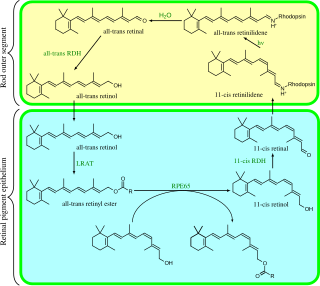
Visual phototransduction is the sensory transduction of the visual system. It is a process by which light is converted into electrical signals in the rod cells, cone cells and photosensitive ganglion cells of the retina of the eye. This cycle was elucidated by George Wald (1906–1997) for which he received the Nobel Prize in 1967. It is so called "Wald's Visual Cycle" after him.

Arrestin Family of proteins

Arrestins are a small family of proteins important for regulating signal transduction at G protein-coupled receptors. Arrestins were first discovered as a part of a conserved two-step mechanism for regulating the activity of G protein-coupled receptors (GPCRs) in the visual rhodopsin system by Hermann Kühn, Scott Hall, and Ursula Wilden and in the β-adrenergic system by Martin J. Lohse and co-workers.

G protein-coupled receptor kinase

G protein-coupled receptor kinases are a family of protein kinases within the AGC group of kinases. Like all AGC kinases, GRKs use ATP to add phosphate to Serine and Threonine residues in specific locations of target proteins. In particular, GRKs phosphorylate intracellular domains of G protein-coupled receptors (GPCRs). GRKs function in tandem with arrestin proteins to regulate the sensitivity of GPCRs for stimulating downstream heterotrimeric G protein and G protein-independent signaling pathways.

Recoverin

Recoverin is a 23 kilodalton (kDa) neuronal calcium-binding protein that is primarily detected in the photoreceptor cells of the eye. It plays a key role in the inhibition of rhodopsin kinase, a molecule which regulates the phosphorylation of rhodopsin. A reduction in this inhibition helps regulate sensory adaptation in the retina, since the light-dependent channel closure in photoreceptors causes calcium levels to decrease, which relieves the inhibition of rhodopsin kinase by calcium-bound recoverin, leading to a more rapid inactivation of metarhodopsin II.

G protein-coupled receptor kinase 2 Enzyme

G-protein-coupled receptor kinase 2 (GRK2) is an enzyme that in humans is encoded by the ADRBK1 gene. GRK2 was initially called Beta-adrenergic receptor kinase, and is a member of the G protein-coupled receptor kinase subfamily of the Ser/Thr protein kinases that is most highly similar to GRK3(βARK2).

Photoreceptor proteins are light-sensitive proteins involved in the sensing and response to light in a variety of organisms. Some examples are rhodopsin in the photoreceptor cells of the vertebrate retina, phytochrome in plants, and bacteriorhodopsin and bacteriophytochromes in some bacteria. They mediate light responses as varied as visual perception, phototropism and phototaxis, as well as responses to light-dark cycles such as circadian rhythm and other photoperiodisms including control of flowering times in plants and mating seasons in animals.

Retinylidene proteins, are proteins that use retinal as a chromophore for light reception. They are the molecular basis for a variety of light-sensing systems from phototaxis in flagellates to eyesight in animals. Retinylidene proteins include all forms of opsin and rhodopsin. While rhodopsin in the narrow sense refers to a dim-light visual pigment found in vertebrates, usually on rod cells, rhodopsin in the broad sense refers any molecule consisting of an opsin and a retinal chromophore in the ground state. When activated by light, the chromophore is isomerized, at which point the molecule as a whole is no longer rhodopsin, but a related molecule such as metarhodopsin. However, it remains a retinylidene protein. The chromophore then separates from the opsin, at which point the bare opsin is a retinylidene protein. Thus, the molecule remains a retinylidene protein throughout the phototransduction cycle.

GRK6

This gene encodes a member of the G protein-coupled receptor kinase subfamily of the Ser/Thr protein kinase family, and is most highly similar to GRK4 and GRK5. The protein phosphorylates the activated forms of G protein-coupled receptors to regulate their signaling.

GRK5

G protein-coupled receptor kinase 5 is a member of the G protein-coupled receptor kinase subfamily of the Ser/Thr protein kinases, and is most highly similar to GRK4 and GRK6. The protein phosphorylates the activated forms of G protein-coupled receptors to regulate their signaling.

SAG (gene)

S-arrestin is a protein that in humans is encoded by the SAG gene.

ARR3 Protein-coding gene in the species Homo sapiens

Arrestin-C also known as retinal cone arrestin-3 is a protein that in humans is encoded by the ARR3 gene.

Retinal degeneration (rhodopsin mutation) Retinopathy

Retinal degeneration is a retinopathy which consists in the deterioration of the retina caused by the progressive death of its cells. There are several reasons for retinal degeneration, including artery or vein occlusion, diabetic retinopathy, R.L.F./R.O.P., or disease. These may present in many different ways such as impaired vision, night blindness, retinal detachment, light sensitivity, tunnel vision, and loss of peripheral vision to total loss of vision. Of the retinal degenerative diseases retinitis pigmentosa (RP) is a very important example.

G-protein-coupled receptor kinase 7 is a serine/threonine-specific protein kinase involved in phototransduction. This enzyme catalyses the phosphorylation of cone (color) photopsins in retinal cones during high acuity color vision primarily in the fovea.

G protein-coupled receptor kinase 3

G-protein-coupled receptor kinase 3 (GRK3) is an enzyme that in humans is encoded by the ADRBK2 gene. GRK3 was initially called Beta-adrenergic receptor kinase 2 (βARK-2), and is a member of the G protein-coupled receptor kinase subfamily of the Ser/Thr protein kinases that is most highly similar to GRK2.

References

  1. Lorenz W, Inglese J, Palczewski K, Onorato JJ, Caron MG, Lefkowitz RJ (October 1991). "The receptor kinase family: primary structure of rhodopsin kinase reveals similarities to the beta-adrenergic receptor kinase". Proceedings of the National Academy of Sciences of the United States of America. 88 (19): 8715–9. Bibcode:1991PNAS...88.8715L. doi: 10.1073/pnas.88.19.8715 . PMC   52580 . PMID   1656454.
  2. Benovic JL, Mayor F, Somers RL, Caron MG, Lefkowitz RJ (1986). "Light-dependent phosphorylation of rhodopsin by beta-adrenergic receptor kinase". Nature. 321 (6073): 869–72. Bibcode:1986Natur.321..869B. doi:10.1038/321869a0. PMID   3014340. S2CID   4346322.
  3. Shichi H, Somers RL (October 1978). "Light-dependent phosphorylation of rhodopsin. Purification and properties of rhodopsin kinase". The Journal of Biological Chemistry. 253 (19): 7040–6. doi: 10.1016/S0021-9258(17)38026-2 . PMID   690139.
  4. Palczewski K, McDowell JH, Hargrave PA (October 1988). "Purification and characterization of rhodopsin kinase". The Journal of Biological Chemistry. 263 (28): 14067–73. doi: 10.1016/S0021-9258(18)68185-2 . PMID   2844754.
  5. Weller M, Virmaux N, Mandel P (January 1975). "Light-stimulated phosphorylation of rhodopsin in the retina: the presence of a protein kinase that is specific for photobleached rhodopsin". Proceedings of the National Academy of Sciences of the United States of America. 72 (1): 381–5. Bibcode:1975PNAS...72..381W. doi: 10.1073/pnas.72.1.381 . PMC   432309 . PMID   164024.
  6. Cha K, Bruel C, Inglese J, Khorana HG (September 1997). "Rhodopsin kinase: expression in baculovirus-infected insect cells, and characterization of post-translational modifications". Proceedings of the National Academy of Sciences of the United States of America. 94 (20): 10577–82. Bibcode:1997PNAS...9410577C. doi: 10.1073/pnas.94.20.10577 . PMC   23407 . PMID   9380677.
  7. Khani SC, Abitbol M, Yamamoto S, Maravic-Magovcevic I, Dryja TP (August 1996). "Characterization and chromosomal localization of the gene for human rhodopsin kinase". Genomics. 35 (3): 571–6. doi:10.1006/geno.1996.0399. PMID   8812493.
  8. Willets JM, Challiss RA, Nahorski SR (December 2003). "Non-visual GRKs: are we seeing the whole picture?". Trends in Pharmacological Sciences. 24 (12): 626–33. doi:10.1016/j.tips.2003.10.003. PMID   14654303.
  9. Yamamoto S, Sippel KC, Berson EL, Dryja TP (February 1997). "Defects in the rhodopsin kinase gene in the Oguchi form of stationary night blindness". Nature Genetics. 15 (2): 175–8. doi:10.1038/ng0297-175. PMID   9020843. S2CID   9317102.
  10. Chen CK, Zhang K, Church-Kopish J, Huang W, Zhang H, Chen YJ, Frederick JM, Baehr W (December 2001). "Characterization of human GRK7 as a potential cone opsin kinase". Molecular Vision. 7: 305–13. PMID   11754336.
  11. Inglese J, Glickman JF, Lorenz W, Caron MG, Lefkowitz RJ (January 1992). "Isoprenylation of a protein kinase. Requirement of farnesylation/alpha-carboxyl methylation for full enzymatic activity of rhodopsin kinase". The Journal of Biological Chemistry. 267 (3): 1422–5. doi: 10.1016/S0021-9258(18)45960-1 . PMID   1730692.
  12. Kutuzov MA, Andreeva AV, Bennett N (December 2012). "Regulation of the methylation status of G protein-coupled receptor kinase 1 (rhodopsin kinase)". Cellular Signalling. 24 (12): 2259–67. doi:10.1016/j.cellsig.2012.07.020. PMID   22846544.
  13. Sakurai K, Chen J, Khani SC, Kefalov VJ (April 2015). "Regulation of mammalian cone phototransduction by recoverin and rhodopsin kinase". The Journal of Biological Chemistry. 290 (14): 9239–50. doi: 10.1074/jbc.M115.639591 . PMC   4423708 . PMID   25673692.
  14. Sakurai K, Young JE, Kefalov VJ, Khani SC (August 2011). "Variation in rhodopsin kinase expression alters the dim flash response shut off and the light adaptation in rod photoreceptors". Investigative Ophthalmology & Visual Science. 52 (9): 6793–800. doi:10.1167/iovs.11-7158. PMC   3176010 . PMID   21474765.
  15. Chen CK, Inglese J, Lefkowitz RJ, Hurley JB (July 1995). "Ca(2+)-dependent interaction of recoverin with rhodopsin kinase". The Journal of Biological Chemistry. 270 (30): 18060–6. doi: 10.1074/jbc.270.30.18060 . PMID   7629115.
  16. Komolov KE, Senin II, Kovaleva NA, Christoph MP, Churumova VA, Grigoriev II, Akhtar M, Philippov PP, Koch KW (July 2009). "Mechanism of rhodopsin kinase regulation by recoverin". Journal of Neurochemistry. 110 (1): 72–9. doi: 10.1111/j.1471-4159.2009.06118.x . PMID   19457073. S2CID   205620698.
  17. Orban, Tivadar, et al. “Substrate-Induced Changes in the Dynamics of Rhodopsin Kinase (G Protein-Coupled Receptor Kinase 1).” Biochemistry, vol. 51, no. 16, 2012, pp. 3404–3411.
  18. Teke MY, Citirik M, Kabacam S, Demircan S, Alikasifoglu M (October 2016). "A novel missense mutation of the GRK1 gene in Oguchi disease". Molecular Medicine Reports. 14 (4): 3129–33. doi: 10.3892/mmr.2016.5620 . PMC   5042745 . PMID   27511724.
  19. Choi S, Hao W, Chen CK, Simon MI (November 2001). "Gene expression profiles of light-induced apoptosis in arrestin/rhodopsin kinase-deficient mouse retinas". Proceedings of the National Academy of Sciences of the United States of America. 98 (23): 13096–101. Bibcode:2001PNAS...9813096C. doi: 10.1073/pnas.201417498 . PMC   60830 . PMID   11687607.
  20. Murakami Y, Notomi S, Hisatomi T, Nakazawa T, Ishibashi T, Miller JW, Vavvas DG (November 2013). "Photoreceptor cell death and rescue in retinal detachment and degenerations". Progress in Retinal and Eye Research. 37 (2013): 114–40. doi:10.1016/j.preteyeres.2013.08.001. PMC   3871865 . PMID   23994436.