
A head injury is any injury that results in trauma to the skull or brain. The terms traumatic brain injury and head injury are often used interchangeably in the medical literature. Because head injuries cover such a broad scope of injuries, there are many causes—including accidents, falls, physical assault, or traffic accidents—that can cause head injuries.

The skull is a bone protective cavity for the brain. The skull is composed of four types of bone i.e., cranial bones, facial bones, ear ossicles and hyoid bone, however two parts are more prominent: the cranium and the mandible. In humans, these two parts are the neurocranium (braincase) and the viscerocranium that includes the mandible as its largest bone. The skull forms the anterior-most portion of the skeleton and is a product of cephalisation—housing the brain, and several sensory structures such as the eyes, ears, nose, and mouth. In humans, these sensory structures are part of the facial skeleton.

The occipital bone is a cranial dermal bone and the main bone of the occiput. It is trapezoidal in shape and curved on itself like a shallow dish. The occipital bone overlies the occipital lobes of the cerebrum. At the base of the skull in the occipital bone, there is a large oval opening called the foramen magnum, which allows the passage of the spinal cord.

The temporal bones are situated at the sides and base of the skull, and lateral to the temporal lobes of the cerebral cortex.
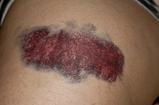
A bruise, also known as a contusion, is a type of hematoma of tissue, the most common cause being capillaries damaged by trauma, causing localized bleeding that extravasates into the surrounding interstitial tissues. Most bruises occur close enough to the epidermis such that the bleeding causes a visible discoloration. The bruise then remains visible until the blood is either absorbed by tissues or cleared by immune system action. Bruises which do not blanch under pressure can involve capillaries at the level of skin, subcutaneous tissue, muscle, or bone.
A HANS device is a type of head restraint and a safety device in motorsports. Head restraints are mandatory when competing with most major motorsports sanctioning bodies. They reduce the likelihood of head or neck injuries, including the often fatal basilar skull fracture, in the event of a crash. There are many such devices on the market today, but the HANS is the original and the most common.

A periorbital hematoma, commonly called a black eye or a shiner, is bruising around the eye commonly due to an injury to the face rather than to the eye. The name refers to the dark-colored bruising which is the result of accumulated blood and fluid in the loose areolar tissue following a blow to the head. This blood tracks freely under the scalp producing a generalised swelling over the dome of the skull but cannot pass into either occipital or the temple regions because of the bony attachments of the occipitofrontalis muscle. But this fluid can, however, track forward into the eyelid because the occipitofrontalis muscle has no bony attachment anteriorly. This leads to formation of hematoma a few hours after the head injury or cranial operation. If injury is more extensive, potentially even a skull fracture, an apparent black eye can sometimes worsen and may require professional medical treatment before it will resolve. This is more likely if the area around both eyes has been injured or if there is a history of prior head injury or fracture around the eye. Though disfiguring, the vast majority of black eyes are not serious, require little or no treatment, and will resolve spontaneously within a week or two.

Mastoiditis is the result of an infection that extends to the air cells of the skull behind the ear. Specifically, it is an inflammation of the mucosal lining of the mastoid antrum and mastoid air cell system inside the mastoid process. The mastoid process is the portion of the temporal bone of the skull that is behind the ear. The mastoid process contains open, air-containing spaces. Mastoiditis is usually caused by untreated acute otitis media and used to be a leading cause of child mortality. With the development of antibiotics, however, mastoiditis has become quite rare in developed countries where surgical treatment is now much less frequent and more conservative, unlike former times.

A skull fracture is a break in one or more of the eight bones that form the cranial portion of the skull, usually occurring as a result of blunt force trauma. If the force of the impact is excessive, the bone may fracture at or near the site of the impact and cause damage to the underlying structures within the skull such as the membranes, blood vessels, and brain.

A basilar skull fracture is a break of a bone in the base of the skull. Symptoms may include bruising behind the ears, bruising around the eyes, or blood behind the ear drum. A cerebrospinal fluid (CSF) leak occurs in about 20% of cases and may result in fluid leaking from the nose or ear. Meningitis occurs in about 14% of cases. Other complications include injuries to the cranial nerves or blood vessels.

Raccoon eyes or periorbital ecchymosis is a sign of basal skull fracture or subgaleal hematoma, a craniotomy that ruptured the meninges, or (rarely) certain cancers. Bilateral hemorrhage occurs when damage at the time of a facial fracture tears the meninges and causes the venous sinuses to bleed into the arachnoid villi and the cranial sinuses. In lay terms, blood from skull fracture seeps into the soft tissue around the eyes. Raccoon eyes may be accompanied by Battle's sign, an ecchymosis behind the ear. These signs may be the only sign of a skull fracture, as it may not show on an X-ray. They normally appear between 48 and 72 hours after the injury. It is recommended that the patient not blow their nose, cough vigorously, or strain, to prevent further tearing of the meninges.

Subgaleal hemorrhage, also known as subgaleal hematoma, is bleeding in the potential space between the skull periosteum and the scalp galea aponeurosis.

The petrous part of the temporal bone is pyramid-shaped and is wedged in at the base of the skull between the sphenoid and occipital bones. Directed medially, forward, and a little upward, it presents a base, an apex, three surfaces, and three angles, and houses in its interior, the components of the inner ear. The petrous portion is among the most basal elements of the skull and forms part of the endocranium. Petrous comes from the Latin word petrosus, meaning "stone-like, hard". It is one of the densest bones in the body. In other mammals, it is a separate bone, the petrosal bone.

The occipital condyles are undersurface protuberances of the occipital bone in vertebrates, which function in articulation with the superior facets of the atlas vertebra.
William Henry Battle was an English surgeon and teacher.
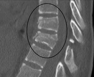
A Chance fracture is a type of vertebral fracture that results from excessive flexion of the spine. Symptoms may include abdominal bruising, or less commonly paralysis of the legs. In around half of cases there is an associated abdominal injury such as a splenic rupture, small bowel injury, pancreatic injury, or mesenteric tear. Injury to the bowel may not be apparent on the first day.
Cerebrospinal fluid rhinorrhoea refers to the drainage of cerebrospinal fluid through the nose (rhinorrhoea). It is typically caused by a basilar skull fracture, which presents complications such as infection. It may be diagnosed using brain scans, and by testing to see if discharge from the nose is cerebrospinal fluid. Treatment may be conservative, but usually involves neurosurgery.

Hemotympanum, or hematotympanum, refers to the presence of blood in the tympanic cavity of the middle ear. Hemotympanum is often the result of basilar skull fracture.

A nasal fracture, commonly referred to as a broken nose, is a fracture of one of the bones of the nose. Symptoms may include bleeding, swelling, bruising, and an inability to breathe through the nose. They may be complicated by other facial fractures or a septal hematoma.

Dislocations occur when two bones that originally met at the joint detach. Dislocations should not be confused with subluxation. Subluxation is when the joint is still partially attached to the bone.
