Related Research Articles

A blister is a small pocket of body fluid within the upper layers of the skin, usually caused by forceful rubbing (friction), burning, touching poison ivy, freezing, chemical exposure or infection. Most blisters are filled with a clear fluid, either serum or plasma. However, blisters can be filled with blood or with pus.
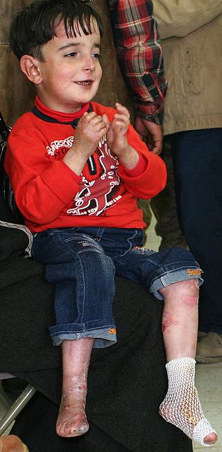
Epidermolysis bullosa (EB) is a group of rare medical conditions that result in easy blistering of the skin and mucous membranes. Blisters occur with minor trauma or friction and are painful. Its severity can range from mild to fatal. Inherited EB is a rare disease with a prevalence in the United States of 8.2 per million live births. Those with mild cases may not develop symptoms until they start to crawl or walk. Complications may include esophageal narrowing, squamous cell skin cancer, and the need for amputations.

Hemidesmosomes are very small stud-like structures found in keratinocytes of the epidermis of skin that attach to the extracellular matrix. They are similar in form to desmosomes when visualized by electron microscopy, however, desmosomes attach to adjacent cells. Hemidesmosomes are also comparable to focal adhesions, as they both attach cells to the extracellular matrix. Instead of desmogleins and desmocollins in the extracellular space, hemidesmosomes utilize integrins. Hemidesmosomes are found in epithelial cells connecting the basal epithelial cells to the lamina lucida, which is part of the basal lamina. Hemidesmosomes are also involved in signaling pathways, such as keratinocyte migration or carcinoma cell intrusion.

Pemphigus is a rare group of blistering autoimmune diseases that affect the skin and mucous membranes. The name is derived from the Greek root pemphix, meaning "blister".

Bullous pemphigoid is an autoimmune pruritic skin disease that typically occurs in people aged over 60, that may involve the formation of blisters (bullae) in the space between the epidermal and dermal skin layers. It is classified as a type II hypersensitivity reaction, which involves formation of anti-hemidesmosome antibodies, causing a loss of keratinocytes to basement membrane adhesion.

Epidermolysis bullosa simplex (EBS) is a disorder resulting from mutations in the genes encoding keratin 5 or keratin 14.
Pemphigoid is a group of rare autoimmune blistering diseases of the skin, and mucous membranes. As its name indicates, pemphigoid is similar in general appearance to pemphigus, but, unlike pemphigus, pemphigoid does not feature acantholysis, a loss of connections between skin cells.

Kindler syndrome is a rare congenital disease of the skin caused by a mutation in the KIND1 gene.

Collagen XVII, previously called BP180, is a transmembrane protein which plays a critical role in maintaining the linkage between the intracellular and the extracellular structural elements involved in epidermal adhesion, identified by Diaz and colleagues in 1990.

Genodermatosis is a hereditary skin disease with three inherited modes including single gene inheritance, multiple gene inheritance and chromosome inheritance. There are many different types of genodermatosis, the prevalence of genodermatosis ranges from 1 per 6000 people to 1 per 500,000 people. Genodermatosis has influence on the texture, color and structure of skin cuticle and connective tissue, specific lesion site and clinical manifestations on the body vary depending on the type. In the spite of the variety and complexity of genodermatosis, there are still some common methods that can help people diagnose. After diagnosis, different types of genodermatosis require different levels of therapy including interventions, nursing interventions and treatments. Among that, research of therapy for some new, complex and rare types are still in the developing stage. The impact of genodermatosis not only can be seen in body but also can be seen in all aspects of patients' life, including but not limited to psychological, family life, economic conditions and social activities. Accordingly, the patients need treatment, support and help in these areas.

Dermatitis herpetiformis (DH) is a chronic autoimmune blistering skin condition, characterised by intensely itchy blisters filled with a watery fluid. DH is a cutaneous manifestation of coeliac disease, although the exact causal mechanism is not known. DH is neither related to nor caused by herpes virus; the name means that it is a skin inflammation having an appearance similar to herpes.
Epidermolysis bullosa acquisita, also known as acquired epidermolysis bullosa, is a longterm autoimmune blistering skin disease. It generally presents with fragile skin that blisters and becomes red with or without trauma. Marked scarring is left with thin skin, milia and nail changes. It typically begins around age 50.
Paraneoplastic pemphigus (PNP) is an autoimmune disorder stemming from an underlying tumor. It is hypothesized that antigens associated with the tumor trigger an immune response resulting in blistering of the skin and mucous membranes.

Linear IgA bullous dermatosis is a rare immune-mediated blistering skin disease frequently associated with medication exposure, especially vancomycin, with men and women being equally affected. It was first described by Tadeusz Chorzelski in 1979 and may be divided into two types:
Junctional epidermolysis bullosa is a skin condition characterized by blister formation within the lamina lucida of the basement membrane zone.
Transient bullous dermolysis of the newborn (TBDN) is a skin condition that presents in newborns. It is characterized by blister formation secondary to even mild trauma.

A vesiculobullous disease is a type of mucocutaneous disease characterized by vesicles and bullae. Both vesicles and bullae are fluid-filled lesions, and they are distinguished by size. In the case of vesiculobullous diseases which are also immune disorders, the term immunobullous is sometimes used. Examples of vesiculobullous diseases include:
Mucous membrane pemphigoid is a rare chronic autoimmune subepithelial blistering disease characterized by erosive lesions of the mucous membranes and skin. It is one of the pemphigoid diseases that can result in scarring.
References
- 1 2 Agarwal A, Bansal M, Conner K (March 2012). "Coma blisters with hypoxemic respiratory failure". Dermatology Online Journal. 18 (3): 10. doi:10.5070/D35175b1pg. PMID 22483521.
- 1 2 3 4 Bosco L, Schena D, Colato C, Biban P, Girolomoni G (December 2013). "Coma blisters in children: case report and review of the literature". Journal of Child Neurology. 28 (12): 1677–1680. doi:10.1177/0883073812464684. PMID 23155203. S2CID 2299594.
- ↑ "Coma blisters. A key to neurological diagnosis". Neurología (English Edition). Elsevier. Retrieved 2022-07-26.
- ↑ Piede J, Wallace E (January 2011). "Coma bullae: associations beyond medications". Mayo Clinic Proceedings. 86 (1): e5. doi:10.4065/mcp.2010.0364. PMC 3012639 . PMID 21193646.
- 1 2 3 4 5 6 7 8 9 10 Rocha J, Pereira T, Ventura F, Pardal F, Brito C (October 2009). "Coma Blisters". Case Reports in Dermatology. 1 (1): 66–70. doi:10.1159/000249150. PMC 2895214 . PMID 20652118.
- 1 2 3 4 Vázquez-Osorio I, Gonzalvo-Rodríguez P, Rodríguez-Díaz E (2017). "Coma Blisters after an Overdose of Central Nervous System Depressants". Actas Dermo-Sifiliograficas. 108 (1): 81–83. doi: 10.1016/j.adengl.2016.11.015 . PMID 27737761.
- 1 2 Dinis-Oliveira RJ (2019). "Drug Overdose-Induced Coma Blisters: Pathophysiology and Clinical and Forensic Diagnosis". Current Drug Research Reviews. 11 (1): 21–25. doi:10.2174/1874473711666180730102343. PMID 30058500. S2CID 51863951.
- ↑ Reilly GD, Harrington CI (December 1983). "Positive immunofluorescence in bullous lesions in drug-induced coma". The British Journal of Dermatology. 109 (6): 720. doi:10.1111/j.1365-2133.1983.tb00554.x. PMID 6360197. S2CID 32953723.
- ↑ Asokan N, Binesh VG, Andrews AM, Jayalakshmi PS (November 2014). "Bullous lesions, sweat gland necrosis and rhabdomyolysis in alcoholic coma". Indian Journal of Dermatology. 59 (6): 632. doi: 10.4103/0019-5154.143576 . PMC 4248528 . PMID 25484420.
- ↑ Ferreli C, Sulica VI, Aste N, Atzori L, Pinna M, Biggio P (July 2003). "Drug-induced sweat gland necrosis in a non-comatose patient: a case presentation". Journal of the European Academy of Dermatology and Venereology. 17 (4): 443–445. doi:10.1046/j.1468-3083.2003.00695.x. PMID 12834457. S2CID 45514949.
- ↑ Arndt KA, Mihm MC, Parrish JA (May 1973). "Bullae: a cutaneous sign of a variety of neurologic diseases". The Journal of Investigative Dermatology. 60 (5): 312–320. doi: 10.1111/1523-1747.ep12723147 . PMID 4758735.
- ↑ Hollenberg SM (April 2011). "Vasoactive drugs in circulatory shock". American Journal of Respiratory and Critical Care Medicine. 183 (7): 847–855. doi:10.1164/rccm.201006-0972CI. PMID 21097695.
- ↑ Knapik JJ, Reynolds KL, Duplantis KL, Jones BH (September 1995). "Friction blisters. Pathophysiology, prevention and treatment". Sports Medicine. 20 (3): 136–147. doi:10.2165/00007256-199520030-00002. PMID 8570998. S2CID 40287087.
- ↑ Chen SX, Cohen PR (October 2017). "Edema Bullae Mimicking Disseminated Herpes Zoster". Cureus. 9 (10): e1780. doi: 10.7759/cureus.1780 . PMC 5732012 . PMID 29255659.
- ↑ Chouk C, Litaiem N (2022). "Bullosis Diabeticorum". StatPearls. Treasure Island (FL): StatPearls Publishing. PMID 30969694 . Retrieved 2022-07-28.
- ↑ Bardhan A, Bruckner-Tuderman L, Chapple IL, Fine JD, Harper N, Has C, et al. (September 2020). "Epidermolysis bullosa". Nature Reviews. Disease Primers. 6 (1): 78. doi:10.1038/s41572-020-0210-0. PMID 32973163. S2CID 221861310.
- ↑ Miyamoto D, Santi CG, Aoki V, Maruta CW (2019-05-09). "Bullous pemphigoid". Anais Brasileiros de Dermatologia. 94 (2): 133–146. doi:10.1590/abd1806-4841.20199007. PMC 6486083 . PMID 31090818.
- 1 2 Waring WS, Sandilands EA (2007). "Coma blisters". Clinical Toxicology. 45 (7): 808–809. doi:10.1080/15563650701709189. PMID 17952753. S2CID 5371293.
- ↑ Northover JM, Pickard JD, Murray-Lyon IM, Presbury DG, Haskell R, Keith DA (June 1972). "Bullous lesions of the skin and mucous membranes in primary amyloidosis". Postgraduate Medical Journal. 48 (560): 351–353. doi:10.1136/pgmj.48.560.351. PMC 2495229 . PMID 5049254.
- ↑ Torres-Navarro I, Pujol-Marco C, Roca-Ginés J, Botella-Estrada R (September 2020). "Coma blisters. A key to neurological diagnosis". Neurologia. 35 (7): 512–513. doi: 10.1016/j.nrleng.2018.11.007 . PMID 30857784. S2CID 226194253.
- ↑ Bluestein D, Javaheri A (November 2008). "Pressure ulcers: prevention, evaluation, and management". American Family Physician. 78 (10): 1186–1194. PMID 19035067.