Cyanobacteria often live in colonial aggregates that can take a multitude of forms.[3] Of particular interest among the many species of cyanobacteria are those that live colonially in elongate hair-like structures, known as trichomes. These filamentous species can contain hundreds to thousands of cells.[3] They often dominate the upper layers of microbial mats found in extreme environments such as hot springs, hypersaline water, deserts and polar regions,[4] as well as being widely distributed in more mundane environments.[3]
Many filamentous species are also motile, gliding along their long axis, and displaying photomovement by which a trichome modulates its gliding according to the incident light. The latter has been found to play an important role in guiding the trichomes to optimal lighting conditions, which can either inhibit the cells if the incident light is too weak, or damage the cells if too strong.[3]
Cellular functions require a well-organized and coordinated internal structure to operate effectively. Cells need to build, sustain, and sometimes modify their shape, which allows them to rapidly change their behaviour in response to external factors. During different life cycle stages, such as cell growth, cell division or cell differentiation, internal structures must dynamically adapt to the current requirements. In eukaryotes, these manifold tasks are fulfilled by the cytoskeleton: proteinaceous polymers that assemble into stable or dynamic filaments or tubulesin vivo and in vitro. The eukaryotic cytoskeleton is historically divided into three classes: the actin filaments (consisting of actinmonomers), the microtubules (consisting of tubulin subunits) and the intermediate filaments (IFs), although other cytoskeletal classes have been identified in recent years.[5][6] Only the collaborative work of all three cytoskeletal systems enables proper cell mechanics.[7][8]
The long-lasting dogma that prokaryotes, based on their simple cell shapes, do not require cytoskeletal elements was finally abolished by the discovery of FtsZ, a prokaryotic tubulin homolog,[9][10][11] and MreB, a bacterial actin homolog.[12][13] These discoveries started an intense search for other cytoskeletal proteins in bacteria and archaea which finally led to the identification of bacterial IF-like proteins such as Crescentin from Caulobacter crescentus[14] and even bacterial-specific cytoskeletal protein classes, including bactofilins.[15] Constant influx of new findings finally established that numerous prokaryotic cellular functions, including cell division, cell elongation or bacterial microcompartment segregation are governed by the prokaryotic cytoskeleton.[16][17][8]
Cyanobacteria are today's only known prokaryotes capable of performing oxygenic photosynthesis. Based on the presence of an outer membrane, cyanobacteria are generally considered Gram-negative bacteria. However, unlike other Gram-negative bacteria, cyanobacteria contain an unusually thick peptidoglycan (PG) layer between the inner and outer membrane, thus containing features of both Gram phenotypes.[18][19][20] Additionally, the degree of PG crosslinking is much higher in cyanobacteria than in other Gram-negative bacteria, although teichoic acids, typically present in Gram-positive bacteria, are absent.[21][8]
While Cyanobacteria are monophyletic,[22] their cellular morphologies are extremely diverse and range from unicellular species to complex cell-differentiating, multicellular species. Based on this observation, cyanobacteria have been classically divided into five subsections.[23] Subsection I cyanobacteria (Chroococcales) are unicellular and divide by binary fission or budding, whereas subsection II cyanobacteria (Pleurocapsales) are also unicellular but can undergo multiple fission events, giving rise to many small daughter cells termed baeocytes. Subsection III comprises multicellular, non-cell differentiating cyanobacteria (Oscillatoriales) and subsection IV and V cyanobacteria (Nostocales and Stigonematales) are multicellular, cell differentiating cyanobacteria that form specialized cell types in the absence of combined nitrogen (heterocysts), during unfavorable conditions (akinetes) or to spread and initiate symbiosis (hormogonia). Whereas subsections III and IV form linear cell filaments (termed trichomes) that are surrounded by a common sheath, subsection V can produce lateral branches and/or divide in multiple planes, establishing multiseriate trichomes.[23] Considering this complex morphology, it was postulated that certain subsection V-specific (cytoskeletal) proteins could be responsible for this phenotype. However, no specific gene was identified whose distribution was specifically correlated with the cell morphology among different cyanobacterial subsections.[24][25] Therefore, it seems more likely that differential expression of cell growth and division genes rather than the presence or absence of a single gene is responsible for the cyanobacterial morphological diversity.[24][26][8]
Morphogenesis is the biological process that causes an organism to develop its shape. Cyanobacteria show a high degree of morphological diversity and can undergo a variety of cellular differentiation processes in order to adapt to certain environmental conditions. This helps them thrive in almost every habitat on Earth, ranging from freshwater to marine and terrestrial habitats, including even symbiotic interactions.[27][8]
One factor which can drive morphological changes in cyanobacteria is light. As cyanobacteria are bacteria that use light to fuel their energy-producing photosynthetic machinery they depend on perceiving light in order to optimize their response and to avoid harmful light that could result in the formation of reactive oxygen species and subsequently in their death.[28] Optimal light conditions may be defined by quantity (irradiance), duration (day–night cycle) and wavelength (color of light). The photosynthetically usable light range of the solar spectrum is generally referred to as PAR (photosynthetically active radiation), but some cyanobacteria may expand on PAR by not only absorbing in the visible spectrum, but also the near-infrared light spectrum. This employs a variety of chlorophylls and allows phototrophic growth up to a wavelength of 750nm.[29] To sense the light across this range of wavelengths, cyanobacteria possess various photoreceptors of the phytochrome superfamily.[30][8]
Morphological plasticity, or the ability of one cell to alternate between different shapes, is a common strategy of many bacteria in response to environmental changes or as part of their normal life cycle.[31][32][33] Bacteria may alter their shape by simpler transitions from rod to coccoid (and vice versa) as in Escherichia coli,[34] by more complex transitions while establishing multicellularity[31] or by the development of specialized cells, structures or appendages where the population presents a pleomorphic lifestyle.[35] The precise molecular circuits that govern those morphological changes are yet to be identified, however, a so-far constant factor is that the cell shape is determined by the rigid PG sacculus which consists of glycan strands crosslinked by peptides. To grow, cells must synthesize new PG while breaking down the existent polymer to insert the newly synthesized material. How cells grow and elongate has been extensively reviewed in model organisms of both, rod-shaped[36][37] and coccoid bacteria.[38] The molecular basis for morphological plasticity and pleomorphism in more complex bacteria, however, is slowly being elucidated as well.[33][8]
Despite their morphological complexity, cyanobacteria contain all conserved and so far known bacterial morphogens.[8] Understanding cyanobacterial morphogenesis is challenging, as there are numerous morphotypes among cyanobacterial taxa, which can also vary within a given strain during its life cycle.[23] Changes in cellular or even trichome morphologies are tasks that would require active cell wall remodelling and thus far no genes attributed to the different morphotypes have been identified in cyanobacteria.[24] Therefore, the most likely scenario is that genes or their products are differentially regulated during these cell morphology transitions,[26] as it has been hypothesized for most bacteria.[33] In multicellular cyanobacteria, division of labor between cells within a trichome is achieved by different cell programing strategies. Thus, gene regulation occurs differentially in these specific cell types [30,97,98].[8]
Diversity of forms
Cyanobacteria present remarkable variability in terms of morphology: from unicellular and colonial to multicellular filamentous forms. Their cell size varies from less than 1µm in diameter (picocyanobacteria) up to 100µm (some tropical forms in the genus Oscillatoria)[39][40][41]
Filamentous forms exhibit functional cell differentiation such as heterocysts (for nitrogen fixation), akinetes (resting stage cells), and hormogonia (reproductive, motile filaments). These, together with the intercellular connections they possess, are considered the first signs of multicellularity.[31][42][43][44]
Many cyanobacteria form motile filaments of cells, called hormogonia, that travel away from the main biomass to bud and form new colonies elsewhere.[45][46] The cells in a hormogonium are often thinner than in the vegetative state, and the cells on either end of the motile chain may be tapered. To break away from the parent colony, a hormogonium often must tear apart a weaker cell in a filament, called a necridium.
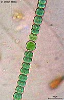
Diversity in cyanobacteria morphology
Unicellular and colonial cyanobacteria scale bars about 10 µm
In aquatic habitats, unicellular cyanobacteria are considered as an important group regarding abundance, diversity, and ecological character.[47] Unicellular cyanobacteria have spherical, ovoid, or cylindrical cells that may aggregate into irregular or regular colonies bound together by the mucous matrix (mucilage) secreted during the growth of the colony.[48] Based on the species, the number of cells in each colony may vary from two to several thousand.[47][1]
Each individual cell (each single cyanobacterium) typically has a thick, gelatinous cell wall.[49] They lack flagella, but hormogonia of some species can move about by gliding along surfaces.[50]
Merismopedia forms rectangular colonies held together by a mucilaginous matrix. Species in this genus divide in only two directions, creating a characteristic grid-like pattern arranged in rows and flats.[51]
Many of the multicellular filamentous forms of Oscillatoria are capable of a waving motion; the filament oscillates back and forth. In water columns, some cyanobacteria float by forming gas vesicles, as in archaea.[56] These vesicles are not organelles as such. They are not bounded by lipid membranes but by a protein sheath.
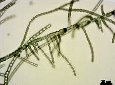
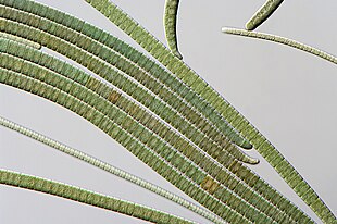
Microphotographs of bundle-forming filamentous cyanobacteria A–C: Microcoleus steenstrupii D–E: Tolypothrix desertorum F: Scytonema cf. calcicola G: S. cf. calcicola H: S. cf. c alcicola Scale bar =10 µm
Lyngbya species form long, unbranching filaments inside rigid mucilaginous sheaths which can form tangles or mats, intermixed with other phytoplankton species
Model components: (A) Trichomes are modeled as thin flexible rods that are discretized into sequences of 50 µm edges. Each edge is loaded with a linear spring. (B) The local bending moment is a function of the radius of curvature. (C) Trichomes can glide along their long axis and reverse their direction of movement photophobically. (D) Trichome collisions are defined between edge-vertex pairs. A vertex that penetrates an edge's volume is repulsed by equal and opposite forces between the pair.
Cyanobacteria are ubiquitous, finding habitats in most water bodies and in extreme environments such as the polar regions, deserts, brine lakes and hot springs.[60][61][62] They have also evolved surprisingly complex collective behaviours that lie at the boundary between single-celled and multicellular life. For example, filamentous cyanobacteria live in long chains of cells that bundle together into larger structures including biofilms, biomats and stromatolites.[63][64] These large colonies provide a rigid, stable and long-term environment for their communities of bacteria. In addition, cyanobacteria-based biofilms can be used as bioreactors to produce a wide range of chemicals, including biofuels like biodiesel and ethanol.[65] However, despite their importance to the history of life on Earth, and their commercial and environmental potentials, there remain basic questions of how filamentous cyanobacteria move, respond to their environment and self-organize into collective patterns and structures.[52]
All known cyanobacteria lack flagella;[66] however, many filamentous species move on surfaces by gliding, a form of locomotion where no physical appendages are seen to aid movement.[67] The actual mechanism behind gliding is not fully understood, although over a century has elapsed since its discovery.[68][69] One theory suggests that gliding motion in cyanobacteria is mediated by the continuous secretion of polysaccharides through pores on individual cells.[70][71][72] Another theory suggests that gliding motion involves the use of type IV pili, polymeric assemblies of the protein pilin,[73] as the driving engines of motion.[74][75][76] However, it is not clear how the action of these pili would lead to motion, with some suggesting they retract,[77] while others suggest they push,[76] to generate forces. Other scholars have suggested surface waves generated by the contraction of a fibril layer as the mechanism behind gliding motion in Oscillatoria.[78][79] Recent work also suggests that shape fluctuations and capillary forces could be involved in gliding motion.[80][52]
Through collective interaction, filamentous cyanobacteria self-organize into colonies or biofilms, symbiotic communities found in a wide variety of ecological niches. Their larger-scale collective structures are characterized by diverse shapes including bundles, vortices and reticulate patterns.[81][82] Similar patterns have been observed in fossil records.[83][82][84] For filamentous cyanobacteria, the mechanics of the filaments is known to contribute to self-organization, for example in determining how one filament will bend when in contact with other filaments or obstacles.[85] Further, biofilms and biomats show some remarkably conserved macro-mechanical properties, typically behaving as viscoelastic materials with a relaxation time of about 20 min.[86][52]
Cyanobacteria have strict light requirements. Too little light can result in insufficient energy production, and in some species may cause the cells to resort to heterotrophic respiration.[4] Too much light can inhibit the cells, decrease photosynthesis efficiency and cause damage by bleaching. UV radiation is especially deadly for cyanobacteria, with normal solar levels being significantly detrimental for these microorganisms in some cases.[87][88][3]
Filamentous cyanobacteria that live in microbial mats often migrate vertically and horizontally within the mat in order to find an optimal niche that balances their light requirements for photosynthesis against their sensitivity to photodamage. For example, the filamentous cyanobacteria Oscillatoria sp. and Spirulina subsalsa found in the hypersaline benthic mats of Guerrero Negro, Mexico migrate downwards into the lower layers during the day in order to escape the intense sunlight and then rise to the surface at dusk.[89] In contrast, the population of Microcoleus chthonoplastes found in hypersaline mats at Salin-de-Giraud, Camargue, France migrate to the upper layer of the mat during the day and are spread homogenously through the mat at night.[90] An in vitro experiment using P. uncinatum also demonstrated this species' tendency to migrate in order to avoid damaging radiation.[87][88] These migrations are usually the result of some sort of photomovement, although other forms of taxis can also play a role.[91][3]
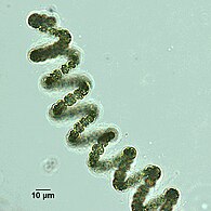
Many species of cyanobacteria are capable of gliding. Gliding is a form of cell movement that differs from crawling or swimming in that it does not rely on any obvious external organ or change in cell shape and it occurs only in the presence of a substrate.[92][93] Gliding in filamentous cyanobacteria appears to be powered by a "slime jet" mechanism, in which the cells extrude a gel that expands quickly as it hydrates providing a propulsion force,[94][95] although some unicellular cyanobacteria use type IV pili for gliding.[96] Individual cells in a trichome have two sets of pores for extruding slime. Each set is organized in a ring at the cell septae and extrudes slime at an acute angle.[97] The sets extrude slime in opposite directions and so only one set is likely to be activated during gliding. An alternative hypothesis is that the cells use contractive elements that produce undulations running over the surface inside the slime tube like an earthworm.[98] The trichomes rotate in a spiral fashion, the angle of which corresponds with the pitch angle of Castenholz's contractile trichomes.[3]
(a) Under ideal conditions active gliding specimens of Oscillatoria lutea appear as long thin curved filaments. (b) When rendered inactive, for example by being briefly cooled, the same filaments adopt a more random shape. (c) Under higher magnification O. lutea is seen to be composed of one-cell-wide strands of connected cells.
The cells appear to coordinate their gliding direction by an electrical potential that establishes polarity in the trichomes, and thus establishes a "head" and the "tail".[99] Trichomes usually reverse their polarity randomly with an average period on the order of minutes to hours.[100][101] Many species also form a semi-rigid sheath that is left behind as a hollow tube as the trichome moves forward. When the trichome reverses direction, it can move back into the sheath or break out.[102][3]
Oscillatoria is a genus of filamentous cyanobacterium named after the oscillation in its movement. Filaments in colonies slide back and forth against each other until the whole mass is reoriented to its light source. Oscillatoria is mainly blue-green or brown-green and is commonly found in watering-troughs. It reproduces by fragmentation forming long filaments of cells which can break into fragments called hormogonia. The hormogonia can then grow into new, longer filaments.
Häder's cyanograph experiment
Häder's cyanograph experiment
Photographic negative projected onto a Petri dish containing a culture of photophobic filamentous cyanobacteria (Phormidium uncinatum). The trichomes cover the lighter areas of the projection while uncovering the darker areas producing a photographic positive.
In 1987, Häder demonstrated that trichomes can position themselves quite precisely within their environment through photomovement. In Häder's cyanograph experiment a photographic negative is projected onto a Petri dish containing a culture of Phormidium uncinatum.[103][104] After a few hours, the trichomes move away from the darker areas onto the lighter areas, forming a photographic positive on the culture. The experiment demonstrates that photomovement is effective not just for discrete light traps, but for minutely patterned, continuously differentiated light fields as well.[3]
Cyanobacteria, also called Cyanobacteriota or Cyanophyta, are a phylum of autotrophic gram-negative bacteria that can obtain biological energy via photosynthesis. The name 'cyanobacteria' refers to their color, which similarly forms the basis of cyanobacteria's common name, blue-green algae, although they are not scientifically classified as algae. They appear to have originated in a freshwater or terrestrial environment.
Heterocysts or heterocytes are specialized nitrogen-fixing cells formed during nitrogen starvation by some filamentous cyanobacteria, such as Nostoc, Cylindrospermum, and Anabaena. They fix nitrogen from dinitrogen (N2) in the air using the enzyme nitrogenase, in order to provide the cells in the filament with nitrogen for biosynthesis.
Algal mats are one of many types of microbial mat that forms on the surface of water or rocks. They are typically composed of blue-green cyanobacteria and sediments. Formation occurs when alternating layers of blue-green bacteria and sediments are deposited or grow in place, creating dark-laminated layers. Stromatolites are prime examples of algal mats. Algal mats played an important role in the Great Oxidation Event on Earth some 2.3 billion years ago. Algal mats can become a significant ecological problem, if the mats grow so expansive or thick as to disrupt the other underwater marine life by blocking the sunlight or producing toxic chemicals.
Oscillatoria is a genus of sugar making microscopic creature.s
Beggiatoa is a genus of Gammaproteobacteria belonging to the order Thiotrichales, in the Pseudomonadota phylum. These bacteria form colorless filaments composed of cells that can be up to 200 µm in diameter, and are one of the largest prokaryotes on Earth. Beggiatoa are chemolithotrophic sulfur-oxidizers, using reduced sulfur species as an energy source. They live in sulfur-rich environments such as soil, both marine and freshwater, in the deep sea hydrothermal vents, and in polluted marine environments. In association with other sulfur bacteria, e.g. Thiothrix, they can form biofilms that are visible to the naked eye as mats of long white filaments; the white color is due to sulfur globules stored inside the cells.
Anabaena circinalis is a species of Gram-negative, photosynthetic cyanobacteria common to freshwater environments throughout the world. Much of the scientific interest in A. circinalis owes to its production of several potentially harmful cyanotoxins, ranging in potency from irritating to lethal. Under favorable conditions for growth, A. circinalis forms large algae-like blooms, potentially harming the flora and fauna of an area.
Bacterial motility is the ability of bacteria to move independently using metabolic energy. Most motility mechanisms that evolved among bacteria also evolved in parallel among the archaea. Most rod-shaped bacteria can move using their own power, which allows colonization of new environments and discovery of new resources for survival. Bacterial movement depends not only on the characteristics of the medium, but also on the use of different appendages to propel. Swarming and swimming movements are both powered by rotating flagella. Whereas swarming is a multicellular 2D movement over a surface and requires the presence of surfactants, swimming is movement of individual cells in liquid environments.
Cyanobionts are cyanobacteria that live in symbiosis with a wide range of organisms such as terrestrial or aquatic plants; as well as, algal and fungal species. They can reside within extracellular or intracellular structures of the host. In order for a cyanobacterium to successfully form a symbiotic relationship, it must be able to exchange signals with the host, overcome defense mounted by the host, be capable of hormogonia formation, chemotaxis, heterocyst formation, as well as possess adequate resilience to reside in host tissue which may present extreme conditions, such as low oxygen levels, and/or acidic mucilage. The most well-known plant-associated cyanobionts belong to the genus Nostoc. With the ability to differentiate into several cell types that have various functions, members of the genus Nostoc have the morphological plasticity, flexibility and adaptability to adjust to a wide range of environmental conditions, contributing to its high capacity to form symbiotic relationships with other organisms. Several cyanobionts involved with fungi and marine organisms also belong to the genera Richelia, Calothrix, Synechocystis, Aphanocapsa and Anabaena, as well as the species Oscillatoria spongeliae. Although there are many documented symbioses between cyanobacteria and marine organisms, little is known about the nature of many of these symbioses. The possibility of discovering more novel symbiotic relationships is apparent from preliminary microscopic observations.
Phototaxis is a kind of taxis, or locomotory movement, that occurs when a whole organism moves towards or away from a stimulus of light. This is advantageous for phototrophic organisms as they can orient themselves most efficiently to receive light for photosynthesis. Phototaxis is called positive if the movement is in the direction of increasing light intensity and negative if the direction is opposite.
Thioploca is a genus of filamentous sulphur-oxidizing bacteria, in the order Thiotrichales. They inhabit both marine and freshwater environments, forming vast communities off the Pacific coast of South America and in other areas with a high organic matter sedimentation and bottom waters rich in nitrate and poor in oxygen. Their cells contain large vacuoles that occupy more than 80% of the cellular volume, used to store nitrate to oxidize sulphur for anaerobic respiration in the absence of oxygen, an important characteristic of the genus. With cell diameters ranging from 15-40 µm, they are some of the largest bacteria known. They provide an important link between the nitrogen and sulphur cycles, because they use both sulfur and nitrogen compounds. They secrete a sheath of mucus which they use as a tunnel to travel between sulphide-containing sediment and nitrate-containing sea water.
Gliding motility is a type of translocation used by microorganisms that is independent of propulsive structures such as flagella, pili, and fimbriae. Gliding allows microorganisms to travel along the surface of low aqueous films. The mechanisms of this motility are only partially known.
Planktothrix is a diverse genus of filamentous cyanobacteria observed to amass in algal blooms in water ecosystems across the globe. Like all Oscillatoriales, Planktothrix species have no heterocysts and no akinetes. Planktothrix are unique because they have trichomes and contain gas vacuoles unlike typical planktonic organisms. Previously, some species of the taxon were grouped within the genus Oscillatoria, but recent work has defined Planktothrix as its own genus. A tremendous body of work on Planktothrix ecology and physiology has been done by Anthony E. Walsby, and the 55.6 kb microcystin synthetase gene which gives these organisms the ability to synthesize toxins has been sequenced. P. agardhii is an example of a type species of the genus. P. agardhii and P. rubescens are commonly observed in lakes of the Northern Hemisphere where they are known producers of potent hepatotoxins called microcystins.
Raphidiopsis raciborskii is a freshwater cyanobacterium.
Bacterial morphological plasticity refers to changes in the shape and size that bacterial cells undergo when they encounter stressful environments. Although bacteria have evolved complex molecular strategies to maintain their shape, many are able to alter their shape as a survival strategy in response to protist predators, antibiotics, the immune response, and other threats.
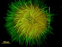
Gloeotrichia is a large (~2 mm) colonial genus of Cyanobacteria, belonging to the order Nostocales. The name Gloeotrichia is derived from its appearance of filamentous body with mucilage matrix. Found in lakes across the globe, gloeotrichia are notable for the important roles that they play in the nitrogen and phosphorus cycles. Gloeotrichia are also a species of concern for lake managers, as they have been shown to push lakes towards eutrophication and produce deadly toxins.
Trichodesmium thiebautii is a cyanobacteria that is often found in open oceans of tropical and subtropical regions and is known to be a contributor to large oceanic surface blooms. This microbial species is a diazotroph, meaning it fixes nitrogen gas (N2), but it does so without the use of heterocysts. T. thiebautii is able to simultaneously perform oxygenic photosynthesis. T. thiebautii was discovered in 1892 by M.A. Gomont. T. thiebautii are important for nutrient cycling in marine habitats because of their ability to fix N2, a limiting nutrient in ocean ecosystems.
Oscillatoria brevis is a species of the genus Oscillatoria first identified in 1892. It is a blue-green filamentous cyanobacterium, which can be found in brackish and fresh waterways. O. brevis can also be isolated from soil.
Marine prokaryotes are marine bacteria and marine archaea. They are defined by their habitat as prokaryotes that live in marine environments, that is, in the saltwater of seas or oceans or the brackish water of coastal estuaries. All cellular life forms can be divided into prokaryotes and eukaryotes. Eukaryotes are organisms whose cells have a nucleus enclosed within membranes, whereas prokaryotes are the organisms that do not have a nucleus enclosed within a membrane. The three-domain system of classifying life adds another division: the prokaryotes are divided into two domains of life, the microscopic bacteria and the microscopic archaea, while everything else, the eukaryotes, become the third domain.
Richelia is a genus of nitrogen-fixing, filamentous, heterocystous and cyanobacteria. It contains the single species Richelia intracellularis. They exist as both free-living organisms as well as symbionts within potentially up to 13 diatoms distributed throughout the global ocean. As a symbiont, Richelia can associate epiphytically and as endosymbionts within the periplasmic space between the cell membrane and cell wall of diatoms.
Aphanizomenon ovalisporum is a filamentous cyanobacteria present in many algal blooms.
↑ Alberts, Bruce; Heald, Rebecca; Johnson, Alexander; Morgan, David Owen; Raff, Martin C.; Roberts, K.; Walter, Peter (2022). Molecular biology of the cell. New York, NY. ISBN978-0-393-88482-1. OCLC1276902141.{{cite book}}: CS1 maint: location missing publisher (link)
↑ Wagstaff, James; Löwe, Jan (2018-01-22). "Prokaryotic cytoskeletons: protein filaments organizing small cells". Nature Reviews Microbiology. Springer Science and Business Media LLC. 16 (4): 187–201. doi:10.1038/nrmicro.2017.153. ISSN1740-1526. PMID29355854. S2CID3537215.
↑ Shih, Patrick M.; Wu, Dongying; Latifi, Amel; Axen, Seth D.; Fewer, David P.; Talla, Emmanuel; Calteau, Alexandra; Cai, Fei; Tandeau de Marsac, Nicole; Rippka, Rosmarie; Herdman, Michael; Sivonen, Kaarina; Coursin, Therese; Laurent, Thierry; Goodwin, Lynne; Nolan, Matt; Davenport, Karen W.; Han, Cliff S.; Rubin, Edward M.; Eisen, Jonathan A.; Woyke, Tanja; Gugger, Muriel; Kerfeld, Cheryl A. (2012-12-31). "Improving the coverage of the cyanobacterial phylum using diversity-driven genome sequencing". Proceedings of the National Academy of Sciences. 110 (3): 1053–1058. doi:10.1073/pnas.1217107110. ISSN0027-8424. PMC3549136. PMID23277585.
↑ Larkum, A. W. D.; Ritchie, R. J.; Raven, J. A. (2018). "Living off the Sun: Chlorophylls, bacteriochlorophylls and rhodopsins". Photosynthetica. 56: 11–43. doi:10.1007/s11099-018-0792-x. S2CID4907693.
↑ Wiltbank, Lisa B.; Kehoe, David M. (2018-11-08). "Diverse light responses of cyanobacteria mediated by phytochrome superfamily photoreceptors". Nature Reviews Microbiology. Springer Science and Business Media LLC. 17 (1): 37–50. doi:10.1038/s41579-018-0110-4. ISSN1740-1526. PMID30410070. S2CID256744429.
↑ Egan, Alexander J. F.; Errington, Jeff; Vollmer, Waldemar (2020-05-18). "Regulation of peptidoglycan synthesis and remodelling". Nature Reviews Microbiology. Springer Science and Business Media LLC. 18 (8): 446–460. doi:10.1038/s41579-020-0366-3. ISSN1740-1526. PMID32424210. S2CID256745837.
↑ Jasser, Iwona; Callieri, Cristiana (2017-02-11). "Picocyanobacteria". Handbook of Cyanobacterial Monitoring and Cyanotoxin Analysis. Chichester, UK: John Wiley & Sons, Ltd. pp.19–27. doi:10.1002/9781119068761.ch3. ISBN9781119068761.
↑ Nürnberg, Dennis J.; Mariscal, Vicente; Parker, Jamie; Mastroianni, Giulia; Flores, Enrique; Mullineaux, Conrad W. (2014). "Branching and intercellular communication in the Section V cyanobacterium Mastigocladus laminosus, a complex multicellular prokaryote". Molecular Microbiology. 91 (5): 935–949. doi:10.1111/mmi.12506. hdl:10261/99110. PMID24383541. S2CID25479970.
1 2 Dvořák, Petr; Casamatta, Dale A.; Hašler, Petr; Jahodářová, Eva; Norwich, Alyson R.; Poulíčková, Aloisie (2017). "Diversity of the Cyanobacteria". Modern Topics in the Phototrophic Prokaryotes. Cham: Springer International Publishing. pp.3–46. doi:10.1007/978-3-319-46261-5_1. ISBN978-3-319-46259-2.
↑ Chorus, Ingrid; Bartram, Jamie (1999). Toxic cyanobacteria in water: a guide to their public health consequences, monitoring, and management. London: E & FN Spon. ISBN0-419-23930-8. OCLC40395794.
↑ Palinska, Katarzyna A.; Liesack, Werner; Rhiel, Erhard; Krumbein, W. E. (17 October 1996). "Phenotype variability of identical genotypes: the need for a combined approach in cyanobacterial taxonomy demonstrated on Merismopedia-like isolates". Archives of Microbiology. 166 (4): 224–233. doi:10.1007/s002030050378. PMID8824145. S2CID3022844.
↑ Wolk, C. Peter; Ernst, Annaliese; Elhai, Jeff (1994). "Heterocyst Metabolism and Development". In Donald A. Bryant (ed.). The Molecular Biology of Cyanobacteria. Advances in Photosynthesis and Respiration. pp.769–823. doi:10.1007/978-94-011-0227-8_27. ISBN978-0-7923-3273-2.
↑ Walter, M.R.; Bauld, J.; Brock, T.D. (1976). "Chapter 6.2 Microbiology and Morphogenesis of Columnar Stromatolites (Conophyton, Vacerrilla) from Hot Springs in Yellowstone National Park". Stromatolites. Developments in Sedimentology. Vol.20. pp.273–310. doi:10.1016/S0070-4571(08)71140-3. ISBN9780444413765.
↑ Wharton, Robert A.; Parker, Bruce C.; Simmons, George M. (1983). "Distribution, species composition and morphology of algal mats in Antarctic dry valley lakes". Phycologia. 22 (4): 355–365. doi:10.2216/i0031-8884-22-4-355.1.
↑ Walsby, A. E. (1968). "Mucilage secretion and the movements of blue-green algae". Protoplasma. 65 (1–2): 223–238. doi:10.1007/BF01666380. S2CID20310025.
↑ Halfen, Lawrence N.; Castenholz, Richard W. (1971). "Gliding Motility in the Blue-Green Alga Oscillatoria Princeps 1". Journal of Phycology. 7 (2): 133–145. doi:10.1111/j.1529-8817.1971.tb01492.x. S2CID86115246.
↑ McBride, Mark J. (2001). "Bacterial Gliding Motility: Multiple Mechanisms for Cell Movement over Surfaces". Annual Review of Microbiology. 55: 49–75. doi:10.1146/annurev.micro.55.1.49. PMID11544349.
↑ Halfen, Lawrence N.; Castenholz, Richard W. (1971). "Gliding Motility in the Blue-Green Alga Oscillatoria Princeps 1". Journal of Phycology. 7 (2): 133–145. doi:10.1111/j.1529-8817.1971.tb01492.x. S2CID86115246.
↑ "Enhanced Model for Photophobic Responses of the Blue-Green Alga, <italic>Phormidium uncinatum</italic>". Plant and Cell Physiology. 1982. doi:10.1093/oxfordjournals.pcp.a076487.
This page is based on this Wikipedia article Text is available under the CC BY-SA 4.0 license; additional terms may apply. Images, videos and audio are available under their respective licenses.