The history of molecular biology begins in the 1930s with the convergence of various, previously distinct biological and physical disciplines: biochemistry, genetics, microbiology, virology and physics. With the hope of understanding life at its most fundamental level, numerous physicists and chemists also took an interest in what would become molecular biology.
In its modern sense, molecular biology attempts to explain the phenomena of life starting from the macromolecular properties that generate them. Two categories of macromolecules in particular are the focus of the molecular biologist: 1) nucleic acids, among which the most famous is deoxyribonucleic acid (or DNA), the constituent of genes, and 2) proteins, which are the active agents of living organisms. One definition of the scope of molecular biology therefore is to characterize the structure, function and relationships between these two types of macromolecules. This relatively limited definition will suffice to allow us to establish a date for the so-called "molecular revolution", or at least to establish a chronology of its most fundamental developments.
General overview
In its earliest manifestations, molecular biology—the name was coined by Warren Weaver of the Rockefeller Foundation in 1938[1]—was an idea of physical and chemical explanations of life, rather than a coherent discipline. Following the advent of the Mendelian-chromosome theory of heredity in the 1910s and the maturation of atomic theory and quantum mechanics in the 1920s, such explanations seemed within reach. Weaver and others encouraged (and funded) research at the intersection of biology, chemistry and physics, while prominent physicists such as Niels Bohr and Erwin Schrödinger turned their attention to biological speculation. However, in the 1930s and 1940s it was by no means clear which—if any—cross-disciplinary research would bear fruit; work in colloid chemistry, biophysics and radiation biology, crystallography, and other emerging fields all seemed promising.
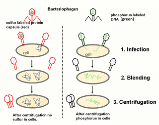
In 1940, George Beadle and Edward Tatum demonstrated the existence of a precise relationship between genes and proteins.[2] In the course of their experiments connecting genetics with biochemistry, they switched from the genetics mainstay Drosophila to a more appropriate model organism, the fungus Neurospora; the construction and exploitation of new model organisms would become a recurring theme in the development of molecular biology. In 1944, Oswald Avery, working at the Rockefeller Institute of New York, demonstrated that genes are made up of DNA[3] (see Avery–MacLeod–McCarty experiment). In 1952, Alfred Hershey and Martha Chase confirmed that the genetic material of the bacteriophage, the virus which infects bacteria, is made up of DNA[4] (see Hershey–Chase experiment). In 1953, James Watson and Francis Crick discovered the double helical structure of the DNA molecule based on the discoveries made by Rosalind Franklin.[5] In 1961, François Jacob and Jacques Monod demonstrated that the products of certain genes regulated the expression of other genes by acting upon specific sites at the edge of those genes. They also hypothesized the existence of an intermediary between DNA and its protein products, which they called messenger RNA.[6] Between 1961 and 1965, the relationship between the information contained in DNA and the structure of proteins was determined: there is a code, the genetic code, which creates a correspondence between the succession of nucleotides in the DNA sequence and a series of amino acids in proteins.
In April 2023, scientists, based on new evidence, concluded that Rosalind Franklin was a contributor and "equal player" in the discovery process of DNA, rather than otherwise, as may have been presented subsequently after the time of the discovery.[7][8][9]
The chief discoveries of molecular biology took place in a period of only about twenty-five years. Another fifteen years were required before new and more sophisticated technologies, united today under the name of genetic engineering, would permit the isolation and characterization of genes, in particular those of highly complex organisms.
The exploration of the molecular dominion
If we evaluate the molecular revolution within the context of biological history, it is easy to note that it is the culmination of a long process which began with the first observations through a microscope. The aim of these early researchers was to understand the functioning of living organisms by describing their organization at the microscopic level. From the end of the 18th century, the characterization of the chemical molecules which make up living beings gained increasingly greater attention, along with the birth of physiological chemistry in the 19th century, developed by the German chemist Justus von Liebig and following the birth of biochemistry at the beginning of the 20th, thanks to another German chemist Eduard Buchner. Between the molecules studied by chemists and the tiny structures visible under the optical microscope, such as the cellular nucleus or the chromosomes, there was an obscure zone, "the world of the ignored dimensions," as it was called by the chemical-physicist Wolfgang Ostwald. This world is populated by colloids, chemical compounds whose structure and properties were not well defined.
The successes of molecular biology derived from the exploration of that unknown world by means of the new technologies developed by chemists and physicists: X-ray diffraction, electron microscopy, ultracentrifugation, and electrophoresis. These studies revealed the structure and function of the macromolecules.
The development of molecular biology is also the encounter of two disciplines which made considerable progress in the course of the first thirty years of the twentieth century: biochemistry and genetics. The first studies the structure and function of the molecules which make up living things. Between 1900 and 1940, the central processes of metabolism were described: the process of digestion and the absorption of the nutritive elements derived from alimentation, such as the sugars. Every one of these processes is catalyzed by a particular enzyme. Enzymes are proteins, like the antibodies present in blood or the proteins responsible for muscular contraction. As a consequence, the study of proteins, of their structure and synthesis, became one of the principal objectives of biochemists.
The second discipline of biology which developed at the beginning of the 20th century is genetics. After the rediscovery of the laws of Mendel through the studies of Hugo de Vries, Carl Correns and Erich von Tschermak in 1900, this science began to take shape thanks to the adoption by Thomas Hunt Morgan, in 1910, of a model organism for genetic studies, the famous fruit fly (Drosophila melanogaster). Shortly after, Morgan showed that the genes are localized on chromosomes. Following this discovery, he continued working with Drosophila and, along with numerous other research groups, confirmed the importance of the gene in the life and development of organisms. Nevertheless, the chemical nature of genes and their mechanisms of action remained a mystery. Molecular biologists committed themselves to the determination of the structure, and the description of the complex relations between, genes and proteins.
The development of molecular biology was not just the fruit of some sort of intrinsic "necessity" in the history of ideas, but was a characteristically historical phenomenon, with all of its unknowns, imponderables and contingencies: the remarkable developments in physics at the beginning of the 20th century highlighted the relative lateness in development in biology, which became the "new frontier" in the search for knowledge about the empirical world. Moreover, the developments of the theory of information and cybernetics in the 1940s, in response to military exigencies, brought to the new biology a significant number of fertile ideas and, especially, metaphors.
The choice of bacteria and of its virus, the bacteriophage, as models for the study of the fundamental mechanisms of life was almost natural—they are the smallest living organisms known to exist—and at the same time the fruit of individual choices. This model owes its success, above all, to the fame and the sense of organization of Max Delbrück, a German physicist, who was able to create a dynamic research group, based in the United States, whose exclusive scope was the study of the bacteriophage: the phage group.[10]
The phage group was an informal network of biologists that carried out basic research mainly on bacteriophage T4 and made numerous seminal contributions to microbial genetics and the origins of molecular biology in the mid-20th century. In 1961, Sydney Brenner, an early member of the phage group, collaborated with Francis Crick, Leslie Barnett and Richard Watts-Tobin at the Cavendish Laboratory in Cambridge to perform genetic experiments that demonstrated the basic nature of the genetic code for proteins.[11] These experiments, carried out with mutants of the rIIB gene of bacteriophage T4, showed, that for a gene that encodes a protein, three sequential bases of the gene's DNA specify each successive amino acid of the protein. Thus the genetic code is a triplet code, where each triplet (called a codon) specifies a particular amino acid. They also found that the codons do not overlap with each other in the DNA sequence encoding a protein, and that such a sequence is read from a fixed starting point. During 1962–1964 phage T4 researchers provided an opportunity to study the function of virtually all of the genes that are essential for growth of the bacteriophage under laboratory conditions.[12][13] These studies were facilitated by the discovery of two classes of conditional lethal mutants. One class of such mutants is known as amber mutants.[14] Another class of conditional lethal mutants is referred to as temperature-sensitive mutants.[15] Studies of these two classes of mutants led to considerable insight into numerous fundamental biologic problems. Thus understanding was gained on the functions and interactions of the proteins employed in the machinery of DNA replication, DNA repair and DNA recombination. Furthermore, understanding was gained on the processes by which viruses are assembled from protein and nucleic acid components (molecular morphogenesis). Also, the role of chain terminating codons was elucidated. One noteworthy study used amber mutants defective in the gene encoding the major head protein of bacteriophage T4.[16] This experiment provided strong evidence for the widely held, but prior to 1964 still unproven, "sequence hypothesis" that the amino acid sequence of a protein is specified by the nucleotide sequence of the gene determining the protein. Thus, this study demonstrated the co-linearity of the gene with its encoded protein.
The geographic panorama of the developments of the new biology was conditioned above all by preceding work. The US, where genetics had developed the most rapidly, and the UK, where there was a coexistence of both genetics and biochemical research of highly advanced levels, were in the avant-garde. Germany, the cradle of the revolutions in physics, with the best minds and the most advanced laboratories of genetics in the world, should have had a primary role in the development of molecular biology. But history decided differently: the arrival of the Nazis in 1933—and, to a less extreme degree, the rigidification of totalitarian measures in fascist Italy—caused the emigration of a large number of Jewish and non-Jewish scientists. The majority of them fled to the US or the UK, providing an extra impulse to the scientific dynamism of those nations. These movements ultimately made molecular biology a truly international science from the very beginnings.
The study of DNA is a central part of molecular biology.
First isolation of DNA
Working in the 19th century, biochemists initially isolated DNA and RNA (mixed together) from cell nuclei. They were relatively quick to appreciate the polymeric nature of their "nucleic acid" isolates, but realized only later that nucleotides were of two types—one containing ribose and the other deoxyribose. It was this subsequent discovery that led to the identification and naming of DNA as a substance distinct from RNA.
Friedrich Miescher (1844–1895) discovered a substance he called "nuclein" in 1869. Somewhat later, he isolated a pure sample of the material now known as DNA from the sperm of salmon, and in 1889 his pupil, Richard Altmann, named it "nucleic acid". This substance was found to exist only in the chromosomes.
In 1919 Phoebus Levene at the Rockefeller Institute identified the components (the four bases, the sugar and the phosphate chain) and he showed that the components of DNA were linked in the order phosphate-sugar-base. He called each of these units a nucleotide and suggested the DNA molecule consisted of a string of nucleotide units linked together through the phosphate groups, which are the 'backbone' of the molecule. However Levene thought the chain was short and that the bases repeated in the same fixed order. Torbjörn Caspersson and Einar Hammersten showed that DNA was a polymer.
Chromosomes and inherited traits
In 1927, Nikolai Koltsov proposed that inherited traits would be inherited via a "giant hereditary molecule" which would be made up of "two mirror strands that would replicate in a semi-conservative fashion using each strand as a template".[17]Max Delbrück, Nikolay Timofeev-Ressovsky, and Karl G. Zimmer published results in 1935 suggesting that chromosomes are very large molecules the structure of which can be changed by treatment with X-rays, and that by so changing their structure it was possible to change the heritable characteristics governed by those chromosomes. In 1937 William Astbury produced the first X-ray diffraction patterns from DNA. He was not able to propose the correct structure but the patterns showed that DNA had a regular structure and therefore it might be possible to deduce what this structure was.
In 1943, Oswald Theodore Avery and a team of scientists discovered that traits proper to the "smooth" form of the Pneumococcus could be transferred to the "rough" form of the same bacteria merely by making the killed "smooth" (S) form available to the live "rough" (R) form. Quite unexpectedly, the living R Pneumococcus bacteria were transformed into a new strain of the S form, and the transferred S characteristics turned out to be heritable. Avery called the medium of transfer of traits the transforming principle; he identified DNA as the transforming principle, and not protein as previously thought. He essentially redid Frederick Griffith's experiment. In 1953, Alfred Hershey and Martha Chase did an experiment (Hershey–Chase experiment) that showed, in T2 phage, that DNA is the genetic material (Hershey shared the Nobel prize with Luria).
Discovery of the structure of DNA
In the 1950s, three groups made it their goal to determine the structure of DNA. The first group to start was at King's College London and was led by Maurice Wilkins and was later joined by Rosalind Franklin. Another group consisting of Francis Crick and James Watson was at Cambridge. A third group was at Caltech and was led by Linus Pauling. Crick and Watson built physical models using metal rods and balls, in which they incorporated the known chemical structures of the nucleotides, as well as the known position of the linkages joining one nucleotide to the next along the polymer. At King's College Maurice Wilkins and Rosalind Franklin examined X-ray diffraction patterns of DNA fibers. Of the three groups, only the London group was able to produce good quality diffraction patterns and thus produce sufficient quantitative data about the structure.
Helix structure
In 1948, Pauling discovered that many proteins included helical (see alpha helix) shapes. Pauling had deduced this structure from X-ray patterns and from attempts to physically model the structures. (Pauling was also later to suggest an incorrect three chain helical DNA structure based on Astbury's data.) Even in the initial diffraction data from DNA by Maurice Wilkins, it was evident that the structure involved helices. But this insight was only a beginning. There remained the questions of how many strands came together, whether this number was the same for every helix, whether the bases pointed toward the helical axis or away, and ultimately what were the explicit angles and coordinates of all the bonds and atoms. Such questions motivated the modeling efforts of Watson and Crick.
Complementary nucleotides
In their modeling, Watson and Crick restricted themselves to what they saw as chemically and biologically reasonable. Still, the breadth of possibilities was very wide. A breakthrough occurred in 1952, when Erwin Chargaff visited Cambridge and inspired Crick with a description of experiments Chargaff had published in 1947. Chargaff had observed that the proportions of the four nucleotides vary between one DNA sample and the next, but that for particular pairs of nucleotides—adenine and thymine, guanine and cytosine—the two nucleotides are always present in equal proportions.
Crick and Watson DNA model built in 1953, was reconstructed largely from its original pieces in 1973 and donated to the National Science Museum in London.
Using X-ray diffraction, as well as other data from Rosalind Franklin and her information that the bases were paired, James Watson and Francis Crick arrived at the first accurate model of DNA's molecular structure in 1953, which was accepted through inspection by Rosalind Franklin.[18] The discovery was announced on February 28, 1953; the first Watson/Crick paper appeared in Nature on April 25, 1953. Sir Lawrence Bragg, the director of the Cavendish Laboratory, where Watson and Crick worked, gave a talk at Guy's Hospital Medical School in London on Thursday, May 14, 1953, which resulted in an article by Ritchie Calder in the News Chronicle of London, on Friday, May 15, 1953, entitled "Why You Are You. Nearer Secret of Life." The news reached readers of The New York Times the next day; Victor K. McElheny, in researching his biography, "Watson and DNA: Making a Scientific Revolution", found a clipping of a six-paragraph New York Times article written from London and dated May 16, 1953, with the headline "Form of 'Life Unit' in Cell Is Scanned." The article ran in an early edition and was then pulled to make space for news deemed more important. (The New York Times subsequently ran a longer article on June 12, 1953). The Cambridge University undergraduate newspaper also ran its own short article on the discovery on Saturday, May 30, 1953. Bragg's original announcement at a Solvay Conference on proteins in Belgium on April 8, 1953, went unreported by the press. In 1962 Watson, Crick, and Maurice Wilkins jointly received the Nobel Prize in Physiology or Medicine for their determination of the structure of DNA.
"Central Dogma"
Watson and Crick's model attracted great interest immediately upon its presentation. Arriving at their conclusion on February 21, 1953, Watson and Crick made their first announcement on February 28. In an influential presentation in 1957, Crick laid out the "central dogma of molecular biology", which foretold the relationship between DNA, RNA, and proteins, and articulated the "sequence hypothesis." A critical confirmation of the replication mechanism that was implied by the double-helical structure followed in 1958 in the form of the Meselson–Stahl experiment. Work by Crick and coworkers showed that the genetic code was based on non-overlapping triplets of bases, called codons, and Har Gobind Khorana and others deciphered the genetic code not long afterward (1966). These findings represent the birth of molecular biology.
The earliest work in RNA structural biology coincided, more or less, with the work being done on DNA in the early 1950s. In their seminal 1953 paper, Watson and Crick suggested that van der Waals crowding by the 2`OH group of ribose would preclude RNA from adopting a double helical structure identical to the model they proposed—what we now know as B-form DNA.[19] This provoked questions about the three-dimensional structure of RNA: could this molecule form some type of helical structure, and if so, how? As with DNA, early structural work on RNA centered around isolation of native RNA polymers for fiber diffraction analysis. In part because of heterogeneity of the samples tested, early fiber diffraction patterns were usually ambiguous and not readily interpretable. In 1955, Marianne Grunberg-Manago and colleagues published a paper describing the enzyme polynucleotide phosphorylase, which cleaved a phosphate group from nucleotide diphosphates to catalyze their polymerization.[20] This discovery allowed researchers to synthesize homogenous nucleotide polymers, which they then combined to produce double stranded molecules. These samples yielded the most readily interpretable fiber diffraction patterns yet obtained, suggesting an ordered, helical structure for cognate, double stranded RNA that differed from that observed in DNA. These results paved the way for a series of investigations into the various properties and propensities of RNA. Through the late 1950s and early 1960s, numerous papers were published on various topics in RNA structure, including RNA-DNA hybridization,[21] triple stranded RNA,[22] and even small-scale crystallography of RNA di-nucleotides—G-C, and A-U—in primitive helix-like arrangements.[23] For a more in-depth review of the early work in RNA structural biology, see the article The Era of RNA Awakening: Structural biology of RNA in the early years by Alexander Rich.[24]
The beginning: crystal structure of tRNAPHE
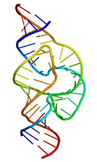
In the mid-1960s, the role of tRNA in protein synthesis was being intensively studied. At this point, ribosomes had been implicated in protein synthesis, and it had been shown that an mRNA strand was necessary for the formation of these structures. In a 1964 publication, Warner and Rich showed that ribosomes active in protein synthesis contained tRNA molecules bound at the A and P sites, and discussed the notion that these molecules aided in the peptidyl transferase reaction.[25] However, despite considerable biochemical characterization, the structural basis of tRNA function remained a mystery. In 1965, Holley et al. purified and sequenced the first tRNA molecule, initially proposing that it adopted a cloverleaf structure, based largely on the ability of certain regions of the molecule to form stem loop structures.[26] The isolation of tRNA proved to be the first major windfall in RNA structural biology. Following Robert W. Holley's publication, numerous investigators began work on isolation tRNA for crystallographic study, developing improved methods for isolating the molecule as they worked. By 1968 several groups had produced tRNA crystals, but these proved to be of limited quality and did not yield data at the resolutions necessary to determine structure.[27] In 1971, Kim et al. achieved another breakthrough, producing crystals of yeast tRNAPHE that diffracted to 2–3 Ångström resolutions by using spermine, a naturally occurring polyamine, which bound to and stabilized the tRNA.[28] Despite having suitable crystals, however, the structure of tRNAPHE was not immediately solved at high resolution; rather it took pioneering work in the use of heavy metal derivatives and a good deal more time to produce a high-quality density map of the entire molecule. In 1973, Kim et al. produced a 4 Ångström map of the tRNA molecule in which they could unambiguously trace the entire backbone.[29] This solution would be followed by many more, as various investigators worked to refine the structure and thereby more thoroughly elucidate the details of base pairing and stacking interactions, and validate the published architecture of the molecule.
The tRNAPHE structure is notable in the field of nucleic acid structure in general, as it represented the first solution of a long-chain nucleic acid structure of any kind—RNA or DNA—preceding Richard E. Dickerson's solution of a B-form dodecamer by nearly a decade.[30] Also, tRNAPHE demonstrated many of the tertiary interactions observed in RNA architecture which would not be categorized and more thoroughly understood for years to come, providing a foundation for all future RNA structural research.
The renaissance: the hammerhead ribozyme and the group I intron: P4-6
For a considerable time following the first tRNA structures, the field of RNA structure did not dramatically advance. The ability to study an RNA structure depended upon the potential to isolate the RNA target. This proved limiting to the field for many years, in part because other known targets—i.e., the ribosome—were significantly more difficult to isolate and crystallize. Further, because other interesting RNA targets had simply not been identified, or were not sufficiently understood to be deemed interesting, there was simply a lack of things to study structurally. As such, for some twenty years following the original publication of the tRNAPHE structure, the structures of only a handful of other RNA targets were solved, with almost all of these belonging to the transfer RNA family.[31] This unfortunate lack of scope would eventually be overcome largely because of two major advancements in nucleic acid research: the identification of ribozymes, and the ability to produce them via in vitro transcription.
Subsequent to Tom Cech's publication implicating the Tetrahymenagroup I intron as an autocatalytic ribozyme,[32] and Sidney Altman's report of catalysis by ribonuclease P RNA,[33] several other catalytic RNAs were identified in the late 1980s,[34] including the hammerhead ribozyme. In 1994, McKay et al. published the structure of a 'hammerhead RNA-DNA ribozyme-inhibitor complex' at 2.6 Ångström resolution, in which the autocatalytic activity of the ribozyme was disrupted via binding to a DNA substrate.[35] The conformation of the ribozyme published in this paper was eventually shown to be one of several possible states, and although this particular sample was catalytically inactive, subsequent structures have revealed its active-state architecture. This structure was followed by Jennifer Doudna's publication of the structure of the P4-P6 domains of the Tetrahymena group I intron, a fragment of the ribozyme originally made famous by Cech.[36] The second clause in the title of this publication—Principles of RNA Packing—concisely evinces the value of these two structures: for the first time, comparisons could be made between well described tRNA structures and those of globular RNAs outside the transfer family. This allowed the framework of categorization to be built for RNA tertiary structure. It was now possible to propose the conservation of motifs, folds, and various local stabilizing interactions. For an early review of these structures and their implications, see RNA FOLDS: Insights from recent crystal structures, by Doudna and Ferre-D'Amare.[37]
In addition to the advances being made in global structure determination via crystallography, the early 1990s also saw the implementation of NMR as a powerful technique in RNA structural biology. Coincident with the large-scale ribozyme structures being solved crystallographically, a number of structures of small RNAs and RNAs complexed with drugs and peptides were solved using NMR.[38] In addition, NMR was now being used to investigate and supplement crystal structures, as exemplified by the determination of an isolated tetraloop-receptor motif structure published in 1997.[39] Investigations such as this enabled a more precise characterization of the base pairing and base stacking interactions which stabilized the global folds of large RNA molecules. The importance of understanding RNA tertiary structural motifs was prophetically well described by Michel and Costa in their publication identifying the tetraloop motif: "...it should not come as a surprise if self-folding RNA molecules were to make intensive use of only a relatively small set of tertiary motifs. Identifying these motifs would greatly aid modeling enterprises, which will remain essential as long as the crystallization of large RNAs remains a difficult task".[40]
The modern era: the age of RNA structural biology
The resurgence of RNA structural biology in the mid-1990s has caused a veritable explosion in the field of nucleic acid structural research. Since the publication of the hammerhead and P4-6 structures, numerous major contributions to the field have been made. Some of the most noteworthy examples include the structures of the Group I and Group II introns,[41] and the Ribosome solved by Nenad Ban and colleagues in the laboratory of Thomas Steitz.[42] The first three structures were produced using in vitro transcription, and that NMR has played a role in investigating partial components of all four structures—testaments to the indispensability of both techniques for RNA research. Most recently, the 2009 Nobel Prize in Chemistry was awarded to Ada Yonath, Venkatraman Ramakrishnan and Thomas Steitz for their structural work on the ribosome, demonstrating the prominent role RNA structural biology has taken in modern molecular biology.
History of protein biochemistry
First isolation and classification
Proteins were recognized as a distinct class of biological molecules in the eighteenth century by Antoine Fourcroy and others. Members of this class (called the "albuminoids", Eiweisskörper, or matières albuminoides) were recognized by their ability to coagulate or flocculate under various treatments such as heat or acid; well-known examples at the start of the nineteenth century included albumen from egg whites, bloodserum albumin, fibrin, and wheatgluten. The similarity between the cooking of egg whites and the curdling of milk was recognized even in ancient times; for example, the name albumen for the egg-white protein was coined by Pliny the Elder from the Latinalbus ovi (egg white).
With the advice of Jöns Jakob Berzelius, the Dutch chemist Gerhardus Johannes Mulder carried out elemental analyses of common animal and plant proteins. To everyone's surprise, all proteins had nearly the same empirical formula, roughly C400H620N100O120 with individual sulfur and phosphorus atoms. Mulder published his findings in two papers (1837,1838) and hypothesized that there was one basic substance (Grundstoff) of proteins, and that it was synthesized by plants and absorbed from them by animals in digestion. Berzelius was an early proponent of this theory and proposed the name "protein" for this substance in a letter dated 10 July 1838
The name protein that he propose for the organic oxide of fibrin and albumin, I wanted to derive from [the Greek word] πρωτειος, because it appears to be the primitive or principal substance of animal nutrition.
Mulder went on to identify the products of protein degradation such as the amino acid, leucine, for which he found a (nearly correct) molecular weight of 131 Da.
Purifications and measurements of mass
The minimum molecular weight suggested by Mulder's analyses was roughly 9kDa, hundreds of times larger than other molecules being studied. Hence, the chemical structure of proteins (their primary structure) was an active area of research until 1949, when Fred Sanger sequenced insulin. The (correct) theory that proteins were linear polymers of amino acids linked by peptide bonds was proposed independently and simultaneously by Franz Hofmeister and Emil Fischer at the same conference in 1902. However, some scientists were sceptical that such long macromolecules could be stable in solution. Consequently, numerous alternative theories of the protein primary structure were proposed, e.g., the colloidal hypothesis that proteins were assemblies of small molecules, the cyclol hypothesis of Dorothy Wrinch, the diketopiperazine hypothesis of Emil Abderhalden and the pyrrol/piperidine hypothesis of Troensgard (1942). Most of these theories had difficulties in accounting for the fact that the digestion of proteins yielded peptides and amino acids. Proteins were finally shown to be macromolecules of well-defined composition (and not colloidal mixtures) by Theodor Svedberg using analytical ultracentrifugation. The possibility that some proteins are non-covalent associations of such macromolecules was shown by Gilbert Smithson Adair (by measuring the osmotic pressure of hemoglobin) and, later, by Frederic M. Richards in his studies of ribonuclease S. The mass spectrometry of proteins has long been a useful technique for identifying posttranslational modifications and, more recently, for probing protein structure.
Most proteins are difficult to purify in more than milligram quantities, even using the most modern methods. Hence, early studies focused on proteins that could be purified in large quantities, e.g., those of blood, egg white, various toxins, and digestive/metabolic enzymes obtained from slaughterhouses. Many techniques of protein purification were developed during World War II in a project led by Edwin Joseph Cohn to purify blood proteins to help keep soldiers alive. In the late 1950s, the Armour Hot Dog Co. purified 1kg (= one million milligrams) of pure bovine pancreatic ribonucleaseA and made it available at low cost to scientists around the world.[43] This generous act made RNaseA the main protein for basic research for the next few decades, resulting in several Nobel Prizes.
Protein folding and first structural models
The study of protein folding began in 1910 with a famous paper by Harriette Chick and C. J. Martin, in which they showed that the flocculation of a protein was composed of two distinct processes: the precipitation of a protein from solution was preceded by another process called denaturation, in which the protein became much less soluble, lost its enzymatic activity and became more chemically reactive. In the mid-1920s, Tim Anson and Alfred Mirsky proposed that denaturation was a reversible process, a correct hypothesis that was initially lampooned by some scientists as "unboiling the egg". Anson also suggested that denaturation was a two-state ("all-or-none") process, in which one fundamental molecular transition resulted in the drastic changes in solubility, enzymatic activity and chemical reactivity; he further noted that the free energy changes upon denaturation were much smaller than those typically involved in chemical reactions. In 1929, Hsien Wu hypothesized that denaturation was protein unfolding, a purely conformational change that resulted in the exposure of amino acid side chains to the solvent. According to this (correct) hypothesis, exposure of aliphatic and reactive side chains to solvent rendered the protein less soluble and more reactive, whereas the loss of a specific conformation caused the loss of enzymatic activity. Although considered plausible, Wu's hypothesis was not immediately accepted, since so little was known of protein structure and enzymology and other factors could account for the changes in solubility, enzymatic activity and chemical reactivity. In the early 1960s, Chris Anfinsen showed that the folding of ribonuclease A was fully reversible with no external cofactors needed, verifying the "thermodynamic hypothesis" of protein folding that the folded state represents the global minimum of free energy for the protein.
The hypothesis of protein folding was followed by research into the physical interactions that stabilize folded protein structures. The crucial role of hydrophobic interactions was hypothesized by Dorothy Wrinch and Irving Langmuir, as a mechanism that might stabilize her cyclol structures. Although supported by J. D. Bernal and others, this (correct) hypothesis was rejected along with the cyclol hypothesis, which was disproven in the 1930s by Linus Pauling (among others). Instead, Pauling championed the idea that protein structure was stabilized mainly by hydrogen bonds, an idea advanced initially by William Astbury (1933). Remarkably, Pauling's incorrect theory about H-bonds resulted in his correct models for the secondary structure elements of proteins, the alpha helix and the beta sheet. The hydrophobic interaction was restored to its correct prominence by a famous article in 1959 by Walter Kauzmann on denaturation, based partly on work by Kaj Linderstrøm-Lang. The ionic nature of proteins was demonstrated by Bjerrum, Weber and Arne Tiselius, but Linderstrom-Lang showed that the charges were generally accessible to solvent and not bound to each other (1949).
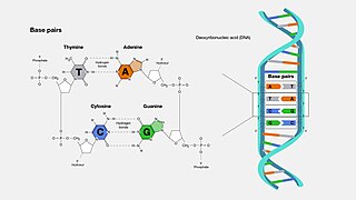
A base pair (bp) is a fundamental unit of double-stranded nucleic acids consisting of two nucleobases bound to each other by hydrogen bonds. They form the building blocks of the DNA double helix and contribute to the folded structure of both DNA and RNA. Dictated by specific hydrogen bonding patterns, "Watson–Crick" base pairs allow the DNA helix to maintain a regular helical structure that is subtly dependent on its nucleotide sequence. The complementary nature of this based-paired structure provides a redundant copy of the genetic information encoded within each strand of DNA. The regular structure and data redundancy provided by the DNA double helix make DNA well suited to the storage of genetic information, while base-pairing between DNA and incoming nucleotides provides the mechanism through which DNA polymerase replicates DNA and RNA polymerase transcribes DNA into RNA. Many DNA-binding proteins can recognize specific base-pairing patterns that identify particular regulatory regions of genes.
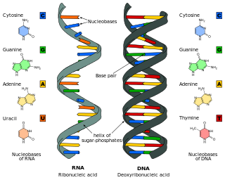
Deoxyribonucleic acid is a polymer composed of two polynucleotide chains that coil around each other to form a double helix. The polymer carries genetic instructions for the development, functioning, growth and reproduction of all known organisms and many viruses. DNA and ribonucleic acid (RNA) are nucleic acids. Alongside proteins, lipids and complex carbohydrates (polysaccharides), nucleic acids are one of the four major types of macromolecules that are essential for all known forms of life.
Francis Harry Compton Crick was an English molecular biologist, biophysicist, and neuroscientist. He, James Watson, Rosalind Franklin, and Maurice Wilkins played crucial roles in deciphering the helical structure of the DNA molecule.
The Hershey–Chase experiments were a series of experiments conducted in 1952 by Alfred Hershey and Martha Chase that helped to confirm that DNA is genetic material.
Molecular biology is a branch of biology that seeks to understand the molecular basis of biological activity in and between cells, including biomolecular synthesis, modification, mechanisms, and interactions.
Nucleic acids are large biomolecules that are crucial in all cells and viruses. They are composed of nucleotides, which are the monomer components: a 5-carbon sugar, a phosphate group and a nitrogenous base. The two main classes of nucleic acids are deoxyribonucleic acid (DNA) and ribonucleic acid (RNA). If the sugar is ribose, the polymer is RNA; if the sugar is deoxyribose, a variant of ribose, the polymer is DNA.
Ribonucleic acid (RNA) is a polymeric molecule that is essential for most biological functions, either by performing the function itself or by forming a template for the production of proteins. RNA and deoxyribonucleic acid (DNA) are nucleic acids. The nucleic acids constitute one of the four major macromolecules essential for all known forms of life. RNA is assembled as a chain of nucleotides. Cellular organisms use messenger RNA (mRNA) to convey genetic information that directs synthesis of specific proteins. Many viruses encode their genetic information using an RNA genome.
The RNA world is a hypothetical stage in the evolutionary history of life on Earth, in which self-replicating RNA molecules proliferated before the evolution of DNA and proteins. The term also refers to the hypothesis that posits the existence of this stage.
The central dogma of molecular biology is an explanation of the flow of genetic information within a biological system. It is often stated as "DNA makes RNA, and RNA makes protein", although this is not its original meaning. It was first stated by Francis Crick in 1957, then published in 1958:
The Central Dogma. This states that once "information" has passed into protein it cannot get out again. In more detail, the transfer of information from nucleic acid to nucleic acid, or from nucleic acid to protein may be possible, but transfer from protein to protein, or from protein to nucleic acid is impossible. Information here means the precise determination of sequence, either of bases in the nucleic acid or of amino acid residues in the protein.
Ribozymes are RNA molecules that have the ability to catalyze specific biochemical reactions, including RNA splicing in gene expression, similar to the action of protein enzymes. The 1982 discovery of ribozymes demonstrated that RNA can be both genetic material and a biological catalyst, and contributed to the RNA world hypothesis, which suggests that RNA may have been important in the evolution of prebiotic self-replicating systems.
Molecular genetics is a branch of biology that addresses how differences in the structures or expression of DNA molecules manifests as variation among organisms. Molecular genetics often applies an "investigative approach" to determine the structure and/or function of genes in an organism's genome using genetic screens.
"Molecular Structure of Nucleic Acids: A Structure for Deoxyribose Nucleic Acid" was the first article published to describe the discovery of the double helix structure of DNA, using X-ray diffraction and the mathematics of a helix transform. It was published by Francis Crick and James D. Watson in the scientific journal Nature on pages 737–738 of its 171st volume.
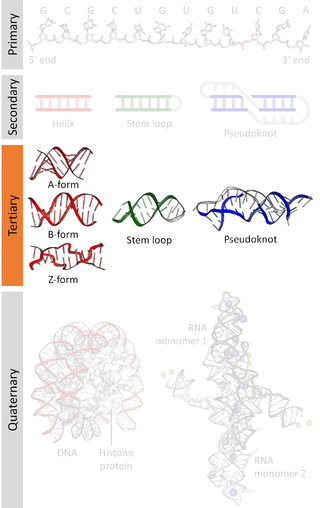
Biomolecular structure is the intricate folded, three-dimensional shape that is formed by a molecule of protein, DNA, or RNA, and that is important to its function. The structure of these molecules may be considered at any of several length scales ranging from the level of individual atoms to the relationships among entire protein subunits. This useful distinction among scales is often expressed as a decomposition of molecular structure into four levels: primary, secondary, tertiary, and quaternary. The scaffold for this multiscale organization of the molecule arises at the secondary level, where the fundamental structural elements are the molecule's various hydrogen bonds. This leads to several recognizable domains of protein structure and nucleic acid structure, including such secondary-structure features as alpha helixes and beta sheets for proteins, and hairpin loops, bulges, and internal loops for nucleic acids. The terms primary, secondary, tertiary, and quaternary structure were introduced by Kaj Ulrik Linderstrøm-Lang in his 1951 Lane Medical Lectures at Stanford University.
The adaptor hypothesis is a theoretical scheme in molecular biology to explain how information encoded in the nucleic acid sequences of messenger RNA (mRNA) is used to specify the amino acids that make up proteins during the process of translation. It was formulated by Francis Crick in 1955 in an informal publication of the RNA Tie Club, and later elaborated in 1957 along with the central dogma of molecular biology and the sequence hypothesis. It was formally published as an article "On protein synthesis" in 1958. The name "adaptor hypothesis" was given by Sydney Brenner.
Molecular models of DNA structures are representations of the molecular geometry and topology of deoxyribonucleic acid (DNA) molecules using one of several means, with the aim of simplifying and presenting the essential, physical and chemical, properties of DNA molecular structures either in vivo or in vitro. These representations include closely packed spheres made of plastic, metal wires for skeletal models, graphic computations and animations by computers, artistic rendering. Computer molecular models also allow animations and molecular dynamics simulations that are very important for understanding how DNA functions in vivo.
Nucleic acid tertiary structure is the three-dimensional shape of a nucleic acid polymer. RNA and DNA molecules are capable of diverse functions ranging from molecular recognition to catalysis. Such functions require a precise three-dimensional structure. While such structures are diverse and seemingly complex, they are composed of recurring, easily recognizable tertiary structural motifs that serve as molecular building blocks. Some of the most common motifs for RNA and DNA tertiary structure are described below, but this information is based on a limited number of solved structures. Many more tertiary structural motifs will be revealed as new RNA and DNA molecules are structurally characterized.
The RNA Tie Club was an informal scientific club, meant partly to be humorous, of select scientists who were interested in how proteins were synthesised from genes, specifically the genetic code. It was created by George Gamow upon a suggestion by James Watson in 1954 when the relationship between nucleic acids and amino acids in genetic information was unknown. The club consisted of 20 full members, each representing an amino acid, and four honorary members, representing the four nucleotides. The function of the club members was to think up possible solutions and share with the other members.
Nucleic acid secondary structure is the basepairing interactions within a single nucleic acid polymer or between two polymers. It can be represented as a list of bases which are paired in a nucleic acid molecule. The secondary structures of biological DNAs and RNAs tend to be different: biological DNA mostly exists as fully base paired double helices, while biological RNA is single stranded and often forms complex and intricate base-pairing interactions due to its increased ability to form hydrogen bonds stemming from the extra hydroxyl group in the ribose sugar.
Numerous key discoveries in biology have emerged from studies of RNA, including seminal work in the fields of biochemistry, genetics, microbiology, molecular biology, molecular evolution and structural biology. As of 2010, 30 scientists have been awarded Nobel Prizes for experimental work that includes studies of RNA. Specific discoveries of high biological significance are discussed in this article.
The history of genetics can be represented on a timeline of events from the earliest work in the 1850s, to the DNA era starting in the 1940s, and the genomics era beginning in the 1970s.
↑ Crick FH, Barnett L, Brenner S, Watts-Tobin RJ (December 1961). "General nature of the genetic code for proteins". Nature. 192 (4809): 1227–32. Bibcode:1961Natur.192.1227C. doi:10.1038/1921227a0. PMID13882203. S2CID 4276146
↑ Edgar RS Conditional lethals: in Phage and the Origins of Molecular Biology (2007) Edited by John Cairns, Gunther S. Stent, and James D. Watson, Cold Spring Harbor Laboratory of Quantitative Biology, Cold Spring Harbor, Long Island, New York ISBN978-0-87969-800-3
↑ Epstein RH, Bolle A, Steinberg CM, Kellenberger E, Boy de la Tour E, Chevalley R, Edgar RS, Susman M, Denhardt GH, Lielausis A (1963). "Physiological studies of conditional lethal mutants of bacteriophage T4D". Cold Spring Harbor Symposia on Quantitative Biology. 28: 375–394. doi:10.1101/SQB.1963.028.01.053. ISSN0091-7451
↑ Edgar RS, Lielausis I (April 1964). "Temperature-sensitive mutants of bacteriophage T4D: Their isolation and Characterization". Genetics. 49: 649–62. doi:10.1093/genetics/49.4.649. PMC 1210603. PMID 14156925
↑ Sarabhai AS, Stretton AO, Brenner S, Bolle A (January 1964). "Co-linearity of the gene with the polypeptide chain". Nature. 201 (4914): 13–7. Bibcode:1964Natur.201...13S. doi:10.1038/201013a0. PMID 14085558. S2CID 10179456
↑ Rich A, Davies DR (July 1956). "A new, two-stranded helical structure: polyadenylic acid and polyuridylic acid". J. Am. Chem. Soc. 78 (14): 3548–3549. doi:10.1021/ja01595a086.
↑ Felsenfeld G, Davies DR, Rich A (April 1957). "Formation of a three-stranded polynucleotide molecule". J. Am. Chem. Soc. 79 (8): 2023–2024. doi:10.1021/ja01565a074.
↑ Cech TR, Zaug AJ, Grabowski PJ (December 1981). "In vitro splicing of the ribosomal RNA precursor of Tetrahymena: involvement of a guanosine nucleotide in the excision of the intervening sequence". Cell. 27 (3 Pt 2): 487–96. doi:10.1016/0092-8674(81)90390-1. PMID6101203. S2CID17674600.
↑ Ramos A, Gubser CC, Varani G (June 1997). "Recent solution structures of RNA and its complexes with drugs, peptides and proteins". Curr. Opin. Struct. Biol. 7 (3): 317–23. doi:10.1016/S0959-440X(97)80046-2. PMID9204272.
This page is based on this Wikipedia article Text is available under the CC BY-SA 4.0 license; additional terms may apply. Images, videos and audio are available under their respective licenses.