Holter monitor

Last updated
Holter monitor
HolterAFT1000.jpg
3-lead Holter monitor
Specialty Cardiology, electrophysiology

In medicine, a Holter monitor (often simply Holter) is a type of ambulatory electrocardiography device, a portable device for cardiac monitoring (the monitoring of the electrical activity of the cardiovascular system) for at least 24 hours.

Contents

The Holter's most common use is for monitoring ECG heart activity (electrocardiography or ECG). Its extended recording period is sometimes useful for observing occasional cardiac arrhythmias which would be difficult to identify in a shorter period. For patients having more transient symptoms, a cardiac event monitor which can be worn for a month or more can be used. [1]

When used to study the heart, much like standard electrocardiography, the Holter monitor records electrical signals from the heart via a series of electrodes attached to the chest. Electrodes are placed over bones to minimize artifacts from muscular activity. The number and position of electrodes varies by model, but most Holter monitors employ between three and eight. These electrodes are connected to a small piece of equipment that is attached to the patient's belt or hung around the neck, keeping a log of the heart's electrical activity throughout the recording period. A 12-lead Holter system is used when precise ECG information is required to analyse the exact origin of the abnormal signals. [2]

History

The Holter monitor was developed at the Holter Research Laboratory in Helena, Montana, USA by experimental physicists Norman J. Holter and Bill Glasscock, [3] [4] who started work on radio telemetry in 1949. Inspired by a suggestion from cardiologist Paul Dudley White in the early 1950s, they redirected their efforts toward development of a wearable cardiac monitoring device. [5] The Holter monitor was released for commercial production in 1962. [5]

Data storage

Atrial fibrillation recorded by a Holter monitor Holter Atrialfibrillation.jpg
Atrial fibrillation recorded by a Holter monitor

Older monitors used reel-to-reel tapes or C90 or C120 audio cassettes, moving at a 1.7 mm/s or 2 mm/s speed to record the data. The recording could be played back and analyzed at 60 times the recording speed, so 24 hours of recording could be analyzed in 24 minutes. Modern monitors record EDF-files on digital flash memory. The information is loaded on a computer, which counts ECG complexes; calculates summary statistics, such as average, minimum, and maximum heart rate; and finds parts of the recording that are worthy of further study.

Components

Each Holter system has hardware (called monitor or recorder) for recording the signal, and software for review and analysis of the record. There may be a "patient button" on the front that the patient can press at specific instants such as on sickness, going to bed, taking pills, etc.; this records a mark that identifies the time of the action on the recording. Advanced Holter recorders are able to display the signal, useful for checking the signal quality.

Recorder

The size of the recorder differs depending on the manufacturer of the device. The average dimensions of today's Holter monitors are about 110x70x30 mm, but some are only 61x46x20 mm and weigh 99 g. [6]

Most Holter monitors the ECG via two or three channels. Depending on manufacturer, different lead systems and numbers of leads are used; the number of leads may be minimised for patient comfort. Two or three channel recording has been used for a long time in the Holter monitoring history; 12-channel Holters were introduced later, using either the standard 12-lead electrocardiograph or the modified (Mason-Likar) exercise lead system. [7] These Holters can occasionally provide information similar to that of an ECG stress test examination. They are also suitable when analyzing patients after myocardial infarction. Recordings from these 12-lead monitors are of a significantly lower resolution than those from a standard 12-lead ECG, and in some cases have been shown to provide misleading ST segment representation, even though some devices allow setting the sampling frequency up to 1000 Hz for special-purpose examinations such as detection of "late potential".

Another innovation is the inclusion of a triaxial movement sensor, which records the patient's physical activity and, on examination and software processing, extracts three movement statuses: sleeping, standing, or walking. Some modern devices can record spoken patient diary entries that can be listened to.

Analyzing software

Screenshot of Holter ECG software Cubeholter.jpg
Screenshot of Holter ECG software

After the recording of ECG signal for typically 24 hours, the signal must be analysed. A person would have to listen for the full 24 hours; instead integrated automatic analysis determines different sorts of heart beats, rhythms, etc. The success of the analysis is closely associated with the signal quality, which mainly depends upon the attachment of the electrodes to the patient's body. Incorrect attachment allows electromagnetic disturbance to add noise to the record, particularly with rapid patient movement, impeding processing. Other factors can also affect signal quality, such as muscle tremors, sampling rate and resolution of the digitized signal (high quality devices offer higher sampling frequency).

The automatic analysis commonly provides the physician with information about heart beat morphology, beat interval measurement, heart rate variability, rhythm overview and patient diary (moments when the patient pressed the patient button). Advanced systems also perform spectral analysis, ischemic burden evaluation, graph of patient's activity or PQ segment analysis. Also possible is the ability to monitor and analyse pacemaker impulse detection, useful for checking pacemaker function.

See also

Related Research Articles

The diagnostic tests in cardiology are methods of identifying heart conditions associated with healthy vs. unhealthy, pathologic heart function.

<span class="mw-page-title-main">Electrocardiography</span> Examination of the hearts electrical activity

Electrocardiography is the process of producing an electrocardiogram, a recording of the heart's electrical activity through repeated cardiac cycles. It is an electrogram of the heart which is a graph of voltage versus time of the electrical activity of the heart using electrodes placed on the skin. These electrodes detect the small electrical changes that are a consequence of cardiac muscle depolarization followed by repolarization during each cardiac cycle (heartbeat). Changes in the normal ECG pattern occur in numerous cardiac abnormalities, including:

<span class="mw-page-title-main">Palpitations</span> Perceived cardiac abnormality in which ones heartbeat can be felt

Palpitations are perceived abnormalities of the heartbeat characterized by awareness of cardiac muscle contractions in the chest, which is further characterized by the hard, fast and/or irregular beatings of the heart.

<span class="mw-page-title-main">Heart rate monitor</span> Personal monitoring device

A heart rate monitor (HRM) is a personal monitoring device that allows one to measure/display heart rate in real time or record the heart rate for later study. It is largely used to gather heart rate data while performing various types of physical exercise. Measuring electrical heart information is referred to as electrocardiography.

A flatline is an electrical time sequence measurement that shows no activity and therefore, when represented, shows a flat line instead of a moving one. It almost always refers to either a flatlined electrocardiogram, where the heart shows no electrical activity (asystole), or to a flat electroencephalogram, in which the brain shows no electrical activity. Both of these specific cases are involved in various definitions of death.

Premature atrial contraction (PAC), also known as atrial premature complexes (APC) or atrial premature beats (APB), are a common cardiac dysrhythmia characterized by premature heartbeats originating in the atria. While the sinoatrial node typically regulates the heartbeat during normal sinus rhythm, PACs occur when another region of the atria depolarizes before the sinoatrial node and thus triggers a premature heartbeat, in contrast to escape beats, in which the normal sinoatrial node fails, leaving a non-nodal pacemaker to initiate a late beat.

<span class="mw-page-title-main">Left bundle branch block</span> Medical condition

Left bundle branch block (LBBB) is a conduction abnormality in the heart that can be seen on an electrocardiogram (ECG). In this condition, activation of the left ventricle of the heart is delayed, which causes the left ventricle to contract later than the right ventricle.

<span class="mw-page-title-main">String galvanometer</span>

A string galvanometer is a sensitive fast-responding measuring instrument that uses a single fine filament of wire suspended in a strong magnetic field to measure small currents. In use, a strong light source is used to illuminate the fine filament, and the optical system magnifies the movement of the filament allowing it to be observed or recorded by photography. The principle of the string galvanometer remained in use for electrocardiograms until the advent of electronic vacuum-tube amplifiers in the 1920s.

Clinical cardiac electrophysiology, is a branch of the medical specialty of cardiology concerned with the study and treatment of rhythm disorders of the heart. Cardiologists with expertise in this area are usually referred to as electrophysiologists. Electrophysiologists are trained in the mechanism, function, and performance of the electrical activities of the heart. Electrophysiologists work closely with other cardiologists and cardiac surgeons to assist or guide therapy for heart rhythm disturbances (arrhythmias). They are trained to perform interventional and surgical procedures to treat cardiac arrhythmia.

A cardiovascular technician, also known as a vascular technician, is health professional that deal with the circulatory system.

A Bioamplifier is an electrophysiological device, a variation of the instrumentation amplifier, used to gather and increase the signal integrity of physiologic electrical activity for output to various sources. It may be an independent unit, or integrated into the electrodes.

<span class="mw-page-title-main">Cardiac monitoring</span>

Cardiac monitoring generally refers to continuous or intermittent monitoring of heart activity to assess a patient's condition relative to their cardiac rhythm. Cardiac monitoring is usually carried out using electrocardiography, which is a noninvasive process that records the heart's electrical activity and displays it in an electrocardiogram. It is different from hemodynamic monitoring, which monitors the pressure and flow of blood within the cardiovascular system. The two may be performed simultaneously on critical heart patients. Cardiac monitoring for ambulatory patients is known as ambulatory electrocardiography and uses a small, wearable device, such as a Holter monitor, wireless ambulatory ECG, or an implantable loop recorder. Data from a cardiac monitor can be transmitted to a distant monitoring station in a process known as telemetry or biotelemetry.

Fabio Badilini is an Italian scientist and business man. He has made major contributions to noninvasive electrocardiography not only through his individual contributions, but also through his truly remarkable ability to foster collaborations across scientific disciplines, academic institutions, governmental agencies, device manufacturers and industries around the world.

Alois A. Langer is an American biomedical engineer best known as one of the co-inventors of the Implantable Cardioverter Defibrillator (ICD).

<span class="mw-page-title-main">Automated ECG interpretation</span>

Automated ECG interpretation is the use of artificial intelligence and pattern recognition software and knowledge bases to carry out automatically the interpretation, test reporting, and computer-aided diagnosis of electrocardiogram tracings obtained usually from a patient.

<span class="mw-page-title-main">Human Research Facility Holter Monitor</span>

The Human Research Facility Holter Monitor (Holter) is a battery-powered, noninvasive electrocardiogram (ECG) device that accurately measures the heart rate of crew members over an extended period of time. ECG information is stored on a Portable Computer Memory Card International Adapter (PCMCIA) card and downlinked to Earth for analysis after monitoring is complete.

<span class="mw-page-title-main">Monitoring (medicine)</span> Observation of a disease, condition or one or several medical parameters over time

In medicine, monitoring is the observation of a disease, condition or one or several medical parameters over time.

<span class="mw-page-title-main">Implantable loop recorder</span>

An implantable loop recorder (ILR), also known as an insertable cardiac monitor (ICM), is a small device that is implanted under the skin of the chest for cardiac monitoring, to record the heart's electrical activity for an extended period.

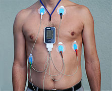
Wireless ambulatory electrocardiography (ECG) is a type of ambulatory electrocardiography with recording devices that use wireless technology, such as Bluetooth and smartphones, for at-home cardiac monitoring (monitoring of heart rhythms). These devices are generally recommended to people who have been previously diagnosed with arrhythmias and want to have them monitored, or for those who have suspected arrhythmias and need to be monitored over an extended period of time in order to be diagnosed.

Bioinstrumentation or Biomedical Instrumentation is an application of biomedical engineering which focuses on development of devices and mechanics used to measure, evaluate, and treat biological systems. The goal of biomedical instrumentation focuses on the use of multiple sensors to monitor physiological characteristics of a human or animal for diagnostic and disease treatment purposes. Such instrumentation originated as a necessity to constantly monitor vital signs of Astronauts during NASA's Mercury, Gemini, and Apollo missions.

References

  1. "Holter Monitor". www.heart.org. Archived from the original on 16 July 2018. Retrieved 8 April 2018.{{cite web}}: CS1 maint: bot: original URL status unknown (link)
  2. Herzschrittmacherther Elektrophysiol. 2013 Jun;24(2):92-96. doi : 10.1007/s00399-013-0268-4. Epub 2013 Jun 19. 12-lead Holter electrocardiography. Review of the literature and clinical application update. Su L1, Borov S, Zrenner B.
  3. Meldrum, Stuart J (1993). "Obituary for Wilford "Bill" Glasscock". Journal of Ambulatory Monitoring. 6 (3): 243. with the passing of Bill Glasscock, we have lost the second half of one of the most successful partnerships ever to exist in the field of biomedical engineering
  4. "At the Heart of the Invention: The development of the Holter Monitor". National Museum of American History. 2011-11-16. Retrieved 13 August 2016.
  5. 1 2 Hilbel, Thomas; Thomas M Helms; Gerd Mikus; Hugo A Katus; Christian Zugck (10 January 2008). "Telemetry in the clinical setting". Herzschrittmachertherapie & Elektrophysiologie . 19 (3): 146–64. doi:10.1007/s00399-008-0017-2. ISSN   0938-7412. PMID   18956161 . Retrieved 4 August 2009.
  6. HeartBug technical specifications Archived 2013-10-04 at the Wayback Machine
  7. Papouchado, M.; Walker, P. R.; James, M. A.; Clarke, L. M. (1 July 1987). "Fundamental differences between the standard 12-lead electrocardiograph and the modified (Mason-Likar) exercise lead system". European Heart Journal. 8 (7). Oxford University Press (OUP): 725–733. doi:10.1093/eurheartj/8.7.725. ISSN   0195-668X. PMID   3653124.