Related Research Articles

Gap junctions are specialized intercellular connections between a multitude of animal cell-types. They directly connect the cytoplasm of two cells, which allows various molecules, ions and electrical impulses to directly pass through a regulated gate between cells.
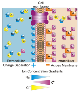
Membrane potential is the difference in electric potential between the interior and the exterior of a biological cell. That is, there is a difference in the energy required for electric charges to move from the internal to exterior cellular environments and vice versa, as long as there is no acquisition of kinetic energy or the production of radiation. The concentration gradients of the charges directly determine this energy requirement. For the exterior of the cell, typical values of membrane potential, normally given in units of millivolts and denoted as mV, range from –80 mV to –40 mV.

In biology, a connexon, also known as a connexin hemichannel, is an assembly of six proteins called connexins that form the pore for a gap junction between the cytoplasm of two adjacent cells. This channel allows for bidirectional flow of ions and signaling molecules. The connexon is the hemichannel supplied by a cell on one side of the junction; two connexons from opposing cells normally come together to form the complete intercellular gap junction channel. However, in some cells, the hemichannel itself is active as a conduit between the cytoplasm and the extracellular space, allowing the transference of ions and small molecules lower than 1-2 KDa. Little is known about this function of connexons besides the new evidence suggesting their key role in intracellular signaling.

The cardiac action potential is a brief change in voltage across the cell membrane of heart cells. This is caused by the movement of charged atoms between the inside and outside of the cell, through proteins called ion channels. The cardiac action potential differs from action potentials found in other types of electrically excitable cells, such as nerves. Action potentials also vary within the heart; this is due to the presence of different ion channels in different cells.

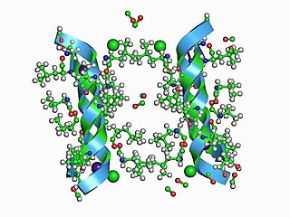
Connexins (Cx), or gap junction proteins, are structurally related transmembrane proteins that assemble to form vertebrate gap junctions. An entirely different family of proteins, the innexins, form gap junctions in invertebrates. Each gap junction is composed of two hemichannels, or connexons, which consist of homo- or heterohexameric arrays of connexins, and the connexon in one plasma membrane docks end-to-end with a connexon in the membrane of a closely opposed cell. The hemichannel is made of six connexin subunits, each of which consist of four transmembrane segments. Gap junctions are essential for many physiological processes, such as the coordinated depolarization of cardiac muscle, proper embryonic development, and the conducted response in microvasculature. For this reason, mutations in connexin-encoding genes can lead to functional and developmental abnormalities.
Voltage-gated calcium channels (VGCCs), also known as voltage-dependent calcium channels (VDCCs), are a group of voltage-gated ion channels found in the membrane of excitable cells (e.g., muscle, glial cells, neurons, etc.) with a permeability to the calcium ion Ca2+. These channels are slightly permeable to sodium ions, so they are also called Ca2+-Na+ channels, but their permeability to calcium is about 1000-fold greater than to sodium under normal physiological conditions.
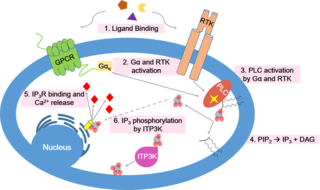
Calcium signaling is the use of calcium ions (Ca2+) to communicate and drive intracellular processes often as a step in signal transduction. Ca2+ is important for cellular signalling, for once it enters the cytosol of the cytoplasm it exerts allosteric regulatory effects on many enzymes and proteins. Ca2+ can act in signal transduction resulting from activation of ion channels or as a second messenger caused by indirect signal transduction pathways such as G protein-coupled receptors.

Neurotransmission is the process by which signaling molecules called neurotransmitters are released by the axon terminal of a neuron, and bind to and react with the receptors on the dendrites of another neuron a short distance away. A similar process occurs in retrograde neurotransmission, where the dendrites of the postsynaptic neuron release retrograde neurotransmitters that signal through receptors that are located on the axon terminal of the presynaptic neuron, mainly at GABAergic and glutamatergic synapses.

Osmotic shock or osmotic stress is physiologic dysfunction caused by a sudden change in the solute concentration around a cell, which causes a rapid change in the movement of water across its cell membrane. Under hypertonic conditions - conditions of high concentrations of either salts, substrates or any solute in the supernatant - water is drawn out of the cells through osmosis. This also inhibits the transport of substrates and cofactors into the cell thus “shocking” the cell. Alternatively, under hypotonic conditions - when concentrations of solutes are low - water enters the cell in large amounts, causing it to swell and either burst or undergo apoptosis.

The ATP-gated P2X receptor cation channel family, or simply P2X receptor family, consists of cation-permeable ligand-gated ion channels that open in response to the binding of extracellular adenosine 5'-triphosphate (ATP). They belong to a larger family of receptors known as the ENaC/P2X superfamily. ENaC and P2X receptors have similar 3-D structures and are homologous. P2X receptors are present in a diverse array of organisms including humans, mouse, rat, rabbit, chicken, zebrafish, bullfrog, fluke, and amoeba.

Pannexins are a family of vertebrate proteins identified by their homology to the invertebrate innexins. While innexins are responsible for forming gap junctions in invertebrates, the pannexins have been shown to predominantly exist as large transmembrane channels connecting the intracellular and extracellular space, allowing the passage of ions and small molecules between these compartments.
Innexins are transmembrane proteins that form gap junctions in invertebrates. Gap junctions are composed of membrane proteins that form a channel permeable to ions and small molecules connecting the cytoplasm of adjacent cells. Although gap junctions provide similar functions in all multicellular organisms, it was not known what proteins invertebrates used for this purpose until the late 1990s. While the connexin family of gap junction proteins was well-characterized in vertebrates, no homologues were found in non-chordates.
P2Y receptors are a family of purinergic G protein-coupled receptors, stimulated by nucleotides such as adenosine triphosphate, adenosine diphosphate, uridine triphosphate, uridine diphosphate and UDP-glucose. To date, 8 P2Y receptors have been cloned in humans: P2Y1, P2Y2, P2Y4, P2Y6, P2Y11, P2Y12, P2Y13 and P2Y14.
Gliotransmitters are chemicals released from glial cells that facilitate neuronal communication between neurons and other glial cells. They are usually induced from Ca2+ signaling, although recent research has questioned the role of Ca2+ in gliotransmitters and may require a revision of the relevance of gliotransmitters in neuronal signalling in general.

P2X purinoceptor 7 is a protein that in humans is encoded by the P2RX7 gene.
P2 receptor may refer to:
P2X purinoceptor 4 is a protein that in humans is encoded by the P2RX4 gene. The product of this gene belongs to the family of purinoceptors for ATP. Multiple alternatively spliced transcript variants have been identified for this gene although their full-length natures have not been determined.
In electrophysiology, the term gating refers to the opening (activation) or closing of ion channels. This change in conformation is a response to changes in transmembrane voltage.
Purinergic signalling is a form of extracellular signalling mediated by purine nucleotides and nucleosides such as adenosine and ATP. It involves the activation of purinergic receptors in the cell and/or in nearby cells, thereby regulating cellular functions.

Gap junction modulation describes the functional manipulation of gap junctions, specialized channels that allow direct electrical and chemical communication between cells without exporting material from the cytoplasm. Gap junctions play an important regulatory role in various physiological processes including signal propagation in cardiac muscles and tissue homeostasis of the liver. Modulation is required, since gap junctions must respond to their environment, whether through an increased expression or permeability. Impaired or altered modulation can have significant health implications and are associated with the pathogenesis of the liver, heart and intestines.
References
- ↑ Labra VC, Santibáñez CA, Gajardo-Gómez R, Díaz EF, Gómez GI, Orellana JA (2018-03-20). "The Neuroglial Dialog Between Cannabinoids and Hemichannels". Frontiers in Molecular Neuroscience. 11: 79. doi: 10.3389/fnmol.2018.00079 . PMC 5890195 . PMID 29662436.
- 1 2 Retamal MA (2014). "Connexin and Pannexin hemichannels are regulated by redox potential". Frontiers in Physiology. 5: 80. doi: 10.3389/fphys.2014.00080 . PMC 3933782 . PMID 24611056.
- ↑ Spray DC, Ye ZC, Ransom BR (November 2006). "Functional connexin "hemichannels": a critical appraisal". Glia. 54 (7): 758–73. CiteSeerX 10.1.1.124.4117 . doi:10.1002/glia.20429. PMID 17006904. S2CID 17677517.
- 1 2 Baroja-Mazo A, Barberà-Cremades M, Pelegrín P (January 2013). "The participation of plasma membrane hemichannels to purinergic signaling". Biochimica et Biophysica Acta (BBA) - Biomembranes. 1828 (1): 79–93. doi: 10.1016/j.bbamem.2012.01.002 . PMID 22266266.