Metastatic breast cancer

Last updated
Common sites of metastasis for breast cancer Diagram showing most common sites breast cancer spreads to CRUK 169.svg
Common sites of metastasis for breast cancer

Metastatic breast cancer, also referred to as metastases, advanced breast cancer, secondary tumors, secondaries or stage IV breast cancer, is a stage of breast cancer where the breast cancer cells have spread to distant sites beyond the axillary lymph nodes. There is no cure for metastatic breast cancer; [1] there is no stage after IV.

Contents

Metastases can occur several years after the primary breast cancer, although it is sometimes diagnosed at the same time as the primary breast cancer or, rarely, before the primary breast cancer has been diagnosed. [2]

Metastatic breast cancer cells frequently differ from the preceding primary breast cancer in properties such as receptor status. The cells have often developed resistance to several lines of previous treatment and have acquired special properties that permit them to metastasize to distant sites. Metastatic breast cancer can be treated, sometimes for many years, but it cannot be cured. [2] Distant metastases are the cause of about 90% of deaths due to breast cancer. [3]

Breast cancer can metastasize anywhere in body but primarily metastasizes to the bone, lungs, regional lymph nodes, liver and brain, with the most common site being the bone. [4] Treatment of metastatic breast cancer depends on location of the metastatic tumors and includes surgery, radiation, chemotherapy, biological, and hormonal therapy. [5]

Typical environmental barriers in a metastatic event include physical (a basement membrane), chemical (reactive oxygen species or ROS, hypoxia and low pH) and biological (immune surveillance, inhibitory cytokines and regulatory extra-cellular matrix (ECM) peptides) components. [6] Organ-specific anatomic considerations also influence metastasis; these include blood-flow patterns from the primary tumor and the homing ability of cancer cells to certain tissues. The targeting by cancer cells of specific organs is probably regulated by chemo-attractant factors and adhesion molecules produced by the target organ, along with cell-surface receptors expressed by the tumor cells.

Symptoms

The symptoms produced by metastatic breast cancer vary by the location of the metastases. [7] For instance:

Bone

Roughly 70% of all patients living with advanced breast cancer have bone metastases [9] Very often bone metastases can be successfully managed for a long time.

Brain

Brain metastasis is observed in 10% of breast cancer patients with metastatic properties [10] Many of the breast cancer therapies (like targeted antibodies) fail to penetrate the blood–brain barrier, hence allowing for tumor recurrence in the central nervous system.

Pathophysiology

The main steps involved in the metastatic cascade of a cancer cell are:

The potential of a tumor cell to metastasize depends on its microenvironment, or “niche” interactions with local factors promoting tumor-cell growth, survival, angiogenesis, invasion and metastasis. [11] This is explained by the seed and soil hypothesis.

Extracellular matrix degradation in cancer

Cell-cell and cell-ECM matrix adhesion, motility, and localised proteolysis are mediated mainly by matrix metalloproteases (MMPs). Degradation of the extracellular matrix begins the process of metastasis. The cell develops structures called invadopodia, which are highly concentrated in several proteases and have a highly dynamic actin cytoskeleton.

Mechanisms of metalloprotease action in cell motility involve:

Most of these processes require a delicate balance between the functions of matrix metalloproteases (MMPs) or metalloprotease-disintegrins (ADAMs) and natural tissue inhibitors of metalloproteases (TIMPs). Regulated proteolysis is an important mechanism to maintain homeostasis. There is increased expression of protease systems in cancer cells, to equip them with the tools necessary to degrade the extracellular matrix and release growth factors or transmembrane receptors. MMP-2 is upregulated in the bone, and increased levels of MMP-1 and MMP-19 are observed in the brain. This in turn, upregulates the signaling pathways necessary to provide increased cell adhesion, cell motility, cell migration, invasion, cancer- cell proliferation and survival.

Extracellular matrix components

ECM-tumor cell interactions play a critical role in each of the events of the metastatic cascade. Interactions of the breast cancer cells with integrins, fibronectin, laminins, collagens, hyaluronan and proteoglycans can contribute to the metastatic process. Some of these proteins are discussed here in relation to breast-cancer metastasis.

Fibrinogen-Integrin

Fibronectin is an extracellular glycoprotein that can bind to integrins and other ECM components like collagen, fibrin and heparan sulphate proteoglycans(HSPGs). Several different integrins bind to fibronectin. Fibronectin-integrin interactions are important in tumor cell migration, invasion, metastasis and cell proliferation by signaling through integrins. Integrin-mediated tumor cell adhesion to ECM proteins can trigger signal transduction and cause upregulation of gene expression, increased tyrosine phosphorylytion of the focal adhesion kinase, and activation and nuclear translocation of mitogen-activated protein (MAP) kinases.

Heparanase

Heparanase cleaves heparin sulfate chains of HSPGs, which have an extensive network with several proteins on the cell surface and ECM. The basic HSPG structure consists of a protein core to which several linear heparin sulfate (HS) chains are covalently O-linked; this acts as an assembly of different ECM proteins, including fibronectin, laminins, interstitial collagens, heparin-binding growth factors, chemokines and lipoproteins. HSPGs are prominent components of blood vessels. [12] to HS stabilizes fibroblast growth factors (FGFs) and vascular endothelial growth factors (VEGFs) and prevents them from inactivation. HS chains function as low-affinity co-receptors which promote dimerization of FGFs, aids in the sequestration of the growth factors (GFs) and causes activation of the signaling tyrosine kinase receptors even under low circulating concentrations of growth factors. Heparanase expressed by cancer cells participates in angiogenesis and neovascularization by degrading the polysaccharide scaffold of the endothelial BM, thereby releasing angiogenic growth factors from the ECM.

Tenascin

The ECM protein tenascin C (TNC) is up-regulated in metastatic breast cancer. TNC is an adhesion-modulating extracellular matrix glycoprotein. It is highly expressed in tumor stroma and stimulates tumor-cell proliferation. It is hypothesised that TNC stimulates invasion via up-regulation of MMP-1 expression through activation of the MAPK pathway. MMP-1 (interstitial collagenase) cleaves collagen type I, II, III, VII and X. Therefore, tenascin C over-expression can significantly alter collagen in the ECM and influence tumor cell migration in cartilaginous tissues.

Endoglin

Endoglin is a cell-surface disulfide-linked homodimeric glycoprotein which binds to integrins and other RGD ligands and is a co-receptor for TGF-beta. Brain-metastatic breast-tumor cells express endoglin in large amounts. Endoglin-overexpressing cells develop large numbers of invadopodia; endoglin is localized in these structures. Endoglin expression in tumor cells contributes to metastasis by upregulating MMP-1 and MMP-19. MMP-19 cleaves components of the basal lamina such as collagen type IV, laminin 5, nidogen (entactin) and other ECM proteins such as tenascin, aggrecan and fibronectin. Therefore, endoglin over-expression alters the proteolytic balance of the cells to greater matrix degradation and increased invasive properties of breast cancer.

Mechanisms in bone metastases

The primary extracellular matrix components and cell-surface receptors which aid in metastasis are:

Integrin signalling

Integrin αvβ3 (a cell-surface adhesion molecule) is important for tumor attachment, cell-to-cell communication between the breast tumor cells and the environment in bone, osteoclast bone resorption and angiogenesis. Integrin-mediated adhesion between cancer cells and osteoclasts in bone metastases induces phosphorylation of extracellular signal-regulated kinases (ERK1/2) in osteoclasts, which in turn induces osteoclast differentiation and survival. [13]

Cancer cell-blood platelet interaction

Metastatic breast-cancer cells excrete lysophosphatidic acid (LPA) which binds to receptors on tumor cells, inducing cell proliferation and release of cytokines(IL-6 and IL-8, potent bone resorptive agents) and stimulating bone resorption. After the breast-cancer cells have left the primary tumor, they interact with the bone microenvironment and secrete osteolytic factors capable of osteoclast formation and bone resorption. Apart from the breast tumor cells, the resident stromal cells also contribute to tumor survival. Growth factors such as epidermal growth factor (EGF), fibroblast growth factor (FGF) and transforming growth factor beta (TGF-β) are implicated in the development and progression of metastatic breast cancer.

Matrix metalloproteinases (MMPs)

MMP-2 is the main metalloprotease secreted by breast-cancer cells or induced in the adjacent bone stroma; it plays an important role in the degradation of the extracellular matrix essential for metastasis. Tumor cells use MMP-2 secreted by bone marrow fibroblasts (BMFs). MMP-2 is stored in an inactive conformation in association with the cell surface (or extracellular matrix) of BMFs. [14] Inactive MMP-2 present on the surface of BMFs is displaced by breast-cancer cells. Cancer cells can then use the proteinase to facilitate tissue invasion, which requires the degradation of connective tissue associated with vascular basement membranes and interstitial connective tissue. MMP-2 is unlike other MMP's as its activity is modulated by metalloproteases called tissue inhibitor of metalloproteases (TIMP) and membrane type 1 MMP (Korhmann et at. 2009)

Mechanism in brain metastasis

The brain is a unique organ for metastasis, since the breast-tumor cells have to pass the blood–brain barrier (BBB) to form micrometastases.

CD44

CD44 (a cell-surface transmembrane glycoprotein) is a receptor for hyaluronic acid, involved in cell adhesion by binding to specific extracellular matrix components. A proposed mechanism for the function of CD44 is to regulate the adhesion of circulating cancer cells in the brain to the endothelium at the secondary site with the help of a hyaluronate matrix ligand or by its cytoplasmic attachments to actin-associated proteins of the merlin/ezrin/radixin/moesin family. [15]

Sialyl transferase (glycosylation modifications of gangliosides)

Cell-surface sialylation has been implicated in cell–to-cell interactions, and over-expression of a brain sialyltransferase in breast-cancer cells is a mechanism highlighting the role of cell-surface glycosylation in organ-specific metastatic interactions. Breast-cancer metastasis to the brain involves mediators of extravasation through non-fenestrated capillaries, complemented by specific enhancers of BBB-crossing and brain colonization. [16]

Seed and soil hypothesis

The "seed and soil" hypothesis states that specific organs harbor metastases from one type of cancer by stimulating their growth better than other types of cancer. This interaction is dynamic and reciprocal, since cancer cells modify the environment they encounter. Tumor embolus = seed and Target organ = soil.

Workup

In the detection of bone metastases, skeletal scintigraphy (bone scan) is very sensitive and is recommended as the first imaging study in asymptomatic individuals with suspected breast-cancer metastases. [17] X-ray radiography is recommended if there is abnormal radionuclide uptake from the bone scan and in assessing the risk of pathological fractures, and is recommended as the initial imaging study in patients with bone pain. [17] MRI or the combination PET-CT may be considered for cases of abnormal radionuclide uptake on bone scan, when radiography does not give an acceptably clear result. [17]

Treatment

Metastasis is a complex and interconnected multi-step process. Each step in the process is a potential target for therapies to prevent or reduce metastasis. Those steps which have a good clinical window are the best targets for therapy. Each event in metastasis is highly regulated and requires a synergistic activation of different ECM proteins, growth factors and so on. Although the occasional patient with metastatic breast cancer benefits from surgical resection of an isolated metastasis and most patients receive radiotherapy (often for palliation alone) during the course of their disease, the treatment of metastatic breast carcinoma typically involves the use of systemic therapy. There is no sufficient evidence on the benefits and risks of breast surgery associated with systemic treatment for women diagnosed with metastatic breast cancer. [18]

Chemotherapy

Chemotherapy is one of the most important components of therapy for metastatic breast cancer. Therapy of choice is based on three variables; 1. the extent, pattern and aggressiveness at first presentation. 2. what stage of menopause the patient is at. 3. What receptor hormone the tumour has. Observation of metastases provides direct feedback on the effectiveness of the treatment and often a number of chemotherapy agents are tried sequentially to determine one that works. Adding one or more chemotherapy drugs to an established regimen in women with metastatic breast cancer gives greater tumour shrinkage on imaging but also increased toxicity. [19]

Combination chemotherapy is often used in patients with metastatic breast cancer. Research suggests no difference in overall survival time between sequential single agent chemotherapy and combination chemotherapy. [20] Sequential single agent chemotherapy may have a more positive effect on progression-free survival. [20]

Taxanes are very active in metastatic breast cancer, and abraxane is approved for patients with metastatic breast cancer who either relapsed within six months of adjuvant chemotherapy or failed to respond to combination chemotherapy. This has a higher response rate than solvent-based paclitaxel (15% vs 8%). Abraxane can also deliver a 49% higher dose of medication than solvent-based paclitaxel; however, the side effects are severe and include chemotherapy-induced peripheral neuropathy. In women with metastatic breast cancer, taxane-containing chemotherapy regimens appear to improve survival and tumour shrinkage and decrease time to progression. [21] Taxanes are associated with increased risk of neuro-toxicity and less nausea and vomiting when contrasted to non-taxane containing regimens. [21]

Vinorelbine is also active in metastatic breast cancer. Eribulin was approved in the US in Nov 2010. A targeted therapy drug, Kadcyla, was approved in February 2013. This antibody-drug conjugate targets only cancerous cells. It works by only releasing its toxic payload when triggered by a protein found in the cancerous cells in HER2+ breast cancer. It has extremely low side effects using this target therapy method. [22]

Platinum-containing chemotherapy regimens are known to be effective for treating a variety of different cancer types. [23] In women with metastatic breast cancer who do not have triple negative disease, there is little or no survival benefit and excess toxicity from platinum-based regimens. [23] However, in women with metastatic triple negative breast cancer, there may be a moderate survival benefit from platinum-based regimens. [23]

Antitumour antibiotics are also used in metastatic breast cancer. Antitumour antibiotics work to prevent cancer cells multiplying by damaging them or stop cell growth. [24] A meta-analysis has demonstrated that women taking antitumour antibiotics as part of their regimen had an advantage in time to progression and tumour shrinkage, but also increased side effect such as cardiotoxicity, leukopenia and nausea. [25]

Tamoxifen and other anti-estrogens

For estrogen-receptor-positive metastatic breast carcinoma the first line of therapy is often tamoxifen or another anti-estrogen drug unless there are liver metastases, significant lung involvement, rapidly progressive disease or severe symptoms requiring immediate palliation.

Radiotherapy

Radiotherapy is used in the treatment of metastatic breast cancer. The most common reasons for a patient with metastatic breast carcinoma to be treated with radiotherapy are:

"Alternative" and "complementary" therapies

Some patients with metastatic breast cancer opt to try alternative therapies that are claimed to achieve healing effects similar to scientifically-tested medical approaches, but lack scientific evidence to support those claims. Approaches that are considered alternative therapies when applied to cancer treatment include vitamin therapies, homeopathic treatments, extreme diets, chiropractic treatment and acupuncture. [28]

Some alternative treatments are harmful or even life-threatening. Amygdalin, an extract derived from apricot kernels, exposes the patient to cyanide. Bee venom can cause a life-threatening allergic reaction. Severe dietary restrictions such as macrobiotic diets can disrupt the body’s metabolism and cause dangerous weight loss. [28] [29] People should be aware that foods, vitamins, and other treatments may interfere with the effectiveness of surgery, chemotherapy or radiation. It is essential that patients work with their doctors and openly discuss possible effects of any treatment they are considering. [28] Alternative and complementary therapies are not regulated by the U.S. federal government and may lack quality controls. [29]

While there is no evidence that any alternative treatments will cure cancer, there are treatments that may alleviate symptoms and improve quality of life for patients. When integrated into a broader medical treatment plan, these are sometimes referred to as "complementary" or "integrative" therapies. [30] For example, patients may find hypnosis, massage, meditation, relaxation techniques, tai chi or yoga to be helpful for issues such as stress, pain, nausea, and difficulty sleeping. Gentle exercise and a healthy diet can contribute to overall well-being and quality of life. [31] Some early research suggests that women who refrain from eating for at least 13 hours overnight are less likely to have a cancer recurrence, possibly due to differences in insulin, ketone or glucose metabolism. As yet, the effects of nutrition and exercise on cancers are not well understood and some theories are controversial. [32]

Patients have been shown to face a greater risk of mortality if they refuse or delay scientifically-proven treatments in favor of alternative therapies. This is also true in the case of complementary therapies. A study published in JAMA Oncology compared the survival of those who used complementary cancer treatments and those who used only the medical cancer treatments recommended by their doctors. They found that those who used complementary treatments during cancer treatment were more likely to refuse some of the conventional cancer treatments their doctors recommended, resulting in a higher risk of dying as compared to those who used no complementary treatments at all. Those most likely to choose complementary treatments were young, affluent, well-educated women with private insurance. [28] [33]

"Complementary medicine can be quite useful when used in addition to all physician-prescribed cancer therapies,” says Dr. Park. “However, what is harmful is when patients believe that they can use it to replace surgery, radiation therapy, chemotherapy, hormonal therapy, or immunotherapy, or if it is used without the knowledge of their cancer physicians." [28]

Experimental therapies

Treatment of metastatic breast cancer is currently an active area of research. Several medications are in development or in phase I/II trials. Typically, new medications and treatments are first tested in metastatic cancer before trials in primary cancer are attempted. Another area of research aims to find combination treatments that provide higher efficacy with reduced toxicity and side effects.

Experimental medications:

Scheduling of drug treatments and impact on results

Scheduling of drug treatments and combination treatment can have substantial impact on treatment efficacy. [34]

Nanotherapies using nanoprobes

Nanomedicine is being studied, and there are several developments involving the targeting of cancer cells using nanoprobes. Some instances where nanoprobes are used to target specific tumor cells (based on the organ to which they have metastasized) are:

Central nervous system metastases

Clinically symptomatic CNS metastases are reported in 10–15% of patients with metastatic breast cancer; in large autopsy studies, up to 40% of women who died of metastatic breast cancer were reported to have at least one brain metastasis. CNS metastases are often viewed by patients and doctors as a late complication of metastatic breast cancer for which few effective treatments exist. In most cases, CNS involvement occurs after metastatic dissemination to the bones, liver and/or lungs has already occurred; for that reason, many patients already have refractory, terminal breast cancer by the time they are diagnosed with brain metastases. The diagnosis of brain metastases from breast cancer relies mainly on patient-reported symptoms and neuroimaging. The role of imaging in patients with suspected brain metastases is a very good modality to aid in diagnosis. According to Weil et al., 2005, neuroimaging such as Computed Tomography (CT) and Magnetic Resonance Imaging (MRI) prove to be very effective in the diagnosis of brain and central nervous system metastases.

Symptoms of brain metastases from breast cancer are:

Of all brain-metastatic patients, those with a controlled extra-cranial tumor, age less than 65 years and a favorable general performance (Karnofsky performance status ≥70) fare best; older patients with a Karnofsky performance status below 70 do poorly. Effective treatments for brain metastases from breast cancer exist, although symptomatic therapy alone may be chosen for those with poor performance status. Corticosteroids are crucial to the treatment of brain metastases from any source (including the breast), and are effective in reducing peri-tumoral edema and providing symptomatic relief. Chemotherapy has not been found to be effective in the treatment of brain metastases from breast cancer, due to the inability of most chemotherapeutic agents to penetrate the blood–brain barrier. Whole-brain radiation may provide a median survival of 4 to 5 months, which can be further extended by months with stereotactic surgery. Several non-randomized studies have suggested that stereotactic surgery may provide a nearly equivalent outcome, compared with surgery followed by whole brain-irradiation. Surgery tends to reduce symptoms quickly and prolong life, with an improved quality of life. Multiple metastases (up to three) may be removed surgically with a risk similar to that of a single lesion, providing similar benefits. Adjuvant radiotherapy follows surgical resection; this combined approach has been shown to prolong median survival up to 12 months, depending on the factors noted above. There is evidence that surgery may be useful in select patients with recurrent brain metastases. Mean survival from diagnosis of a brain metastasis varies between studies, ranging from 2 to 16 months (depending on involvement of the CNS, the extent of the extra-cranial metastatic disease, and the treatment applied). The mean 1-year survival is estimated at 20%. Improvements in the treatment of brain metastases are clearly needed. [35]

See also

Related Research Articles

<span class="mw-page-title-main">Metastasis</span> Spread of a disease inside a body

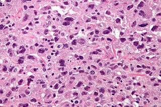
Metastasis is a pathogenic agent's spread from an initial or primary site to a different or secondary site within the host's body; the term is typically used when referring to metastasis by a cancerous tumor. The newly pathological sites, then, are metastases (mets). It is generally distinguished from cancer invasion, which is the direct extension and penetration by cancer cells into neighboring tissues.

<span class="mw-page-title-main">Small-cell carcinoma</span> Type of malignant cancer

Small-cell carcinoma is a type of highly malignant cancer that most commonly arises within the lung, although it can occasionally arise in other body sites, such as the cervix, prostate, and gastrointestinal tract. Compared to non-small cell carcinoma, small cell carcinoma is more aggressive, with a shorter doubling time, higher growth fraction, and earlier development of metastases.

<span class="mw-page-title-main">Invasive carcinoma of no special type</span> Medical condition

Invasive carcinoma of no special type, invasive breast carcinoma of no special type (IBC-NST), invasive ductal carcinoma (IDC), infiltrating ductal carcinoma (IDC) or invasive ductal carcinoma, not otherwise specified (NOS) is a disease. For international audiences this article will use "invasive carcinoma NST" because it is the preferred term of the World Health Organization (WHO).

Adjuvant therapy, also known as adjunct therapy, adjuvant care, or augmentation therapy, is a therapy that is given in addition to the primary or initial therapy to maximize its effectiveness. The surgeries and complex treatment regimens used in cancer therapy have led the term to be used mainly to describe adjuvant cancer treatments. An example of such adjuvant therapy is the additional treatment usually given after surgery where all detectable disease has been removed, but where there remains a statistical risk of relapse due to the presence of undetected disease. If known disease is left behind following surgery, then further treatment is not technically adjuvant.

<span class="mw-page-title-main">Undifferentiated pleomorphic sarcoma</span> Medical condition

Undifferentiated pleomorphic sarcoma (UPS), also termed pleomorphic myofibrosarcoma, high-grade myofibroblastic sarcoma, and high-grade myofibrosarcoma, is characterized by the World Health Organization (WHO), 2020, as a rare, poorly differentiated neoplasm, i.e. an abnormal growth of cells that have an unclear identity and/or cell of origin. WHO classified it as one of the undifferentiated/unclassified sarcomas in the category of tumors of uncertain differentiation. Sarcomas are cancers known or thought to derive from mesenchymal stem cells that typically develop in bone, muscle, fat, blood vessels, lymphatic vessels, tendons, and ligaments. More than 70 sarcoma subtypes have been described. The UPS subtype of these sarcomas consists of tumor cells that are poorly differentiated and may appear as spindle-shaped cells, histiocytes, and giant cells. UPS is considered a diagnosis that defies formal sub-classification after thorough histologic, immunohistochemical, and ultrastructural examinations fail to identify the type of cells involved.

<span class="mw-page-title-main">RANKL</span> Mammalian protein found in Homo sapiens

Receptor activator of nuclear factor kappa-Β ligand (RANKL), also known as tumor necrosis factor ligand superfamily member 11 (TNFSF11), TNF-related activation-induced cytokine (TRANCE), osteoprotegerin ligand (OPGL), and osteoclast differentiation factor (ODF), is a protein that in humans is encoded by the TNFSF11 gene.

Triple-negative breast cancer (TNBC) is any breast cancer that either lacks or shows low levels of estrogen receptor (ER), progesterone receptor (PR) and human epidermal growth factor receptor 2 (HER2) overexpression and/or gene amplification. Triple-negative is sometimes used as a surrogate term for basal-like.

<span class="mw-page-title-main">MMP2</span> Protein-coding gene in the species Homo sapiens

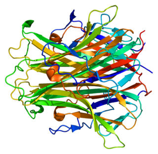
72 kDa type IV collagenase also known as matrix metalloproteinase-2 (MMP-2) and gelatinase A is an enzyme that in humans is encoded by the MMP2 gene. The MMP2 gene is located on chromosome 16 at position 12.2.

Breast cancer management takes different approaches depending on physical and biological characteristics of the disease, as well as the age, over-all health and personal preferences of the patient. Treatment types can be classified into local therapy and systemic treatment. Local therapy is most efficacious in early stage breast cancer, while systemic therapy is generally justified in advanced and metastatic disease, or in diseases with specific phenotypes.

A metastasis suppressor is a protein that acts to slow or prevent metastases from spreading in the body of an organism with cancer. Metastasis is one of the most lethal cancer processes. This process is responsible for about ninety percent of human cancer deaths. Proteins that act to slow or prevent metastases are different from those that act to suppress tumor growth. Genes for about a dozen such proteins are known in humans and other animals.

<span class="mw-page-title-main">Leptomeningeal cancer</span> Medical condition

Leptomeningeal cancer is a rare complication of cancer in which the disease spreads from the original tumor site to the meninges surrounding the brain and spinal cord. This leads to an inflammatory response, hence the alternative names neoplastic meningitis (NM), malignant meningitis, or carcinomatous meningitis. The term leptomeningeal describes the thin meninges, the arachnoid and the pia mater, between which the cerebrospinal fluid is located. The disorder was originally reported by Eberth in 1870. It is also known as leptomeningeal carcinomatosis, leptomeningeal disease (LMD), leptomeningeal metastasis, meningeal metastasis and meningeal carcinomatosis.

<span class="mw-page-title-main">Invadopodia</span>

Invadopodia are actin-rich protrusions of the plasma membrane that are associated with degradation of the extracellular matrix in cancer invasiveness and metastasis. Very similar to podosomes, invadopodia are found in invasive cancer cells and are important for their ability to invade through the extracellular matrix, especially in cancer cell extravasation. Invadopodia are generally visualized by the holes they create in ECM -coated plates, in combination with immunohistochemistry for the invadopodia localizing proteins such as cortactin, actin, Tks5 etc. Invadopodia can also be used as a marker to quantify the invasiveness of cancer cell lines in vitro using a hyaluronic acid hydrogel assay.

<span class="mw-page-title-main">Bone metastasis</span> Medical condition

Bone metastasis, or osseous metastatic disease, is a category of cancer metastases that result from primary tumor invasions into bones. Bone-originating primary tumors such as osteosarcoma, chondrosarcoma, and Ewing sarcoma are rare; the most common bone tumor is a metastasis. Bone metastases can be classified as osteolytic, osteoblastic, or both. Unlike hematologic malignancies which originate in the blood and form non-solid tumors, bone metastases generally arise from epithelial tumors and form a solid mass inside the bone. Bone metastases, especially in a state of advanced disease, can cause severe pain, characterized by a dull, constant ache with periodic spikes of incident pain.

<span class="mw-page-title-main">Brain metastasis</span> Cancer that has metastasized (spread) to the brain from another location in the body

A brain metastasis is a cancer that has metastasized (spread) to the brain from another location in the body and is therefore considered a secondary brain tumor. The metastasis typically shares a cancer cell type with the original site of the cancer. Metastasis is the most common cause of brain cancer, as primary tumors that originate in the brain are less common. The most common sites of primary cancer which metastasize to the brain are lung, breast, colon, kidney, and skin cancer. Brain metastases can occur in patients months or even years after their original cancer is treated. Brain metastases have a poor prognosis for cure, but modern treatments are allowing patients to live months and sometimes years after the diagnosis.

Neuro-oncology is the study of brain and spinal cord neoplasms, many of which are very dangerous and life-threatening. Among the malignant brain cancers, gliomas of the brainstem and pons, glioblastoma multiforme, and high-grade astrocytoma/oligodendroglioma are among the worst. In these cases, untreated survival usually amounts to only a few months, and survival with current radiation and chemotherapy treatments may extend that time from around a year to a year and a half, possibly two or more, depending on the patient's condition, immune function, treatments used, and the specific type of malignant brain neoplasm. Surgery may in some cases be curative, but, as a general rule, malignant brain cancers tend to regenerate and emerge from remission easily, especially highly malignant cases. In such cases, the goal is to excise as much of the mass and as much of the tumor margin as possible without endangering vital functions or other important cognitive abilities. The Journal of Neuro-Oncology is the longest continuously published journal in the field and serves as a leading reference to those practicing in the area of neuro-oncology.

<span class="mw-page-title-main">Vasculogenic mimicry</span>

Vasculogenic mimicry (VM) is a strategy used by tumors to ensure sufficient blood supply is brought to its cells through establishing new tumor vascularization. This process is similar to tumor angiogenesis; on the other hand vascular mimicry is unique in that this process occurs independent of endothelial cells. Vasculature is instead developed de novo by cancer cells, which under stress conditions such as hypoxia, express similar properties to stem cells, capable of differentiating to mimic the function of endothelial cells and form vasculature-like structures. The ability of tumors to develop and harness nearby vasculature is considered one of the hallmarks of cancer disease development and is thought to be closely linked to tumor invasion and metastasis. Vascular mimicry has been observed predominantly in aggressive and metastatic cancers and has been associated with negative tumor characteristics such as increased metastasis, increased tissue invasion, and overall poor outcomes for patient survival. Vascular mimicry poses a serious problem for current therapeutic strategies due to its ability to function in the presence of Anti-angiogenic therapeutic agents. In fact, such therapeutics have been found to actually drive VM formation in tumors, causing more aggressive and difficult to treat tumors to develop.

A pre-metastatic niche is an environment in a secondary organ that can be conducive to the metastasis of a primary tumor. Such a niche provides favorable conditions for growth, and eventual metastasis, in an otherwise foreign and hostile environment for the primary tumor cells. This concept demonstrated the fundamental role of the microenvironment in regulating tumor growth and metastasis. The discovery of the pre-metastatic niche has fostered new research regarding the potential treatment of metastases, including targeting myeloid derived suppressor cells, and stromal cell plasticity including fibroblasts and pericytes and perivascular smooth muscle cells and (attempts to stop the flow of vesicles from primary tumors to pre-metastatic niches in secondary organs and different combinations of microenvironment targeted therapies.

<span class="mw-page-title-main">Invasion (cancer)</span> Direct extension and penetration by cancer cells into neighboring tissues

Invasion is the process by which cancer cells directly extend and penetrate into neighboring tissues in cancer. It is generally distinguished from metastasis, which is the spread of cancer cells through the circulatory system or the lymphatic system to more distant locations. Yet, lymphovascular invasion is generally the first step of metastasis.

The host response to cancer therapy is defined as a physiological response of the non-malignant cells of the body to a specific cancer therapy. The response is therapy-specific, occurring independently of cancer type or stage.

Mammary secretory carcinoma (MSC), also termed secretory carcinoma of the breast, is a rare form of the breast cancers. MSC usually affects women but in a significant percentage of cases also occurs in men and children. Indeed, McDvitt and Stewart first described MSC in 1966 and termed it juvenile breast carcinoma because an increased number of cases were at that time diagnosed in juvenile females. MSC is the most common form of breast cancer in children, representing 80% of childhood breast cancers, although it accounts for less than 0.15% of all breast cancers.

References

  1. John’s Hopkins. "Metastatic Breast Cancer". Hopkins medicine. Retrieved 6 January 2021.
  2. 1 2 "Secondary (metastatic) breast cancer". Breast Cancer Care. Retrieved 22 October 2013.
  3. Fouad TM, Kogawa T, Liu DD, Shen Y, Masuda H, El-Zein R, et al. (July 2015). "Overall survival differences between patients with inflammatory and noninflammatory breast cancer presenting with distant metastasis at diagnosis". Breast Cancer Research and Treatment. 152 (2): 407–16. doi:10.1007/s10549-015-3436-x. PMC   4492876 . PMID   26017070.
  4. Lee YT (July 1983). "Breast carcinoma: pattern of metastasis at autopsy". Journal of Surgical Oncology. 23 (3): 175–80. doi:10.1002/jso.2930230311. PMID   6345937. S2CID   21869121.
  5. "Metastatic cancer overview". Canadian Cancer Society. Retrieved 8 September 2014.
  6. Suva LJ, Griffin RJ, Makhoul I (September 2009). "Mechanisms of bone metastases of breast cancer". Endocrine-Related Cancer. Bioscientifica. 16 (3): 703–13. doi:10.1677/ERC-09-0012. PMC   2914697 . PMID   19443538 . Retrieved 15 January 2010.
  7. "Metastatic Cancer". National Cancer Institute. 2015-05-12. Retrieved 2019-05-06.
  8. "Breast Cancer - Metastatic - Symptoms and Signs". Cancer.Net. 2017-05-19. Retrieved 2019-10-28.
  9. Macedo F, Ladeira K, Pinho F, Saraiva N, Bonito N, Pinto L, Goncalves F (March 2017). "Bone Metastases: An Overview". Oncology Reviews. 11 (1): 321. doi:10.4081/oncol.2017.321. PMC   5444408 . PMID   28584570.
  10. Berghoff AS, Preusser M (2018). "New developments in brain metastases". Therapeutic Advances in Neurological Disorders. 11: 1756286418785502. doi:10.1177/1756286418785502. PMC   6048670 . PMID   30034538.
  11. Shaffrey ME, Mut M, Asher AL, Burri SH, Chahlavi A, Chang SM, Farace E, Fiveash JB, Lang FF, Lopes MB, Markert JM, Schiff D, Siomin V, Tatter SB, Vogelbaum MA (August 2004). "Brain metastases". Current Problems in Surgery. 41 (8): 665–741. doi:10.1067/j.cpsurg.2004.06.001. PMID   15354117.
  12. Vlodavsky I, Goldshmidt O, Zcharia E, Atzmon R, Rangini-Guatta Z, Elkin M, Peretz T, Friedmann Y (April 2002). "Mammalian heparanase: involvement in cancer metastasis, angiogenesis and normal development". Seminars in Cancer Biology. 12 (2): 121–9. doi:10.1006/scbi.2001.0420. PMID   12027584.
  13. MetaBre Archived January 6, 2008, at the Wayback Machine
  14. Saad S, Gottlieb DJ, Bradstock KF, Overall CM, Bendall LJ (January 2002). "Cancer cell-associated fibronectin induces release of matrix metalloproteinase-2 from normal fibroblasts". Cancer Research. American Association for Cancer Research. 62 (1): 283–9. PMID   11782389 . Retrieved January 15, 2010.
  15. Nathoo N, Chahlavi A, Barnett GH, Toms SA (March 2005). "Pathobiology of brain metastases". Journal of Clinical Pathology. 58 (3): 237–42. doi:10.1136/jcp.2003.013623. PMC   1770599 . PMID   15735152.
  16. Bos PD, Zhang XH, Nadal C, Shu W, Gomis RR, Nguyen DX, Minn AJ, van de Vijver MJ, Gerald WL, Foekens JA, Massagué J (June 2009). "Genes that mediate breast cancer metastasis to the brain". Nature. 459 (7249): 1005–9. Bibcode:2009Natur.459.1005B. doi:10.1038/nature08021. PMC   2698953 . PMID   19421193.
  17. 1 2 3 Costelloe CM, Rohren EM, Madewell JE, Hamaoka T, Theriault RL, Yu TK, Lewis VO, Ma J, Stafford RJ, Tari AM, Hortobagyi GN, Ueno NT (June 2009). "Imaging bone metastases in breast cancer: techniques and recommendations for diagnosis". The Lancet. Oncology. 10 (6): 606–14. doi:10.1016/S1470-2045(09)70088-9. PMID   19482249.
  18. Tosello G, Torloni MR, Mota BS, Neeman T, Riera R (March 2018). "Breast surgery for metastatic breast cancer". The Cochrane Database of Systematic Reviews. 2018 (3): CD011276. doi:10.1002/14651858.CD011276.pub2. PMC   6494198 . PMID   29542106.
  19. Butters DJ, Ghersi D, Wilcken N, Kirk SJ, Mallon PT (November 2010). "Addition of drug/s to a chemotherapy regimen for metastatic breast cancer". The Cochrane Database of Systematic Reviews. 2021 (11): CD003368. doi:10.1002/14651858.cd003368.pub3. PMC   7154379 . PMID   21069675.
  20. 1 2 Dear RF, McGeechan K, Jenkins MC, Barratt A, Tattersall MH, Wilcken N (December 2013). "Combination versus sequential single agent chemotherapy for metastatic breast cancer". The Cochrane Database of Systematic Reviews. 2021 (12): CD008792. doi:10.1002/14651858.cd008792.pub2. PMC   8094913 . PMID   24347031.
  21. 1 2 Ghersi D, Willson ML, Chan MM, Simes J, Donoghue E, Wilcken N (June 2015). "Taxane-containing regimens for metastatic breast cancer". The Cochrane Database of Systematic Reviews. 2015 (6): CD003366. doi:10.1002/14651858.cd003366.pub3. PMC   6464903 . PMID   26058962.
  22. Cancer, Cleveland Clinic. "Kadcyla - Chemotherapy Drug Information - Chemocare.com". Chemocare.com. Retrieved 21 March 2017.
  23. 1 2 3 Egger SJ, Willson ML, Morgan J, Walker HS, Carrick S, Ghersi D, Wilcken N (June 2017). "Platinum-containing regimens for metastatic breast cancer". The Cochrane Database of Systematic Reviews. 6 (8): CD003374. doi:10.1002/14651858.cd003374.pub4. PMC   6481538 . PMID   28643430.
  24. Shaaban, Saeed M.; Gaber, Zeinab; Semary, Samah; Dewidar, Ahmad M. (2023-03-31). "Impact of Vitamin B12 on outcome of Early Stage Luminal A and B Breast Cancer, single center experience". Medical and Pharmaceutical Journal. 2 (1). ISSN   2957-6067.
  25. Lord S, Ghersi D, Gattellari M, Wortley S, Wilcken N, Simes J (October 2004). "Antitumour antibiotic containing regimens for metastatic breast cancer". The Cochrane Database of Systematic Reviews. 2021 (4): CD003367. doi:10.1002/14651858.cd003367.pub2. PMC   6999796 . PMID   15495049.
  26. Garsa, Adam; Jang, Julie K.; Baxi, Sangita; Chen, Christine; Akinniranye, Olamigoke; Hall, Owen; Larkin, Jody; Motala, Aneesa; Newberry, Sydne (2021-06-09). Radiation Therapy for Brain Metasases (Report). Agency for Healthcare Research and Quality (AHRQ). doi:10.23970/ahrqepccer242.
  27. http url www.emedicine.net
  28. 1 2 3 4 5 Moriarty, Colleen (July 1, 2019). "Natural Cancer 'Cures': What Are the Risks?". Yale Medicine. Retrieved 29 June 2021.
  29. 1 2 "Complementary and Integrative Therapies". Susan G. Komen Foundation. Retrieved 29 June 2021.
  30. "Complementary and Alternative Therapies for Breast Cancer". The Bridge. July 15, 2020. Retrieved 29 June 2021.
  31. "Alternative cancer treatments: 10 options to consider". Mayo Clinic. Retrieved 29 June 2021.
  32. von Bubnoff, Andreas (January 29, 2021). "Could fasting or ketone diets help quash cancer?". Knowable Magazine. doi: 10.1146/knowable-012821-2 .{{cite journal}}: CS1 maint: date and year (link)
  33. Johnson, Skyler B.; Park, Henry S.; Gross, Cary P.; Yu, James B. (2018). "Use of Alternative Medicine for Cancer and Its Impact on Survival". JNCI: Journal of the National Cancer Institute. 110 (1): 121–124. doi: 10.1093/jnci/djx145 . PMID   28922780.
  34. "News: Loyola University Chicago Health Sciences Division".
  35. Weil RJ, Palmieri DC, Bronder JL, Stark AM, Steeg PS (October 2005). "Breast cancer metastasis to the central nervous system". The American Journal of Pathology. 167 (4): 913–20. doi:10.1016/S0002-9440(10)61180-7. PMC   1603675 . PMID   16192626.