Mu wave

Last updated
One second sample of an EEG alpha oscillations . This rhythm occurs at frequencies similar to the mu rhythm, although alpha oscillations are detected over a different part of the brain. Eeg alpha.svg
One second sample of an EEG alpha oscillations . This rhythm occurs at frequencies similar to the mu rhythm, although alpha oscillations are detected over a different part of the brain.
The left motor cortex, or BA4, is highlighted in green on this left lateral view of the brain. This is the area over which mu rhythms are detected bilaterally. Motor cortex.PNG
The left motor cortex, or BA4, is highlighted in green on this left lateral view of the brain. This is the area over which mu rhythms are detected bilaterally.

The sensorimotor mu rhythm, also known as mu wave, comb or wicket rhythms or arciform rhythms, are synchronized patterns of electrical activity involving large numbers of neurons, probably of the pyramidal type, in the part of the brain that controls voluntary movement. [1] These patterns as measured by electroencephalography (EEG), magnetoencephalography (MEG), or electrocorticography (ECoG), repeat at a frequency of 7.5–12.5 (and primarily 9–11) Hz, and are most prominent when the body is physically at rest. [1] Unlike the alpha wave, which occurs at a similar frequency over the resting visual cortex at the back of the scalp, the mu rhythm is found over the motor cortex, in a band approximately from ear to ear. People suppress mu rhythms when they perform motor actions or, with practice, when they visualize performing motor actions. This suppression is called desynchronization of the wave because EEG wave forms are caused by large numbers of neurons firing in synchrony. The mu rhythm is even suppressed when one observes another person performing a motor action or an abstract motion with biological characteristics. Researchers such as V. S. Ramachandran and colleagues have suggested that this is a sign that the mirror neuron system is involved in mu rhythm suppression, [2] [3] although others disagree. [4]

Contents

The mu rhythm is of interest to a variety of scholars. Scientists who study neural development are interested in the details of the development of the mu rhythm in infancy and childhood and its role in learning. [5] Since a group of researchers believe that autism spectrum disorder (ASD) is strongly influenced by an altered mirror neuron system [2] [6] [7] and that mu rhythm suppression is a downstream indication of mirror neuron activity, [3] many of these scientists have kindled a more popular interest in investigating the mu wave in people with ASD. Assorted investigators are also in the process of using mu rhythms to develop a new technology: the brain–computer interface (BCI). With the emergence of BCI systems, clinicians hope to give the severely physically disabled population new methods of communication and a means to manipulate and navigate their environments. [8]

Mirror neurons

The mirror neuron system consists of a class of neurons that was first studied in the 1990s in macaque monkeys. [7] Studies have found sets of neurons that fire when these monkeys perform simple tasks and also when the monkeys view others performing the same simple tasks. [9] This suggests they play a role in mapping others' movements into the brain without actually physically performing the movements. These sets of neurons are called mirror neurons and together make up the mirror neuron system. Mu waves are suppressed when these neurons fire, a phenomenon which allows researchers to study mirror neuron activity in humans. [10] There is evidence that mirror neurons exist in humans as well as in non-human animals. The right fusiform gyrus, left inferior parietal lobule, right anterior parietal cortex, and left inferior frontal gyrus are of particular interest. [7] [11] [12] Some researchers believe that mu wave suppression can be a consequence of mirror neuron activity throughout the brain, and represents a higher-level integrative processing of mirror neuron activity. [3] Tests in both monkeys (using invasive measuring techniques) and humans (using EEG and fMRI) have found that these mirror neurons not only fire during basic motor tasks, but also have components that deal with intention. [13] There is evidence of an important role for mirror neurons in humans, and mu waves may represent a high level coordination of those mirror neurons. [3]

Development

A fruitful conceptualization of mu waves in pediatric use is that mu wave suppression is a representation of activity going on in the world, and is detectable in the frontal and parietal networks. [3] A resting oscillation becomes suppressed during the observation of sensory information such as sounds or sights, usually within the frontoparietal (motor) cortical region. [3] The mu wave is detectable during infancy as early as four to six months, when the peak frequency the wave reaches can be as low as 5.4 Hz. [5] [14] There is a rapid increase in peak frequency in the first year of life, [14] and by age two frequency typically reaches 7.5 Hz. [11] The peak frequency of the mu wave increases with age until maturation into adulthood, when it reaches its final and stable frequency of 8–13 Hz. [5] [11] [14] These varying frequencies are measured as activity around the central sulcus, within the Rolandic cortex. [3]

Mu waves are thought to be indicative of an infant’s developing ability to imitate. This is important because the ability to imitate plays a vital role in the development of motor skills, tool use, and understanding causal information through social interaction. [11] Mimicking is integral in the development of social skills and understanding nonverbal cues. [5] Causal relationships can be made through social learning without requiring experience firsthand. In action execution, mu waves are present in both infants and adults before and after the execution of a motor task and its accompanying desynchronization. While executing a goal-oriented action, however, infants exhibit a higher degree of desynchronization than do adults. Just as with an action execution, during action observation infants’ mu waves not only show a desynchronization, but show a desynchronization greater in degree than the one evidenced in adults. [5] This tendency for changes in degree of desynchronization, rather than actual changes in frequency, becomes the measure for mu wave development throughout adulthood, although the most changes take place during the first year of life. [14] Understanding the mechanisms that are shared between action perception and execution in the earliest years of life has implications for language development. Learning and understanding through social interaction comes from imitating movements as well as vowel sounds. Sharing the experience of attending to an object or event with another person can be a powerful force in the development of language. [15]

Autism

Autism is a disorder that is associated with social and communicative deficits. A single cause of autism has yet to be identified, but the mu wave and mirror neuron system have been studied specifically for their role in the disorder. In a typically developing individual, the mirror neuron system responds when they either watch someone perform a task or perform the task themself. In individuals with autism, mirror neurons become active (and consequently mu waves are suppressed) only when the individual performs the task themself. [6] [16] This finding has led some scientists, notably V. S. Ramachandran and colleagues, to view autism as disordered understanding of other individuals' intentions and goals due to problems with the mirror neuron system. [7] This deficiency would explain the difficulty people with autism have in communicating with and understanding others. While most studies of the mirror neuron system and mu waves in people with autism have focused on simple motor tasks, some scientists speculate that these tests can be expanded to show that problems with the mirror neuron system underlie overarching cognitive and social deficits. [2] [6]

fMRI activation magnitudes in the inferior frontal gyrus increase with age in people with autism, but not in typically developing individuals. Furthermore, greater activation was associated with greater amounts of eye contact and better social functioning. [17] Scientists believe the inferior frontal gyrus is one of the main neural correlates with the mirror neuron system in humans and is often related to deficits associated with autism. [12] These findings suggest that the mirror neuron system may not be non-functional in individuals with autism, but simply abnormal in its development. This information is significant to the present discussion because mu waves may be integrating different areas of mirror neuron activity in the brain. [3] Other studies have assessed attempts to consciously stimulate the mirror neuron system and suppress mu waves using neurofeedback (a type of biofeedback given through computers that analyze real time recordings of brain activity, in this case EEGs of mu waves). This type of therapy is still in its early phases of implementation for individuals with autism, and has conflicting forecasts for success. [18] [19]

Brain–computer interfaces

Brain–computer interfaces (BCIs) are a developing technology that clinicians hope will one day bring more independence and agency to the severely physically disabled. This technology has the potential to help include people with near-total or total paralysis, such as those with tetraplegia (quadriplegia) or advanced amyotrophic lateral sclerosis (ALS); BCIs are intended to help them to communicate or even move objects such as motorized wheelchairs, neuroprostheses, or robotic grasping tools. [8] [20] Few of these technologies are currently in regular use by people with disabilities, but a diverse array are in development at an experimental level. [8] [21] [22] One type of BCI uses event-related desynchronization (ERD) of the mu wave in order to control the computer. [8] This method of monitoring brain activity takes advantage of the fact that when a group of neurons is at rest they tend to fire in synchrony with each other. When a participant is cued to imagine movement (an "event"), the resulting desynchronization (the group of neurons that was firing in synchronous waves now firing in complex and individualized patterns) can be reliably detected and analyzed by a computer. Users of such an interface are trained in visualizing movements, typically of the foot, hand, and/or tongue, which are each in different locations on the cortical homunculus and thus distinguishable by an electroencephalograph (EEG) or electrocorticograph (ECoG) recording of electrical activity over the motor cortex. [8] [21] In this method, computers monitor for a typical pattern of mu wave ERD contralateral to the visualized movement combined with event-related synchronization (ERS) in the surrounding tissue. [21] This paired pattern intensifies with training, [8] [21] [22] [23] and the training increasingly takes the form of games, some of which utilize virtual reality. [8] [21] [23] Some researchers have found that the feedback from virtual reality games is particularly effective in giving the user tools to improve control of his or her mu wave patterns. [8] [23] The ERD method can be combined with one or more other methods of monitoring the brain's electrical activity to create hybrid BCIs, which often offer more flexibility than a BCI that uses any single monitoring method. [8] [21]

History

Mu waves have been studied since the 1930s, and are referred to as the wicket rhythm because the rounded EEG waves resemble croquet wickets. In 1950, Henri Gastaut and his coworkers reported desynchronization of these waves not only during active movements of their subjects, but also while the subjects observed actions executed by someone else. [24] [25] These results were later confirmed by additional research groups, [26] [27] [28] [29] including a study using subdural electrode grids in epileptic patients. [30] The latter study showed mu suppression while the patients observed moving body parts in somatic areas of the cortex that corresponded to the body part moved by the actor. Further studies have shown that the mu waves can also be desynchronized by imagining actions [31] [32] and by passively viewing point-light biological motion. [33]

See also

Brain waves

Related Research Articles

<span class="mw-page-title-main">Delta wave</span> High amplitude low frequency brain wave

Delta waves are high amplitude neural oscillations with a frequency between 0.5 and 4 hertz. Delta waves, like other brain waves, can be recorded with electroencephalography (EEG) and are usually associated with the deep stage 3 of NREM sleep, also known as slow-wave sleep (SWS), and aid in characterizing the depth of sleep. Suppression of delta waves leads to inability of body rejuvenation, brain revitalization and poor sleep.

<span class="mw-page-title-main">Brain–computer interface</span> Direct communication pathway between an enhanced or wired brain and an external device

A brain–computer interface (BCI), sometimes called a brain–machine interface (BMI), is a direct communication pathway between the brain's electrical activity and an external device, most commonly a computer or robotic limb. BCIs are often directed at researching, mapping, assisting, augmenting, or repairing human cognitive or sensory-motor functions. Implementations of BCIs range from non-invasive and partially invasive to invasive, based on how close electrodes get to brain tissue.

A mirror neuron is a neuron that fires both when an animal acts and when the animal observes the same action performed by another. Thus, the neuron "mirrors" the behavior of the other, as though the observer were itself acting. Such neurons have been directly observed in human and primate species, and in birds.

<span class="mw-page-title-main">Reticular formation</span> Spinal trigeminal nucleus

The reticular formation is a set of interconnected nuclei that are located throughout the brainstem. It is not anatomically well defined, because it includes neurons located in different parts of the brain. The neurons of the reticular formation make up a complex set of networks in the core of the brainstem that extend from the upper part of the midbrain to the lower part of the medulla oblongata. The reticular formation includes ascending pathways to the cortex in the ascending reticular activating system (ARAS) and descending pathways to the spinal cord via the reticulospinal tracts.

A gamma wave or gamma Rhythm is a pattern of neural oscillation in humans with a frequency between 25 and 140 Hz, the 40-Hz point being of particular interest. Gamma rhythms are correlated with large scale brain network activity and cognitive phenomena such as working memory, attention, and perceptual grouping, and can be increased in amplitude via meditation or neurostimulation. Altered gamma activity has been observed in many mood and cognitive disorders such as Alzheimer's disease, epilepsy, and schizophrenia.

The sensorimotor rhythm (SMR) is a brain wave. It is an oscillatory idle rhythm of synchronized electric brain activity. It appears in spindles in recordings of EEG, MEG, and ECoG over the sensorimotor cortex. For most individuals, the frequency of the SMR is in the range of 13 to 15 Hz.

Beta waves, or beta rhythm, are a neural oscillation (brainwave) in the brain with a frequency range of between 12.5 and 30 Hz. Beta waves can be split into three sections: Low Beta Waves ; Beta Waves ; and High Beta Waves. Beta states are the states associated with normal waking consciousness.

Alpha waves, or the alpha rhythm, are neural oscillations in the frequency range of 8–12 Hz likely originating from the synchronous and coherent electrical activity of thalamic pacemaker cells in humans. Historically, they are also called "Berger's waves" after Hans Berger, who first described them when he invented the EEG in 1924.

Brainwave entrainment, also referred to as brainwave synchronization or neural entrainment, refers to the observation that brainwaves will naturally synchronize to the rhythm of periodic external stimuli, such as flickering lights, speech, music, or tactile stimuli.

<span class="mw-page-title-main">Neural oscillation</span> Brainwaves, repetitive patterns of neural activity in the central nervous system

Neural oscillations, or brainwaves, are rhythmic or repetitive patterns of neural activity in the central nervous system. Neural tissue can generate oscillatory activity in many ways, driven either by mechanisms within individual neurons or by interactions between neurons. In individual neurons, oscillations can appear either as oscillations in membrane potential or as rhythmic patterns of action potentials, which then produce oscillatory activation of post-synaptic neurons. At the level of neural ensembles, synchronized activity of large numbers of neurons can give rise to macroscopic oscillations, which can be observed in an electroencephalogram. Oscillatory activity in groups of neurons generally arises from feedback connections between the neurons that result in the synchronization of their firing patterns. The interaction between neurons can give rise to oscillations at a different frequency than the firing frequency of individual neurons. A well-known example of macroscopic neural oscillations is alpha activity.

Theta waves generate the theta rhythm, a neural oscillation in the brain that underlies various aspects of cognition and behavior, including learning, memory, and spatial navigation in many animals. It can be recorded using various electrophysiological methods, such as electroencephalogram (EEG), recorded either from inside the brain or from electrodes attached to the scalp.

<span class="mw-page-title-main">Electrocorticography</span>

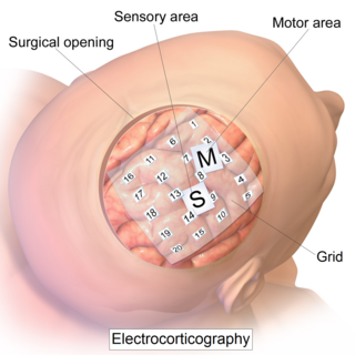
Electrocorticography (ECoG), or intracranial electroencephalography (iEEG), is a type of electrophysiological monitoring that uses electrodes placed directly on the exposed surface of the brain to record electrical activity from the cerebral cortex. In contrast, conventional electroencephalography (EEG) electrodes monitor this activity from outside the skull. ECoG may be performed either in the operating room during surgery or outside of surgery. Because a craniotomy is required to implant the electrode grid, ECoG is an invasive procedure.

The phi complex is a brain rhythm in the awake human brain that appears to serve various social functions. Phi is one of several brain rhythms in the awake human brain that coordinate human behavior. "Phi" operates in the 10-Hz band, and is located above the right centro-parietal cortex. It consists of two components, one favoring independent behaviors, and the other favoring interpersonal coordination between people.

Recurrent thalamo-cortical resonance is an observed phenomenon of oscillatory neural activity between the thalamus and various cortical regions of the brain. It is proposed by Rodolfo Llinas and others as a theory for the integration of sensory information into the whole of perception in the brain. Thalamocortical oscillation is proposed to be a mechanism of synchronization between different cortical regions of the brain, a process known as temporal binding. This is possible through the existence of thalamocortical networks, groupings of thalamic and cortical cells that exhibit oscillatory properties.

Ponto-geniculo-occipital waves or PGO waves are distinctive wave forms of propagating activity between three key brain regions: the pons, lateral geniculate nucleus, and occipital lobe; specifically, they are phasic field potentials. These waves can be recorded from any of these three structures during and immediately before REM sleep. The waves begin as electrical pulses from the pons, then move to the lateral geniculate nucleus residing in the thalamus, and end in the primary visual cortex of the occipital lobe. The appearances of these waves are most prominent in the period right before REM sleep, albeit they have been recorded during wakefulness as well. They are theorized to be intricately involved with eye movement of both wake and sleep cycles in many different animals.

<span class="mw-page-title-main">Electroencephalography</span> Electrophysiological monitoring method to record electrical activity of the brain

Electroencephalography (EEG) is a method to record an electrogram of the electrical activity on the scalp that has been shown to represent the macroscopic activity of the surface layer of the brain underneath. It is typically non-invasive, with the electrodes placed along the scalp. Electrocorticography, involving invasive electrodes, is sometimes called "intracranial EEG".

The neuroscience of music is the scientific study of brain-based mechanisms involved in the cognitive processes underlying music. These behaviours include music listening, performing, composing, reading, writing, and ancillary activities. It also is increasingly concerned with the brain basis for musical aesthetics and musical emotion. Scientists working in this field may have training in cognitive neuroscience, neurology, neuroanatomy, psychology, music theory, computer science, and other relevant fields.

<span class="mw-page-title-main">Brain activity and meditation</span>

Meditation and its effect on brain activity and the central nervous system became a focus of collaborative research in neuroscience, psychology and neurobiology during the latter half of the 20th century. Research on meditation sought to define and characterize various practices. Meditation's effect on the brain can be broken up into two categories: state changes and trait changes, respectively alterations in brain activities during the act of meditating and changes that are the outcome of long-term practice.

Pain empathy is a specific variety of empathy that involves recognizing and understanding another person's pain.

<span class="mw-page-title-main">Fetal EEG</span>

Fetal electroencephalography, also known as prenatal EEG includes any recording of electrical fluctuations arising from the brain of a fetus. Doctors and scientists use EEGs to detect and characterize brain activity, such as sleep states, potential seizures, or levels of a coma. EEG captures the electrical activity in the vicinity of the recording electrodes. The majority of the neural electrical activity arises from the flow of current from the cell bodies of pyramidal neurons to their apical dendrites, which become depolarized by excitatory inputs from other neurons. To record the most accurate signals, scientists try to minimize the distance between the recording electrode and the neural activity that they want to detect. Given the difficulty of attaching electrodes to a fetus inside a uterus, doctors and scientists use a variety of techniques to record fetal brain activity.

References

  1. 1 2 Amzica, Florin; Fernando Lopes da Silva (2010). "Cellular Substrates of Brain Rhythms". In Schomer, Donald L.; Fernando Lopes da Silva (eds.). Niedermeyer's Electroencephalography: Basic Principles, Clinical Applications, and Related Fields (6th ed.). Philadelphia, Pa.: Lippincott Williams & Wilkins. pp. 33–63. ISBN   978-0-7817-8942-4.
  2. 1 2 3 Oberman, Lindsay M.; Edward M. Hubbarda; Eric L. Altschulera; Vilayanur S. Ramachandran; Jaime A. Pineda (July 2005). "EEG evidence for mirror neuron dysfunction in autism spectrum disorders". Cognitive Brain Research. 24 (2): 190–198. doi:10.1016/j.cogbrainres.2005.01.014. PMID   15993757.
  3. 1 2 3 4 5 6 7 8 Pineda, Jaime A. (1 December 2005). "The functional significance of mu rhythms: Translating "seeing" and "hearing" into "doing"". Brain Research Reviews. 50 (1): 57–68. doi:10.1016/j.brainresrev.2005.04.005. PMID   15925412. S2CID   16278949.
  4. Churchland, Patricia (2011). Braintrust: What Neuroscience Tells Us About Morality . Princeton, NJ: Princeton University Press. p.  156. ISBN   978-0-691-13703-2.
  5. 1 2 3 4 5 Nyström, Pär; Ljunghammar, Therese; Rosander, Kerstin; Von Hofsten, Claes (2011). "Using mu rhythm desynchronization to measure mirror neuron activity in infants". Developmental Science. 14 (2): 327–335. doi:10.1111/j.1467-7687.2010.00979.x. PMID   22213903.
  6. 1 2 3 Bernier, R.; Dawson, G.; Webb, S.; Murias, M. (2007). "EEG mu rhythm and imitation impairments in individuals with autism spectrum disorder". Brain and Cognition. 64 (3): 228–237. doi:10.1016/j.bandc.2007.03.004. PMC   2709976 . PMID   17451856.
  7. 1 2 3 4 Williams, Justin H.G.; Waiter, Gordon D.; Gilchrist, Anne; Perrett, David I.; Murray, Alison D.; Whiten, Andrew (1 January 2006). "Neural mechanisms of imitation and 'mirror neuron' functioning in autistic spectrum disorder" (PDF). Neuropsychologia. 44 (4): 610–621. doi:10.1016/j.neuropsychologia.2005.06.010. PMID   16140346. S2CID   39014561. Archived from the original (PDF) on 11 June 2012. Retrieved 6 January 2013.
  8. 1 2 3 4 5 6 7 8 9 Pfurtscheller, Gert; Christa Neuper (2010). "EEG-Based Brain–Computer Interfaces". In Schomer, Donald L.; Fernando H. Lopes da Silva (eds.). Niedermeyer's Electroencephalography: Basic Principles, Clinical Applications, and Related Fields (6th ed.). Philadelphia, Pa.: Lippincott Williams & Wilkins. pp. 1227–1236. ISBN   978-0-7817-8942-4.
  9. di Pellegrino, G.; Fadiga, L.; Fogassi, L.; Gallese, F.; Rizzolatti, G. (1992). "Understanding motor events: A neurophysiological study". Experimental Brain Research. 91 (1): 176–180. doi:10.1007/bf00230027. PMID   1301372. S2CID   206772150.
  10. Rizzolatti, G; Fogassi, L; Gallese, V (September 2001). "Neurophysiological mechanisms underlying the understanding and imitation of action". Nature Reviews. Neuroscience. 2 (9): 661–70. doi:10.1038/35090060. PMID   11533734. S2CID   6792943.
  11. 1 2 3 4 Marshall, Peter J.; Meltzoff, Andrew N. (2011). "Neural mirroring systems: Exploring the EEG mu rhythm in human infancy". Developmental Cognitive Neuroscience. 1 (2): 110–123. doi:10.1016/j.dcn.2010.09.001. PMC   3081582 . PMID   21528008.
  12. 1 2 Keuken, M.C.; Hardie, A.; Dorn, B. T.; Dev, S.; Paulus, M.P.; Jonas, K.J.; Den Wildenberg, W.P.; Pineda, J.A. (April 2011). "The role of the left inferior frontal gyrus in social perception: an rTMS study". Brain Research. 1383: 196–205. doi:10.1016/j.brainres.2011.01.073. PMID   21281612. S2CID   16125324.
  13. Sinigaglia, C; Rizzolatti, G (March 2011). "Through the looking glass: self and others". Consciousness and Cognition. 20 (1): 64–74. doi:10.1016/j.concog.2010.11.012. PMID   21220203. S2CID   21955734.
  14. 1 2 3 4 Berchicci, M.; Zhang, T.; Romero, L.; Peters, A.; Annett, R.; Teuscher, U.; Bertollo, M.; Okada, Y.; Stephen, J.; Comani, S. (21 July 2011). "Development of Mu Rhythm in Infants and Preschool Children". Developmental Neuroscience. 33 (2): 130–143. doi:10.1159/000329095. PMC   3221274 . PMID   21778699.
  15. Meltzoff, A. N.; Kuhl, P. K.; Movellan, J.; Sejnowski, T. J. (17 July 2009). "Foundations for a New Science of Learning". Science. 325 (5938): 284–288. Bibcode:2009Sci...325..284M. doi:10.1126/science.1175626. PMC   2776823 . PMID   19608908.
  16. Oberman, L.M.; Pineda, J. A.; Ramachandran, V.S. (2006). "The human mirror neuron system: A link between action observation and social skills". Social Cognitive and Affective Neuroscience. 2 (1): 62–66. doi:10.1093/scan/nsl022. PMC   2555434 . PMID   18985120.
  17. Bastiaansen, JA; Thioux, M; Nanetti, L; van der Gaag, C; Ketelaars, C; Minderaa, R; Keysers, C (May 1, 2011). "Age-related increase in inferior frontal gyrus activity and social functioning in autism spectrum disorder". Biological Psychiatry. 69 (9): 832–838. doi:10.1016/j.biopsych.2010.11.007. PMID   21310395. S2CID   32646657.
  18. Holtmann, Martin; Steiner, Sabina; Hohmann, Sarah; Poustka, Luise; Banaschewski, Tobias; Bölte, Sven (November 2011) [14 July 2011]. "Neurofeedback in autism spectrum disorders". Developmental Medicine & Child Neurology. 53 (11): 986–993. doi: 10.1111/j.1469-8749.2011.04043.x . PMID   21752020. S2CID   40250064.
  19. Coben, Robert; Linden, Michael; Myers, Thomas E. (March 2010) [24 Oct 2009]. "Neurofeedback for Autistic Spectrum Disorder: A Review of the Literature". Applied Psychophysiology and Biofeedback. 35 (1): 83–105. doi:10.1007/s10484-009-9117-y. PMID   19856096. S2CID   8425504.
  20. Machado, S; Araújo, F; Paes, F; Velasques, B; Cunha, M; Budde, H; Basile, LF; Anghinah, R; Arias-Carrión, O; Cagy, M; Piedade, R; de Graaf, TA; Sack, AT; Ribeiro, P (2010). "EEG-based brain–computer interfaces: an overview of basic concepts and clinical applications in neurorehabilitation". Reviews in the Neurosciences. 21 (6): 451–68. doi:10.1515/REVNEURO.2010.21.6.451. PMID   21438193. S2CID   24562098.
  21. 1 2 3 4 5 6 Pfurtscheller, Gert; McFarland, Dennis J. (2012). "BCIs that use sensorimotor rhythms". In Wolpaw, Jonathan R.; Wolpaw, Elizabeth Winter (eds.). Brain–Computer Interfaces: Principles and Practice. Oxford: Oxford University Press. pp. 227–240. ISBN   9780195388855.
  22. 1 2 Leuthardt, Eric C.; Schalk, Gerwin; Roland, Jarod; Rouse, Adam; Moran, Daniel W. (2009). "Evolution of brain–computer interfaces: going beyond classic motor physiology". Neurosurgical Focus. 27 (1): E4. doi:10.3171/2009.4.FOCUS0979. PMC   2920041 . PMID   19569892.
  23. 1 2 3 Allison, B Z; Leeb, R; Brunner, C; Müller-Putz, G R; Bauernfeind, G; Kelly, J W; Neuper, C (February 2012) [7 Dec 2011]. "Toward smarter BCIs: extending BCIs through hybridization and intelligent control". Journal of Neural Engineering. 9 (1): 013001. Bibcode:2012JNEng...9a3001A. doi:10.1088/1741-2560/9/1/013001. PMID   22156029.
  24. Cohen-Seat, G.; Gastaut, H.; Faure, J. & Heuyer, G. (1954). "Etudes experimentales de l'activite nerveuse pendant la projection cinematographique". Rev. Int. Filmologie. 5: 7–64.
  25. Gastaut, H. J. & Bert, J. (1954). "EEG changes during cinematographic presentation". Electroencephalogr. Clin. Neurophysiol. 6 (3): 433–444. doi:10.1016/0013-4694(54)90058-9. PMID   13200415.
  26. Cochin, S.; Barthelemy, C.; Lejeune, B.; Roux, S. & Martineau, J. (1998). "Perception of motion and qEEG activity in human adults". Electroencephalogr Clin Neurophysiol. 107 (4): 287–295. doi:10.1016/S0013-4694(98)00071-6. PMID   9872446.
  27. Cochin, S.; Barthelemy, C.; Roux, S. & Martineau, J. (1999). "Observation and execution of movement: similarities demonstrated by quantified electroencephalography" (PDF). Eur J Neurosci. 11 (5): 1839–1842. doi:10.1046/j.1460-9568.1999.00598.x. PMID   10215938. S2CID   5825692.
  28. Muthukumaraswamy, S. D.; Johnson, B. W. & McNair, N. A. (2004). "Mu rhythm modulation during observation of an object-directed grasp". Brain Res Cogn Brain Res. 19 (2): 195–201. doi:10.1016/j.cogbrainres.2003.12.001. PMID   15019715.
  29. Arroyo, S.; Lesser, R. P.; Gordon, B.; Uematsu, S.; Jackson, D. & Webber, R. (1993). "Functional significance of the mu rhythm of human cortex: an electrophysiologic study with subdural electrodes". Electroencephalography and Clinical Neurophysiology. 87 (3): 76–87. doi:10.1016/0013-4694(93)90114-B. PMID   7691544.
  30. Pfurtscheller, G.; Brunner, C.; Schlogl, A. & Lopes da Silva, F. H. (2006). "Mu rhythm (de)synchronization and EEG single-trial classification of different motor imagery tasks". NeuroImage. 31 (1): 153–159. doi:10.1016/j.neuroimage.2005.12.003. PMID   16443377. S2CID   21387597.
  31. Pineda, J. A.; Allison, B. Z. & Vankov, A. (2000). "The effects of self-movement, observation, and imagination on mu rhythms and readiness potentials (RP's): toward a brain–computer interface (BCI)" (PDF). IEEE Trans Rehabil Eng. 8 (2): 219–222. doi:10.1109/86.847822. PMID   10896193. S2CID   17015946.
  32. Ulloa, E. R. & Pineda, J. A. (2007). "Recognition of point-light biological motion: mu rhythms and mirror neuron activity". Behav Brain Res. 183 (2): 188–194. doi:10.1016/j.bbr.2007.06.007. PMID   17658625. S2CID   13428201.
  33. Hobson, HM; Bishop, DV (March 2017). "The interpretation of mu suppression as an index of mirror neuron activity: past, present and future". Royal Society Open Science. 4 (3): 160662. Bibcode:2017RSOS....460662H. doi:10.1098/rsos.160662. PMC   5383811 . PMID   28405354.