Electrocorticography

Last updated
Electrocorticography
Intracranial electrode grid for electrocorticography.png
Intracranial electrode grid for electrocorticography.
Synonyms Intracranial electroencephalography
Purposerecord electrical activity from the cerebral cortex.(invasive)

Electrocorticography (ECoG), a type of intracranial electroencephalography (iEEG), is a type of electrophysiological monitoring that uses electrodes placed directly on the exposed surface of the brain to record electrical activity from the cerebral cortex. In contrast, conventional electroencephalography (EEG) electrodes monitor this activity from outside the skull. ECoG may be performed either in the operating room during surgery (intraoperative ECoG) or outside of surgery (extraoperative ECoG). Because a craniotomy (a surgical incision into the skull) is required to implant the electrode grid, ECoG is an invasive procedure.

Contents

History

ECoG was pioneered in the early 1950s by Wilder Penfield and Herbert Jasper, neurosurgeons at the Montreal Neurological Institute. [1] The two developed ECoG as part of their groundbreaking Montreal procedure, a surgical protocol used to treat patients with severe epilepsy. The cortical potentials recorded by ECoG were used to identify epileptogenic zones – regions of the cortex that generate epileptic seizures. These zones would then be surgically removed from the cortex during resectioning, thus destroying the brain tissue where epileptic seizures had originated. Penfield and Jasper also used electrical stimulation during ECoG recordings in patients undergoing epilepsy surgery under local anesthesia. [2] This procedure was used to explore the functional anatomy of the brain, mapping speech areas and identifying the somatosensory and somatomotor cortex areas to be excluded from surgical removal. A doctor named Robert Galbraith Heath was also an early researcher of the brain at the Tulane University School of Medicine. [3] [4]

Electrophysiological basis

ECoG signals are composed of synchronized postsynaptic potentials (local field potentials), recorded directly from the exposed surface of the cortex. The potentials occur primarily in cortical pyramidal cells, and thus must be conducted through several layers of the cerebral cortex, cerebrospinal fluid (CSF), pia mater, and arachnoid mater before reaching subdural recording electrodes placed just below the dura mater (outer cranial membrane). However, to reach the scalp electrodes of a conventional electroencephalogram (EEG), electrical signals must also be conducted through the skull, where potentials rapidly attenuate due to the low conductivity of bone. For this reason, the spatial resolution of ECoG is much higher than EEG, a critical imaging advantage for presurgical planning. [5] ECoG offers a temporal resolution of approximately 5 ms and spatial resolution as low as 1-100 μm. [6]

Using depth electrodes, the local field potential gives a measure of a neural population in a sphere with a radius of 0.5–3 mm around the tip of the electrode. [7] With a sufficiently high sampling rate (more than about 10 kHz), depth electrodes can also measure action potentials. [8] In which case the spatial resolution is down to individual neurons, and the field of view of an individual electrode is approximately 0.05–0.35 mm. [7]

Procedure

The ECoG recording is performed from electrodes placed on the exposed cortex. In order to access the cortex, a surgeon must first perform a craniotomy, removing a part of the skull to expose the brain surface. This procedure may be performed either under general anesthesia or under local anesthesia if patient interaction is required for functional cortical mapping. Electrodes are then surgically implanted on the surface of the cortex, with placement guided by the results of preoperative EEG and magnetic resonance imaging (MRI). Electrodes may either be placed outside the dura mater (epidural) or under the dura mater (subdural). ECoG electrode arrays typically consist of sixteen sterile, disposable stainless steel, carbon tip, platinum, Platinum-iridium alloy or gold ball electrodes, each mounted on a ball and socket joint for ease in positioning. These electrodes are attached to an overlying frame in a "crown" or "halo" configuration. [9] Subdural strip and grid electrodes are also widely used in various dimensions, having anywhere from 4 to 256 [10] electrode contacts. The grids are transparent, flexible, and numbered at each electrode contact. Standard spacing between grid electrodes is 1 cm; individual electrodes are typically 5 mm in diameter. The electrodes sit lightly on the cortical surface, and are designed with enough flexibility to ensure that normal movements of the brain do not cause injury. A key advantage of strip and grid electrode arrays is that they may be slid underneath the dura mater into cortical regions not exposed by the craniotomy. Strip electrodes and crown arrays may be used in any combination desired. Depth electrodes may also be used to record activity from deeper structures such as the hippocampus.

DCES

Direct cortical electrical stimulation (DCES), also known as cortical stimulation mapping, is frequently performed in concurrence with ECoG recording for functional mapping of the cortex and identification of critical cortical structures. [9] When using a crown configuration, a handheld wand bipolar stimulator may be used at any location along the electrode array. However, when using a subdural strip, stimulation must be applied between pairs of adjacent electrodes due to the nonconductive material connecting the electrodes on the grid. Electrical stimulating currents applied to the cortex are relatively low, between 2 and 4 mA for somatosensory stimulation, and near 15 mA for cognitive stimulation. [9] The stimulation frequency is usually 60 Hz in North America and 50 Hz in Europe, and any charge density more than 150 μC/cm2 causes tissue damage. [11] [12]

The functions most commonly mapped through DCES are primary motor, primary sensory, and language. The patient must be alert and interactive for mapping procedures, though patient involvement varies with each mapping procedure. Language mapping may involve naming, reading aloud, repetition, and oral comprehension; somatosensory mapping requires that the patient describe sensations experienced across the face and extremities as the surgeon stimulates different cortical regions. [9]

Clinical applications

Since its development in the 1950s, ECoG has been used to localize epileptogenic zones during presurgical planning, map out cortical functions, and to predict the success of epileptic surgical resectioning. ECoG offers several advantages over alternative diagnostic modalities:

Limitations of ECoG include:

Intractable epilepsy

Epilepsy is currently ranked as the third most commonly diagnosed neurological disorder, afflicting approximately 2.5 million people in the United States alone. [13] Epileptic seizures are chronic and unrelated to any immediately treatable causes, such as toxins or infectious diseases, and may vary widely based on etiology, clinical symptoms, and site of origin within the brain. For patients with intractable epilepsy – epilepsy that is unresponsive to anticonvulsants – surgical treatment may be a viable treatment option. Partial epilepsy [14] is the common intractable epilepsy and the partial seizure is difficult to locate.Treatment for such epilepsy is limited to attachment of vagus nerve stimulator. Epilepsy surgery is the cure for partial epilepsy provided that the brain region generating seizure is carefully and accurately removed.

Extraoperative ECoG

Before a patient can be identified as a candidate for resectioning surgery, MRI must be performed to demonstrate the presence of a structural lesion within the cortex, supported by EEG evidence of epileptogenic tissue. [2] Once a lesion has been identified, ECoG may be performed to determine the location and extent of the lesion and surrounding irritative region. The scalp EEG, while a valuable diagnostic tool, lacks the precision necessary to localize the epileptogenic region. ECoG is considered to be the gold standard for assessing neuronal activity in patients with epilepsy, and is widely used for presurgical planning to guide surgical resection of the lesion and epileptogenic zone. [15] [16] The success of the surgery depends on accurate localization and removal of the epileptogenic zone. ECoG data is assessed with regard to ictal spike activity – "diffuse fast wave activity" recorded during a seizure – and interictal epileptiform activity (IEA), brief bursts of neuronal activity recorded between epileptic events. ECoG is also performed following the resectioning surgery to detect any remaining epileptiform activity, and to determine the success of the surgery. Residual spikes on the ECoG, unaltered by the resection, indicate poor seizure control, and incomplete neutralization of the epileptogenic cortical zone. Additional surgery may be necessary to completely eradicate seizure activity. Extraoperative ECoG is also used to localize functionally-important areas (also known as eloquent cortex) to be preserved during epilepsy surgery. [17] Motor, sensory, cognitive tasks during extraoperative ECoG are reported to increase the amplitude of high-frequency activity at 70–110 Hz in areas involved in execution of given tasks. [17] [18] [19] Task-related high-frequency activity can animate 'when' and 'where' cerebral cortex is activated and inhibited in a 4D manner with a temporal resolution of 10 milliseconds or below and a spatial resolution of 10 mm or below. [18] [19]

Intraoperative ECoG

The objective of the resectioning surgery is to remove the epileptogenic tissue without causing unacceptable neurological consequences. In addition to identifying and localizing the extent of epileptogenic zones, ECoG used in conjunction with DCES is also a valuable tool for functional cortical mapping. It is vital to precisely localize critical brain structures, identifying which regions the surgeon must spare during resectioning (the "eloquent cortex") in order to preserve sensory processing, motor coordination, and speech. Functional mapping requires that the patient be able to interact with the surgeon, and thus is performed under local rather than general anesthesia. Electrical stimulation using cortical and acute depth electrodes is used to probe distinct regions of the cortex in order to identify centers of speech, somatosensory integration, and somatomotor processing. During the resectioning surgery, intraoperative ECoG may also be performed to monitor the epileptic activity of the tissue and ensure that the entire epileptogenic zone is resectioned.

Although the use of extraoperative and intraoperative ECoG in resectioning surgery has been an accepted clinical practice for several decades, recent studies have shown that the usefulness of this technique may vary based on the type of epilepsy a patient exhibits. Kuruvilla and Flink reported that while intraoperative ECoG plays a critical role in tailored temporal lobectomies, in multiple subpial transections (MST), and in the removal of malformations of cortical development (MCDs), it has been found impractical in standard resection of medial temporal lobe epilepsy (TLE) with MRI evidence of mesial temporal sclerosis (MTS). [2] A study performed by Wennberg, Quesney, and Rasmussen demonstrated the presurgical significance of ECoG in frontal lobe epilepsy (FLE) cases. [20]

Research applications

ECoG has recently emerged as a promising recording technique for use in brain-computer interfaces (BCI). [21] BCIs are direct neural interfaces that provide control of prosthetic, electronic, or communication devices via direct use of the individual's brain signals. Brain signals may be recorded either invasively, with recording devices implanted directly into the cortex, or noninvasively, using EEG scalp electrodes. ECoG serves to provide a partially invasive compromise between the two modalities – while ECoG does not penetrate the blood–brain barrier like invasive recording devices, it features a higher spatial resolution and higher signal-to-noise ratio than EEG. [21] ECoG has gained attention recently for decoding imagined speech or music, which could lead to "literal" BCIs [22] in which users simply imagine words, sentences, or music that the BCI can directly interpret. [23] [24]

In addition to clinical applications to localize functional regions to support neurosurgery, real-time functional brain mapping with ECoG has gained attention to support research into fundamental questions in neuroscience. For example, a 2017 study explored regions within face and color processing areas and found that these subregions made highly specific contributions to different aspects of vision. [25] Another study found that high-frequency activity from 70 to 200 Hz reflected processes associated with both transient and sustained decision-making. [26] Other work based on ECoG presented a new approach to interpreting brain activity, suggesting that both power and phase jointly influence instantaneous voltage potential, which directly regulates cortical excitability. [27] Like the work toward decoding imagined speech and music, these research directions involving real-time functional brain mapping also have implications for clinical practice, including both neurosurgery and BCI systems. The system that was used in most of these real-time functional mapping publications, "CortiQ". has been used for both research and clinical applications.

Recent advances

The electrocorticogram is still considered to be the "gold standard" for defining epileptogenic zones; however, this procedure is risky and highly invasive. Recent studies have explored the development of a noninvasive cortical imaging technique for presurgical planning that may provide similar information and resolution of the invasive ECoG.

In one novel approach, Lei Ding et al. [28] seek to integrate the information provided by a structural MRI and scalp EEG to provide a noninvasive alternative to ECoG. This study investigated a high-resolution subspace source localization approach, FINE (first principle vectors) to image the locations and estimate the extents of current sources from the scalp EEG. A thresholding technique was applied to the resulting tomography of subspace correlation values in order to identify epileptogenic sources. This method was tested in three pediatric patients with intractable epilepsy, with encouraging clinical results. Each patient was evaluated using structural MRI, long-term video EEG monitoring with scalp electrodes, and subsequently with subdural electrodes. The ECoG data were then recorded from implanted subdural electrode grids placed directly on the surface of the cortex. MRI and computed tomography images were also obtained for each subject.

The epileptogenic zones identified from preoperative EEG data were validated by observations from postoperative ECoG data in all three patients. These preliminary results suggest that it is possible to direct surgical planning and locate epileptogenic zones noninvasively using the described imaging and integrating methods. EEG findings were further validated by the surgical outcomes of all three patients. After surgical resectioning, two patients are seizure-free and the third has experienced a significant reduction in seizures. Due to its clinical success, FINE offers a promising alternative to preoperative ECoG, providing information about both the location and extent of epileptogenic sources through a noninvasive imaging procedure.

See also

Related Research Articles

<span class="mw-page-title-main">Magnetoencephalography</span> Mapping brain activity by recording magnetic fields produced by currents in the brain

Magnetoencephalography (MEG) is a functional neuroimaging technique for mapping brain activity by recording magnetic fields produced by electrical currents occurring naturally in the brain, using very sensitive magnetometers. Arrays of SQUIDs are currently the most common magnetometer, while the SERF magnetometer is being investigated for future machines. Applications of MEG include basic research into perceptual and cognitive brain processes, localizing regions affected by pathology before surgical removal, determining the function of various parts of the brain, and neurofeedback. This can be applied in a clinical setting to find locations of abnormalities as well as in an experimental setting to simply measure brain activity.

<span class="mw-page-title-main">Deep brain stimulation</span> Neurosurgical treatment involving implantation of a brain pacemaker

Deep brain stimulation (DBS) is a surgical procedure that implants a neurostimulator and electrodes which sends electrical impulses to specified targets in the brain responsible for movement control. The treatment is designed for a range of movement disorders such as Parkinson's disease, essential tremor, and dystonia, as well as for certain neuropsychiatric conditions like obsessive-compulsive disorder (OCD) and epilepsy. The exact mechanisms of DBS are complex and not entirely clear, but it is known to modify brain activity in a structured way.

<span class="mw-page-title-main">Brain–computer interface</span> Direct communication pathway between an enhanced or wired brain and an external device

A brain–computer interface (BCI), sometimes called a brain–machine interface (BMI), is a direct communication pathway between the brain's electrical activity and an external device, most commonly a computer or robotic limb. BCIs are often directed at researching, mapping, assisting, augmenting, or repairing human cognitive or sensory-motor functions. They are often conceptualized as a human–machine interface that skips the intermediary component of the physical movement of body parts, although they also raise the possibility of the erasure of the discreteness of brain and machine. Implementations of BCIs range from non-invasive and partially invasive to invasive, based on how close electrodes get to brain tissue.

Neurotechnology encompasses any method or electronic device which interfaces with the nervous system to monitor or modulate neural activity.

Eloquent cortex is a name used by neurologists for areas of cortex that—if removed—will result in loss of sensory processing or linguistic ability, or paralysis. The most common areas of eloquent cortex are in the left temporal and frontal lobes for speech and language, bilateral occipital lobes for vision, bilateral parietal lobes for sensation, and bilateral motor cortex for movement.

Intraoperative neurophysiological monitoring (IONM) or intraoperative neuromonitoring is the use of electrophysiological methods such as electroencephalography (EEG), electromyography (EMG), and evoked potentials to monitor the functional integrity of certain neural structures during surgery. The purpose of IONM is to reduce the risk to the patient of iatrogenic damage to the nervous system, and/or to provide functional guidance to the surgeon and anesthesiologist.

Long-term or "continuous" video-electroencephalography (EEG) monitoring is a diagnostic technique commonly used in patients with epilepsy. It involves the long-term hospitalization of the patient, typically for days or weeks, during which brain waves are recorded via EEG and physical actions are continuously monitored by video. In epileptic patients, this technique is typically used to capture brain activity during seizures. The information gathered can be used for initial prognosis or long-term care management.

Frontal lobe epilepsy (FLE) is a neurological disorder that is characterized by brief, recurring seizures arising in the frontal lobes of the brain, that often occur during sleep. It is the second most common type of epilepsy after temporal lobe epilepsy (TLE), and is related to the temporal form in that both forms are characterized by partial (focal) seizures.

<span class="mw-page-title-main">Mu wave</span> Electrical activity in the part of the brain controlling voluntary movement

The sensorimotor mu rhythm, also known as mu wave, comb or wicket rhythms or arciform rhythms, are synchronized patterns of electrical activity involving large numbers of neurons, probably of the pyramidal type, in the part of the brain that controls voluntary movement. These patterns as measured by electroencephalography (EEG), magnetoencephalography (MEG), or electrocorticography (ECoG), repeat at a frequency of 7.5–12.5 Hz, and are most prominent when the body is physically at rest. Unlike the alpha wave, which occurs at a similar frequency over the resting visual cortex at the back of the scalp, the mu rhythm is found over the motor cortex, in a band approximately from ear to ear. People suppress mu rhythms when they perform motor actions or, with practice, when they visualize performing motor actions. This suppression is called desynchronization of the wave because EEG wave forms are caused by large numbers of neurons firing in synchrony. The mu rhythm is even suppressed when one observes another person performing a motor action or an abstract motion with biological characteristics. Researchers such as V. S. Ramachandran and colleagues have suggested that this is a sign that the mirror neuron system is involved in mu rhythm suppression, although others disagree.

Stereoelectroencephalography (SEEG) is the practice of recording electroencephalographic signals via depth electrodes. It may be used in patients with epilepsy not responding to medical treatment, and who are potential candidates to receive brain surgery in order to control seizures.

Epilepsy surgery involves a neurosurgical procedure where an area of the brain involved in seizures is either resected, ablated, disconnected or stimulated. The goal is to eliminate seizures or significantly reduce seizure burden. Approximately 60% of all people with epilepsy have focal epilepsy syndromes. In 15% to 20% of these patients, the condition is not adequately controlled with anticonvulsive drugs. Such patients are potential candidates for surgical epilepsy treatment.

<span class="mw-page-title-main">Electroencephalography</span> Electrophysiological monitoring method to record electrical activity of the brain

Electroencephalography (EEG) is a method to record an electrogram of the spontaneous electrical activity of the brain. The biosignals detected by EEG have been shown to represent the postsynaptic potentials of pyramidal neurons in the neocortex and allocortex. It is typically non-invasive, with the EEG electrodes placed along the scalp using the International 10–20 system, or variations of it. Electrocorticography, involving surgical placement of electrodes, is sometimes called "intracranial EEG". Clinical interpretation of EEG recordings is most often performed by visual inspection of the tracing or quantitative EEG analysis.

<span class="mw-page-title-main">Epilepsy in children</span>

Epilepsy is a neurological condition of recurrent episodes of unprovoked epileptic seizures. A seizure is an abnormal neuronal brain activity that can cause intellectual, emotional, and social consequences. Epilepsy affects children and adults of all ages and races, and is one of the most common neurological disorders of the nervous system. Epilepsy is more common among children than adults, affecting about 6 out of 1000 US children that are between the age of 0 to 5 years old. The epileptic seizures can be of different types depending on the part of the brain that was affected, seizures are classified in 2 main types partial seizure or generalized seizure.

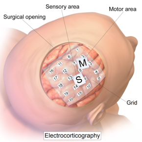
Cortical stimulation mapping (CSM) is a type of electrocorticography that involves a physically invasive procedure and aims to localize the function of specific brain regions through direct electrical stimulation of the cerebral cortex. It remains one of the earliest methods of analyzing the brain and has allowed researchers to study the relationship between cortical structure and systemic function. Cortical stimulation mapping is used for a number of clinical and therapeutic applications, and remains the preferred method for the pre-surgical mapping of the motor cortex and language areas to prevent unnecessary functional damage. There are also some clinical applications for cortical stimulation mapping, such as the treatment of epilepsy.

Awake craniotomy is a neurosurgical technique and type of craniotomy that allows a surgeon to remove a brain tumor while the patient is awake to avoid brain damage. During the surgery, the neurosurgeon performs cortical mapping to identify vital areas, called the "eloquent brain", that should not be disturbed while removing the tumor.

A cortical implant is a subset of neuroprosthetics that is in direct connection with the cerebral cortex of the brain. By directly interfacing with different regions of the cortex, the cortical implant can provide stimulation to an immediate area and provide different benefits, depending on its design and placement. A typical cortical implant is an implantable microelectrode array, which is a small device through which a neural signal can be received or transmitted.

Drug-resistant epilepsy (DRE), also known as refractory epilepsy, intractable epilepsy, or pharmacoresistant epilepsy, is diagnosed following a failure of adequate trials of two tolerated and appropriately chosen and used antiepileptic drugs (AEDs) to achieve sustained seizure freedom. The probability that the next medication will achieve seizure freedom drops with every failed AED. For example, after two failed AEDs, the probability that the third will achieve seizure freedom is around 4%. Drug-resistant epilepsy is commonly diagnosed after several years of uncontrolled seizures, however, in most cases, it is evident much earlier. Approximately 30% of people with epilepsy have a drug-resistant form.

<span class="mw-page-title-main">Hal Blumenfeld</span>

Hal Blumenfeld is a Professor of Neurology, Neuroscience, and Neurosurgery at Yale University. He is an expert on brain mechanisms of consciousness and on altered consciousness in epilepsy. As director of the Yale Clinical Neuroscience Imaging Center (CNIC) he leads multi-disciplinary research and is also well known for his teaching contributions in neuroanatomy and clinical neuroscience.

<span class="mw-page-title-main">Fabrice Bartolomei</span> French neurophysiologist

Fabrice Bartolomei is a French neurophysiologist, and University Professor at Aix-Marseille University (AMU), leading the Service de Neurophysiologie Clinique of the Timone Hospital at the Assistance Publique - Hôpitaux de Marseille, and he is the medical director of the ‘Centre Saint-Paul - Hopital Henri Gastaut’. He is the coordinator of the clinical network CINAPSE that is dedicated to the management of adult and pediatric cases of severe epilepsy and leader of the Federation Hospitalo-Universitaire Epinext. He is also member of the research unit Institut de Neurosciences des Systèmes.

Musicogenic seizure, also known as music-induced seizure, is a rare type of seizure, with an estimated prevalence of 1 in 10,000,000 individuals, that arises from disorganized or abnormal brain electrical activity when a person hears or is exposed to a specific type of sound or musical stimuli. There are challenges when diagnosing a music-induced seizure due to the broad scope of triggers, and time delay between a stimulus and seizure. In addition, the causes of musicogenic seizures are not well-established as solely limited cases and research have been discovered and conducted respectively. Nevertheless, the current understanding of the mechanism behind musicogenic seizure is that music triggers the part of the brain that is responsible for evoking an emotion associated with that music. Dysfunction in this system leads to an abnormal release of dopamine, eventually inducing seizure.

References

  1. Palmini, A (2006). "The concept of the epileptogenic zone: A modern look at Penfield and Jasper's views on the role of interictal spikes" . Epileptic Disorders . 8 (Suppl 2): S10–5. doi:10.1684/j.1950-6945.2006.tb00205.x. hdl: 10923/21709 . PMID   17012068.
  2. 1 2 3 4 5 Kuruvilla, A; Flink, R (2003). "Intraoperative electrocorticography in epilepsy surgery: Useful or not?". Seizure . 12 (8): 577–84. doi: 10.1016/S1059-1311(03)00095-5 . PMID   14630497. S2CID   15643130.
  3. Baumeister AA (2000). "The Tulane Electrical Brain Stimulation Program a historical case study in medical ethics". J Hist Neurosci. 9 (3): 262–78. doi:10.1076/jhin.9.3.262.1787. PMID   11232368. S2CID   38336466.
  4. Marwan Hariz; Patric Blomstedt; Ludvic Zrinzo (2016). "Deep Brain Stimulation between 1947 and 1987: The Untold Story". Neurosurg Focus. 29 (2). e1 via Medscape.
  5. Hashiguchi, K; Morioka, T; Yoshida, F; Miyagi, Y; et al. (2007). "Correlation between scalp-recorded electroencephalographic and electrocorticographic activities during ictal period". Seizure . 16 (3): 238–247. doi: 10.1016/j.seizure.2006.12.010 . PMID   17236792. S2CID   1728557.
  6. Fallegger, Florian; Schiavone, Giuseppe; Pirondini, Elvira; Wagner, Fabien B.; Vachicouras, Nicolas; Serex, Ludovic; Zegarek, Gregory; May, Adrien; Constanthin, Paul; Palma, Marie; Khoshnevis, Mehrdad; Van Roost, Dirk; Yvert, Blaise; Courtine, Grégoire; Schaller, Karl (March 2021). "MRI-Compatible and Conformal Electrocorticography Grids for Translational Research". Advanced Science. 8 (9). doi:10.1002/advs.202003761. ISSN   2198-3844. PMC   8097365 . PMID   33977054.
  7. 1 2 Logothetis, NK (2003). "The underpinning of the BOLD functional magnetic resonance imaging signal". The Journal of Neuroscience . 23 (10): 3963–71. doi:10.1523/JNEUROSCI.23-10-03963.2003. PMC   6741096 . PMID   12764080.
  8. Ulbert, I; Halgren, E; Heit, G; Karmos, G (2001). "Multiple microelectrode-recording system for human intracortical applications". Journal of Neuroscience Methods . 106 (1): 69–79. doi:10.1016/S0165-0270(01)00330-2. PMID   11248342. S2CID   12203755.
  9. 1 2 3 4 Schuh, L; Drury, I (1996). "Intraoperative electrocorticography and direct cortical electrical stimulation". Seminars in Anesthesia . 16: 46–55. doi:10.1016/s0277-0326(97)80007-4.
  10. Mesgarani, N; Chang, EF (2012). "Selective cortical representation of attended speaker in multi-talker speech perception". Nature . 485 (7397): 233–6. Bibcode:2012Natur.485..233M. doi:10.1038/nature11020. PMC   3870007 . PMID   22522927.
  11. Boyer A, Duffau H, Vincent M, Ramdani S, Mandonnet E, Guiraud D, Bonnetblanc F (2018). "Electrophysiological Activity Evoked by Direct Electrical Stimulation of the Human Brain: Interest of the P0 Component" (PDF). 2018 40th Annual International Conference of the IEEE Engineering in Medicine and Biology Society (EMBC). Vol. 2018. pp. 2210–2213. doi:10.1109/EMBC.2018.8512733. ISBN   978-1-5386-3646-6. PMID   30440844. S2CID   53097668.
  12. Ritaccio, Anthony L.; Brunner, Peter; Schalk, Gerwin (March 2018). "Electrical Stimulation Mapping of the Brain: Basic Principles and Emerging Alternatives". Journal of Clinical Neurophysiology. 35 (2): 86–97. doi:10.1097/WNP.0000000000000440. ISSN   0736-0258. PMC   5836484 . PMID   29499015."frequency" on page 6, "damage" on page 3 of pdf
  13. Kohrman, M (2007). "What is epilepsy? Clinical perspectives in the diagnosis and treatment". Journal of Clinical Neurophysiology . 24 (2): 87–95. doi:10.1097/WNP.0b013e3180415b51. PMID   17414964. S2CID   35146214.
  14. Wetjen, Nicholas M.; Marsh, W. Richard; Meyer, Fredric B.; Cascino, Gregory D.; So, Elson; Britton, Jeffrey W.; Stead, S. Matthew; Worrell, Gregory A. (June 2009). "Intracranial electroencephalography seizure onset patterns and surgical outcomes in nonlesional extratemporal epilepsy: Clinical article". Journal of Neurosurgery. 110 (6): 1147–1152. doi:10.3171/2008.8.JNS17643. ISSN   0022-3085. PMC   2841508 . PMID   19072306.
  15. Sugano, H; Shimizu, H; Sunaga, S (2007). "Efficacy of intraoperative electrocorticography for assessing seizure outcomes in intractable epilepsy patients with temporal-lobe-mass lesions". Seizure . 16 (2): 120–127. doi: 10.1016/j.seizure.2006.10.010 . PMID   17158074.
  16. Miller, KJ; denNijs, M; Shenoy, P; Miller, JW; et al. (2007). "Real-time functional brain mapping using electrocorticography". NeuroImage . 37 (2): 504–507. doi:10.1016/j.neuroimage.2007.05.029. PMID   17604183. S2CID   3362496.
  17. 1 2 Crone, NE; Miglioretti, DL; Gordon, B; Lesser, RP (1998). "Functional mapping of human sensorimotor cortex with electrocorticographic spectral analysis. II. Event-related synchronization in the gamma band". Brain. 121 (12): 2301–15. doi: 10.1093/brain/121.12.2301 . PMID   9874481.
  18. 1 2 Nakai, Y; Jeong, JW; Brown, EC; Rothermel, R; Kojima, K; Kambara, T; Shah, A; Mittal, S; Sood, S; Asano, E (2017). "Three- and four-dimensional mapping of speech and language in patients with epilepsy". Brain. 140 (5): 1351–1370. doi:10.1093/brain/awx051. PMC   5405238 . PMID   28334963.
  19. 1 2 Nakai, Ya; Nagashima, A; Hayakawa, A; Osuki, T; Jeong, JW; Sugiura, A; Brown, EC; Asano, E (2018). "Four-dimensional map of the human early visual system". Clin Neurophysiol. 129 (1): 188–197. doi:10.1016/j.clinph.2017.10.019. PMC   5743586 . PMID   29190524.
  20. Wennberg, R; Quesney, F; Olivier, A; Rasmussen, T (1998). "Electrocorticography and outcome in frontal lobe epilepsy". Electroencephalography and Clinical Neurophysiology . 106 (4): 357–68. doi:10.1016/S0013-4694(97)00148-X. PMID   9741764.
  21. 1 2 Shenoy, P; Miller, KJ; Ojemann, JG; Rao, RPN (2007). "Generalized features for electrocorticographic BCIs" (PDF). IEEE Transactions on Biomedical Engineering . 55 (1): 273–80. CiteSeerX   10.1.1.208.7298 . doi:10.1109/TBME.2007.903528. PMID   18232371. S2CID   3034381. Archived (PDF) from the original on 2014-12-14.
  22. Allison, Brendan Z. (2009). "Chapter 2: Toward Ubiquitous BCIs.". Brain-Computer Interfaces. Springer. pp. 357–387. ISBN   978-3-642-02091-9.
  23. Swift, James; Coon, William; Guger, Christoph; Brunner, Peter; Bunch, M; Lynch, T; Frawley, T; Ritaccio, Anthony; Schalk, Gerwin (2018). "Passive functional mapping of receptive language areas using electrocorticographic signals". Clinical Neurophysiology. 6 (12): 2517–2524. doi:10.1016/j.clinph.2018.09.007. PMC   6414063 . PMID   30342252.
  24. Martin, Stephanie; Iturrate, Iñaki; Millán, José del R.; Knight, Robert; Pasley, Brian N. (2018). "Decoding Inner Speech Using Electrocorticography: Progress and Challenges Toward a Speech Prosthesis". Frontiers in Neuroscience. 12: 422. doi: 10.3389/fnins.2018.00422 . PMC   6021529 . PMID   29977189.
  25. Schalk, Gerwin; Kapeller, Christoph; Guger, Christoph; Ogawa, H; Hiroshima, S; Lafer-Sousa, R; Saygin, Zenyip M.; Kamada, Kyousuke; Kanwisher, Nancy (2017). "Facephenes and rainbows: Causal evidence for functional and anatomical specificity of face and color processing in the human brain". Proc Natl Acad Sci U S A. 114 (46): 12285–12290. Bibcode:2017PNAS..11412285S. doi: 10.1073/pnas.1713447114 . PMC   5699078 . PMID   29087337.
  26. Saez, I; Lin, J; Stolk, A; Chang, E; Parvizi, J; Schalk, Gerwin; Knight, Robert T.; Hsu, M. (2018). "Encoding of Multiple Reward-Related Computations in Transient and Sustained High-Frequency Activity in Human OFC". Current Biology. 28 (18): 2889–2899.e3. doi:10.1016/j.cub.2018.07.045. PMC   6590063 . PMID   30220499.
  27. Schalk, Gerwin; Marple, J.; Knight, Robert T.; Coon, William G. (2017). "Instantaneous voltage as an alternative to power- and phase-based interpretation of oscillatory brain activity". NeuroImage. 157: 545–554. doi:10.1016/j.neuroimage.2017.06.014. PMC   5600843 . PMID   28624646.
  28. Ding, L; Wilke, C; Xu, B; Xu, X; et al. (2007). "EEG source imaging: Correlating source locations and extents with electrocorticography and surgical resections in epilepsy patients". Journal of Clinical Neurophysiology . 24 (2): 130–136. doi:10.1097/WNP.0b013e318038fd52. PMC   2758789 . PMID   17414968.