Neuroregeneration

Last updated

Neuroregeneration involves the regrowth or repair of nervous tissues, cells or cell products. Neuroregenerative mechanisms may include generation of new neurons, glia, axons, myelin, or synapses. Neuroregeneration differs between the peripheral nervous system (PNS) and the central nervous system (CNS) by the functional mechanisms involved, especially in the extent and speed of repair. When an axon is damaged, the distal segment undergoes Wallerian degeneration, losing its myelin sheath. The proximal segment can either die by apoptosis or undergo the chromatolytic reaction, which is an attempt at repair. In the CNS, synaptic stripping occurs as glial foot processes invade the dead synapse. [1]

Contents

Nervous system injuries affect over 90,000 people every year. [2] Spinal cord injuries alone affect an estimated 10,000 people each year. [3] As a result of this high incidence of neurological injuries, nerve regeneration and repair, a subfield of neural tissue engineering, is becoming a rapidly growing field dedicated to the discovery of new ways to recover nerve functionality after injury.

The nervous system is divided by neurologists into two parts: the central nervous system (which consists of the brain and spinal cord) and the peripheral nervous system (which consists of cranial and spinal nerves along with their associated ganglia). While the peripheral nervous system has an intrinsic ability for repair and regeneration, the central nervous system is, for the most part, incapable of self-repair and regeneration. There is currently no treatment for recovering human nerve-function after injury to the central nervous system. [4] Multiple attempts at nerve re-growth across the PNS-CNS transition have not been successful. [4] There is simply not enough knowledge about regeneration in the central nervous system. In addition, although the peripheral nervous system has the capability for regeneration, much research still needs to be done to optimize the environment for maximum regrowth potential. Neuroregeneration is important clinically, as it is part of the pathogenesis of many diseases, including multiple sclerosis.

Peripheral nervous system regeneration

Guillain-Barre syndrome - nerve damage Guillain-barre syndrome - Nerve Damage.gif
Guillain–Barré syndrome – nerve damage

Neuroregeneration in the peripheral nervous system (PNS) occurs to a significant degree. [5] [6] After an injury to the axon, peripheral neurons activate a variety of signaling pathways which turn on pro-growth genes, leading to reformation of a functional growth cone and regeneration. The growth of these axons is also governed by chemotactic factors secreted from Schwann cells. Injury to the peripheral nervous system immediately elicits the migration of phagocytes, Schwann cells, and macrophages to the lesion site in order to clear away debris such as damaged tissue which is inhibitory to regeneration. When a nerve axon is severed, the end still attached to the cell body is labeled the proximal segment, while the other end is called the distal segment. After injury, the proximal end swells and experiences some retrograde degeneration, but once the debris is cleared, it begins to sprout axons and the presence of growth cones can be detected. The proximal axons are able to regrow as long as the cell body is intact, and they have made contact with the Schwann cells in the endoneurium (also known as the endoneurial tube or channel). Human axon growth rates can reach 2 mm/day in small nerves and 5 mm/day in large nerves. [4] The distal segment, however, experiences Wallerian degeneration within hours of the injury; the axons and myelin degenerate, but the endoneurium remains. In the later stages of regeneration the remaining endoneurial tube directs axon growth back to the correct targets. During Wallerian degeneration, Schwann cells grow in ordered columns along the endoneurial tube, creating a band of Büngner cells that protects and preserves the endoneurial channel. Also, macrophages and Schwann cells release neurotrophic factors that enhance re-growth.

Central nervous system regeneration

Unlike peripheral nervous system injury, injury to the central nervous system is not followed by extensive regeneration. It is limited by the inhibitory influences of the glial and extracellular environment. The hostile, non-permissive growth environment is, in part, created by the migration of myelin-associated inhibitors, astrocytes, oligodendrocytes, oligodendrocyte precursors, and microglia. The environment within the CNS, especially following trauma, counteracts the repair of myelin and neurons. Growth factors are not expressed or re-expressed; for instance, the extracellular matrix is lacking laminins. Glial scars rapidly form, and the glia actually produce factors that inhibit remyelination and axon repair; for instance, NOGO and NI-35. [6] [7] [8] The axons themselves also lose the potential for growth with age, due to a decrease in GAP43 expression, among others.

Slower degeneration of the distal segment than that which occurs in the peripheral nervous system also contributes to the inhibitory environment because inhibitory myelin and axonal debris are not cleared away as quickly. All these factors contribute to the formation of what is known as a glial scar, which axons cannot grow across. [9] The proximal segment attempts to regenerate after injury, but its growth is hindered by the environment. It is important to note that central nervous system axons have been proven to regrow in permissive environments; therefore, the primary problem to central nervous system axonal regeneration is crossing or eliminating the inhibitory lesion site. [4] Another problem is that the morphology and functional properties of central nervous system neurons are highly complex, for this reason a neuron functionally identical cannot be replaced by one of another type (Llinás' law). [10]

Inhibition of axonal regrowth

Glial cell scar formation is induced following damage to the nervous system. In the central nervous system, this glial scar formation significantly inhibits nerve regeneration, which leads to a loss of function. Several families of molecules are released that promote and drive glial scar formation. For instance, transforming growth factors B-1 and -2, interleukins, and cytokines play a role in the initiation of scar formation. The accumulation of reactive astrocytes at the site of injury and the up regulation of molecules that are inhibitory for neurite outgrowth contribute to the failure of neuroregeneration. [11] The up-regulated molecules alter the composition of the extracellular matrix in a way that has been shown to inhibit neurite outgrowth extension. This scar formation involves several cell types and families of molecules.

Chondroitin sulfate proteoglycan

In response to scar-inducing factors, astrocytes up regulate the production of chondroitin sulfate proteoglycans. Astrocytes are a predominant type of glial cell in the central nervous system that provide many functions including damage mitigation, repair, and glial scar formation. [12] The RhoA pathway is involved. Chondroitin sulfate proteoglycans (CSPGs) have been shown to be up regulated in the central nervous system (CNS) following injury. Repeating disaccharides of glucuronic acid and galactosamine, glycosaminoglycans (CS-GAGs), are covalently coupled to the protein core CSPGs. CSPGs have been shown to inhibit regeneration in vitro and in vivo, but the role that the CSPG core protein vs. CS-GAGs had not been studied until recently.

Keratan sulfate proteoglycans

Like the chondroitin sulfate proteoglycans, keratan sulfate proteoglycan (KSPG) production is up regulated in reactive astrocytes as part of glial scar formation. KSPGs have also been shown to inhibit neurite outgrowth extension, limiting nerve regeneration. Keratan sulfate, also called keratosulfate, is formed from repeating disaccharide galactose units and N-acetylglucosamines. It is also 6-sulfated. This sulfation is crucial to the elongation of the keratan sulfate chain. A study was done using N-acetylglucosamine 6-O-sulfotransferase-1 deficient mice. The wild type mouse showed a significant up regulation of mRNA expressing N-acetylglucosamine 6-O-sulfotransferase-1 at the site of cortical injury. However, in the N-acetylglucosamine 6-O-sulfotransferase-1 deficient mice, the expression of keratan sulfate was significantly decreased when compared to the wild type mice. Similarly, glial scar formation was significantly reduced in the N-acetylglucosamine 6-O-sulfotransferase-1 mice, and as a result, nerve regeneration was less inhibited. [11]

Other inhibitory factors

Proteins of oligodendritic or glial debris origin that influence neuroregeneration:

Clinical treatments

Neurons replacement

in vivo glias to neurons reprogramming

Transcription factors, activation of genes (using CRISPR activation [16] ) or small molecules are used to reprogram glias into neurons.

The most commonly targeted glias are astrocytes (usually using GFAP) because they share the same lineage as neurons and region—specific transcription signatures, [16] while the vector used is typically an adeno-associated virus because some serotypes pass the blood brain barrier and it does not cause disease.

Targeted genes usually depend on the type of neuron sought; (NGN2 is known to produce glutamatergic, ASCL1: GABAergic...); RBPJ-k blocks the Notch pathway and elicits a neurogenic program [17] and Sox2 can also increase reprograming efficiency by causing a dedifferentiation and self-amplification phase before maturating as neurons.

While theses techniques show lot of promise in animal models for many otherwise incurable neurodegenerative diseases and brain injuries, no clinical trials have started as of 2023.

Neural stem cells grafting

Tissue regrowth

Peripheral

Surgery

Surgery can be done in case a peripheral nerve has become cut or otherwise divided. This is called peripheral nerve reconstruction. The injured nerve is identified and exposed so that normal nerve tissue can be examined above and below the level of injury, usually with magnification, using either loupes or an operating microscope. If a large segment of nerve is harmed, as can happen in a crush or stretch injury, the nerve will need to be exposed over a larger area. Injured portions of the nerve are removed. The cut nerve endings are then carefully reapproximated using very small sutures. The nerve repair must be covered by healthy tissue, which can be as simple as closing the skin or it can require moving skin or muscle to provide healthy padded coverage over the nerve. [18] The type of anesthesia used depends on the complexity of the injury. A surgical tourniquet is almost always used. [18]

Prognosis

The expectations after surgical repair of a divided peripheral nerve depends on several factors:

  • Age: Recovery of a nerve after surgical repair depends mainly on the age of the patient. Young children can recover close-to-normal nerve function. In contrast, a patient over 60 years old with a cut nerve in the hand would expect to recover only protective sensation; that is, the ability to distinguish hot/cold or sharp/dull. [18]
  • The mechanism of injury: Sharp injuries, such as a knife wound, damage only a very short segment of the nerve, availing for direct suture. In contrast, nerves that are divided by stretch or crush may be damaged over long segments. These nerve injuries are more difficult to treat and generally have a poorer outcome. In addition, associated injuries, like injury to bone, muscle and skin, can make nerve recovery more difficult. [18]
  • The level of injury: After a nerve is repaired, the regenerating nerve endings must grow all the way to their target. For example, a nerve injured at the wrist that normally provides sensation to the thumb must grow to the end of the thumb in order to provide sensation. The return of function decreases with increased distance over which a nerve must grow. [18]
Autologous nerve grafting

Currently, autologous nerve grafting, or a nerve autograft, is known as the gold standard for clinical treatments used to repair large lesion gaps in the peripheral nervous system. It is important that nerves are not repaired under tension, [18] which could otherwise happen if cut ends are reapproximated across a gap. Nerve segments are taken from another part of the body (the donor site) and inserted into the lesion to provide endoneurial tubes for axonal regeneration across the gap. However, this is not a perfect treatment; often the outcome is only limited function recovery. Also, partial de-innervation is frequently experienced at the donor site, and multiple surgeries are required to harvest the tissue and implant it.

When appropriate, a nearby donor may be used to supply innervation to lesioned nerves. Trauma to the donor can be minimized by utilizing a technique known as end-to-side repair. In this procedure, an epineurial window is created in the donor nerve and the proximal stump of the lesioned nerve is sutured over the window. Regenerating axons are redirected into the stump. Efficacy of this technique is partially dependent upon the degree of partial neurectomy performed on the donor, with increasing degrees of neurectomy giving rise to increasing axon regeneration within the lesioned nerve, but with the consequence of increasing deficit to the donor. [19]

Some evidence suggests that local delivery of soluble neurotrophic factors at the site of autologous nerve grafting may enhance axon regeneration within the graft and help expedite functional recovery of a paralyzed target. [20] [21] Other evidence suggests that gene-therapy induced expression of neurotrophic factors within the target muscle itself can also help enhance axon regeneration. [22] [23] Accelerating neuroregeneration and the reinnervation of a denervated target is critically important in order to reduce the possibility of permanent paralysis due to muscular atrophy.

Allografts and xenografts

Variations on the nerve autograft include the allograft and the xenograft. In allografts, the tissue for the graft is taken from another person, the donor, and implanted in the recipient. Xenografts involve taking donor tissue from another species. Allografts and xenografts have the same disadvantages as autografts, but in addition, tissue rejection from immune responses must also be taken into account. Often immunosuppression is required with these grafts. Disease transmission also becomes a factor when introducing tissue from another person or animal. Overall, allografts and xenografts do not match the quality of outcomes seen with autografts, but they are necessary when there is a lack of autologous nerve tissue.

Nerve guidance conduit

Because of the limited functionality received from autografts, the current gold standard for nerve regeneration and repair, recent neural tissue engineering research has focused on the development of bioartificial nerve guidance conduits in order to guide axonal regrowth. The creation of artificial nerve conduits is also known as entubulation because the nerve ends and intervening gap are enclosed within a tube composed of biological or synthetic materials. [24]

Immunisation

A direction of research is towards the use of drugs that target remyelinating inhibitor proteins, or other inhibitors. Possible strategies include vaccination against these proteins (active immunisation), or treatment with previously created antibodies (passive immunisation). These strategies appear promising on animal models with experimental autoimmune encephalomyelitis (EAE), a model of MS. [25] Monoclonal antibodies have also been used against inhibitory factors such as NI-35 and NOGO. [26]

See also

Related Research Articles

<span class="mw-page-title-main">Axon</span> Long projection on a neuron that conducts signals to other neurons

An axon, or nerve fiber, is a long, slender projection of a nerve cell, or neuron, in vertebrates, that typically conducts electrical impulses known as action potentials away from the nerve cell body. The function of the axon is to transmit information to different neurons, muscles, and glands. In certain sensory neurons, such as those for touch and warmth, the axons are called afferent nerve fibers and the electrical impulse travels along these from the periphery to the cell body and from the cell body to the spinal cord along another branch of the same axon. Axon dysfunction can be the cause of many inherited and acquired neurological disorders that affect both the peripheral and central neurons. Nerve fibers are classed into three types – group A nerve fibers, group B nerve fibers, and group C nerve fibers. Groups A and B are myelinated, and group C are unmyelinated. These groups include both sensory fibers and motor fibers. Another classification groups only the sensory fibers as Type I, Type II, Type III, and Type IV.

<span class="mw-page-title-main">Myelin</span> Fatty substance that surrounds nerve cell axons to insulate them and increase transmission speed

Myelin is a lipid-rich material that surrounds nerve cell axons to insulate them and increase the rate at which electrical impulses pass along the axon. The myelinated axon can be likened to an electrical wire with insulating material (myelin) around it. However, unlike the plastic covering on an electrical wire, myelin does not form a single long sheath over the entire length of the axon. Rather, myelin ensheaths the axon segmentally: in general, each axon is encased in multiple long sheaths with short gaps between, called nodes of Ranvier. At the nodes of Ranvier, which are approximately one thousandth of a mm in length, the axon's membrane is bare of myelin.

<span class="mw-page-title-main">Neuron</span> Electrically excitable cell found in the nervous system of animals

Within a nervous system, a neuron, neurone, or nerve cell is an electrically excitable cell that fires electric signals called action potentials across a neural network. Neurons communicate with other cells via synapses, which are specialized connections that commonly use minute amounts of chemical neurotransmitters to pass the electric signal from the presynaptic neuron to the target cell through the synaptic gap.

<span class="mw-page-title-main">Schwann cell</span> Glial cell type

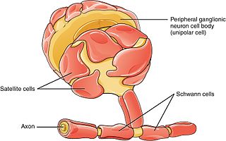
Schwann cells or neurolemmocytes are the principal glia of the peripheral nervous system (PNS). Glial cells function to support neurons and in the PNS, also include satellite cells, olfactory ensheathing cells, enteric glia and glia that reside at sensory nerve endings, such as the Pacinian corpuscle. The two types of Schwann cells are myelinating and nonmyelinating. Myelinating Schwann cells wrap around axons of motor and sensory neurons to form the myelin sheath. The Schwann cell promoter is present in the downstream region of the human dystrophin gene that gives shortened transcript that are again synthesized in a tissue-specific manner.

<span class="mw-page-title-main">Nervous tissue</span> Main component of the nervous system

Nervous tissue, also called neural tissue, is the main tissue component of the nervous system. The nervous system regulates and controls body functions and activity. It consists of two parts: the central nervous system (CNS) comprising the brain and spinal cord, and the peripheral nervous system (PNS) comprising the branching peripheral nerves. It is composed of neurons, also known as nerve cells, which receive and transmit impulses, and neuroglia, also known as glial cells or glia, which assist the propagation of the nerve impulse as well as provide nutrients to the neurons.

<span class="mw-page-title-main">Motor nerve</span> Nerve located in the central nervous system

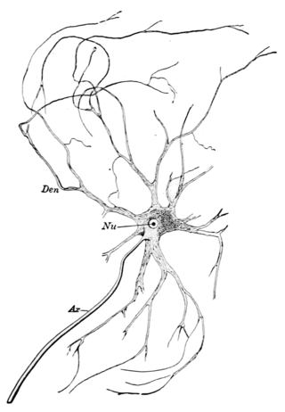
A motor nerve is a nerve that transmits motor signals from the central nervous system (CNS) to the muscles of the body. This is different from the motor neuron, which includes a cell body and branching of dendrites, while the nerve is made up of a bundle of axons. Motor nerves act as efferent nerves which carry information out from the CNS to muscles, as opposed to afferent nerves, which transfer signals from sensory receptors in the periphery to the CNS. Efferent nerves can also connect to glands or other organs/issues instead of muscles. In addition, there are nerves that serve as both sensory and motor nerves called mixed nerves.

<span class="mw-page-title-main">Glia</span> Support cells in the nervous system

Glia, also called glial cells(gliocytes) or neuroglia, are non-neuronal cells in the central nervous system (brain and spinal cord) and the peripheral nervous system that do not produce electrical impulses. The neuroglia make up more than one half the volume of neural tissue in our body. They maintain homeostasis, form myelin in the peripheral nervous system, and provide support and protection for neurons. In the central nervous system, glial cells include oligodendrocytes, astrocytes, ependymal cells and microglia, and in the peripheral nervous system they include Schwann cells and satellite cells.

<span class="mw-page-title-main">Wallerian degeneration</span> Biological process of axonal degeneration

Wallerian degeneration is an active process of degeneration that results when a nerve fiber is cut or crushed and the part of the axon distal to the injury degenerates. A related process of dying back or retrograde degeneration known as 'Wallerian-like degeneration' occurs in many neurodegenerative diseases, especially those where axonal transport is impaired such as ALS and Alzheimer's disease. Primary culture studies suggest that a failure to deliver sufficient quantities of the essential axonal protein NMNAT2 is a key initiating event.

<span class="mw-page-title-main">Astrogliosis</span> Increase in astrocytes in response to brain injury

Astrogliosis is an abnormal increase in the number of astrocytes due to the destruction of nearby neurons from central nervous system (CNS) trauma, infection, ischemia, stroke, autoimmune responses or neurodegenerative disease. In healthy neural tissue, astrocytes play critical roles in energy provision, regulation of blood flow, homeostasis of extracellular fluid, homeostasis of ions and transmitters, regulation of synapse function and synaptic remodeling. Astrogliosis changes the molecular expression and morphology of astrocytes, in response to infection for example, in severe cases causing glial scar formation that may inhibit axon regeneration.

Gliosis is a nonspecific reactive change of glial cells in response to damage to the central nervous system (CNS). In most cases, gliosis involves the proliferation or hypertrophy of several different types of glial cells, including astrocytes, microglia, and oligodendrocytes. In its most extreme form, the proliferation associated with gliosis leads to the formation of a glial scar.

<span class="mw-page-title-main">Myelin-associated glycoprotein</span> Protein-coding gene in the species Homo sapiens

Myelin-associated glycoprotein is a type 1 transmembrane protein glycoprotein localized in periaxonal Schwann cell and oligodendrocyte membranes, where it plays a role in glial-axonal interactions. MAG is a member of the SIGLEC family of proteins and is a functional ligand of the NOGO-66 receptor, NgR. MAG is believed to be involved in myelination during nerve regeneration in the PNS and is vital for the long-term survival of the myelinated axons following myelinogenesis. In the CNS MAG is one of three main myelin-associated inhibitors of axonal regeneration after injury, making it an important protein for future research on neurogenesis in the CNS.

<span class="mw-page-title-main">Nerve injury</span> Damage to nervous tissue

Nerve injury is an injury to a nerve. There is no single classification system that can describe all the many variations of nerve injuries. In 1941, Seddon introduced a classification of nerve injuries based on three main types of nerve fiber injury and whether there is continuity of the nerve. Usually, however, nerve injuries are classified in five stages, based on the extent of damage to both the nerve and the surrounding connective tissue, since supporting glial cells may be involved.

A nerve guidance conduit is an artificial means of guiding axonal regrowth to facilitate nerve regeneration and is one of several clinical treatments for nerve injuries. When direct suturing of the two stumps of a severed nerve cannot be accomplished without tension, the standard clinical treatment for peripheral nerve injuries is autologous nerve grafting. Due to the limited availability of donor tissue and functional recovery in autologous nerve grafting, neural tissue engineering research has focused on the development of bioartificial nerve guidance conduits as an alternative treatment, especially for large defects. Similar techniques are also being explored for nerve repair in the spinal cord but nerve regeneration in the central nervous system poses a greater challenge because its axons do not regenerate appreciably in their native environment.

Nerve tissue is a biological molecule related to the function and maintenance of normal nervous tissue. An example would include, for example, the generation of myelin which insulates and protects nerves. These are typically calcium-binding proteins.

<span class="mw-page-title-main">Glial scar</span> Mass formed in response to injury to the nervous system

A glial scar formation (gliosis) is a reactive cellular process involving astrogliosis that occurs after injury to the central nervous system. As with scarring in other organs and tissues, the glial scar is the body's mechanism to protect and begin the healing process in the nervous system.

<span class="mw-page-title-main">Reticulon 4</span> Protein-coding gene in humans

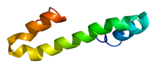
Reticulon 4, also known as Neurite outgrowth inhibitor or Nogo, is a protein that in humans is encoded by the RTN4 gene that has been identified as an inhibitor of neurite outgrowth specific to the central nervous system. During neural development Nogo is expressed mainly by neurons and provides an inhibitory signal for the migration and sprouting of CNS endothelial (tip) cells, thereby restricting blood vessel density.

<span class="mw-page-title-main">LINGO1</span> Protein-coding gene in the species Homo sapiens

Leucine rich repeat and Immunoglobin-like domain-containing protein 1 also known as LINGO-1 is a protein which is encoded by the LINGO1 gene in humans. It belongs to the family of leucine-rich repeat proteins which are known for playing key roles in the biology of the central nervous system. LINGO-1 is a functional component of the Nogo receptor also known as the reticulon 4 receptor.

<span class="mw-page-title-main">Chondroitin sulfate proteoglycan</span>

Chondroitin sulfate proteoglycans (CSPGs) are proteoglycans consisting of a protein core and a chondroitin sulfate side chain. They are known to be structural components of a variety of human tissues, including cartilage, and also play key roles in neural development and glial scar formation. They are known to be involved in certain cell processes, such as cell adhesion, cell growth, receptor binding, cell migration, and interaction with other extracellular matrix constituents. They are also known to interact with laminin, fibronectin, tenascin, and collagen. CSPGs are generally secreted from cells.

<span class="mw-page-title-main">Olfactory ensheathing cell</span> Type of macroglia that ensheath unmyelinated olfactory neurons

Olfactory ensheathing cells (OECs), also known as olfactory ensheathing glia or olfactory ensheathing glial cells, are a type of macroglia found in the nervous system. They are also known as olfactory Schwann cells, because they ensheath the non-myelinated axons of olfactory neurons in a similar way to which Schwann cells ensheath non-myelinated peripheral neurons. They also share the property of assisting axonal regeneration.

Endogenous regeneration in the brain is the ability of cells to engage in the repair and regeneration process. While the brain has a limited capacity for regeneration, endogenous neural stem cells, as well as numerous pro-regenerative molecules, can participate in replacing and repairing damaged or diseased neurons and glial cells. Another benefit that can be achieved by using endogenous regeneration could be avoiding an immune response from the host.

References

  1. Kandel ER, Schwartz JH, Jessell TM (2003). "Chapter 55: The formation and regeneration of synapses". Principles of neural Science (fourth ed.). Cambridge: McGraw Hill. ISBN   978-0-8385-7701-1.
  2. Stabenfeldt SE, García AJ, LaPlaca MC (June 2006). "Thermoreversible laminin-functionalized hydrogel for neural tissue engineering". Journal of Biomedical Materials Research Part A. 77 (4): 718–25. doi:10.1002/jbm.a.30638. PMID   16555267.
  3. Prang P, Müller R, Eljaouhari A, Heckmann K, Kunz W, Weber T, Faber C, Vroemen M, Bogdahn U, Weidner N (July 2006). "The promotion of oriented axonal regrowth in the injured spinal cord by alginate-based anisotropic capillary hydrogels". Biomaterials. 27 (19): 3560–9. doi:10.1016/j.biomaterials.2006.01.053. PMID   16500703.
  4. 1 2 3 4 Recknor JB, Mallapragada SK (2006). "Nerve Regeneration: Tissue Engineering Strategies". In Bronzino JD (ed.). The biomedical engineering handbook (third ed.). Boca Raton, Fla.: CRC Taylor & Francis. ISBN   978-0-8493-2123-8.
  5. Mahar M, Cavalli V (June 2018). "Intrinsic mechanisms of neuronal axon regeneration". Nature Reviews. Neuroscience. 19 (6): 323–337. doi:10.1038/s41583-018-0001-8. PMC   5987780 . PMID   29666508.
  6. 1 2 3 4 5 Yiu G, He Z (August 2006). "Glial inhibition of CNS axon regeneration". Nature Reviews. Neuroscience. 7 (8): 617–27. doi:10.1038/nrn1956. PMC   2693386 . PMID   16858390.
  7. Bradbury EJ, McMahon SB (August 2006). "Spinal cord repair strategies: why do they work?". Nature Reviews. Neuroscience. 7 (8): 644–53. doi:10.1038/nrn1964. PMID   16858392. S2CID   11890502.
  8. Bregman BS, Kunkel-Bagden E, Schnell L, Dai HN, Gao D, Schwab ME (November 1995). "Recovery from spinal cord injury mediated by antibodies to neurite growth inhibitors". Nature. 378 (6556): 498–501. Bibcode:1995Natur.378..498B. doi:10.1038/378498a0. PMID   7477407. S2CID   4352534.
  9. Shafqat, Areez; Albalkhi, Ibrahem; Magableh, Hamzah M.; Saleh, Tariq; Alkattan, Khaled; Yaqinuddin, Ahmed (2023). "Tackling the glial scar in spinal cord regeneration: new discoveries and future directions". Frontiers in Cellular Neuroscience. 17. doi: 10.3389/fncel.2023.1180825 . ISSN   1662-5102. PMC   10244598 . PMID   37293626.
  10. Llinás RR (November 2014). "Intrinsic electrical properties of mammalian neurons and CNS function: a historical perspective". Frontiers in Cellular Neuroscience. 8: 320. doi: 10.3389/fncel.2014.00320 . PMC   4219458 . PMID   25408634.
  11. 1 2 Zhang H, Uchimura K, Kadomatsu K (November 2006). "Brain keratan sulfate and glial scar formation". Annals of the New York Academy of Sciences. 1086 (1): 81–90. Bibcode:2006NYASA1086...81Z. doi:10.1196/annals.1377.014. PMID   17185507. S2CID   27885790.
  12. Song I, Dityatev A (January 2018). "Crosstalk between glia, extracellular matrix and neurons". Brain Research Bulletin. 136: 101–108. doi:10.1016/j.brainresbull.2017.03.003. PMID   28284900. S2CID   3287589.
  13. De Winter F, Oudega M, Lankhorst AJ, Hamers FP, Blits B, Ruitenberg MJ, Pasterkamp RJ, Gispen WH, Verhaagen J (May 2002). "Injury-induced class 3 semaphorin expression in the rat spinal cord". Experimental Neurology. 175 (1): 61–75. doi:10.1006/exnr.2002.7884. PMID   12009760. S2CID   39940363.
  14. Mecollari V, Nieuwenhuis B, Verhaagen J (2014). "A perspective on the role of class III semaphorin signaling in central nervous system trauma". Frontiers in Cellular Neuroscience. 8: 328. doi: 10.3389/fncel.2014.00328 . PMC   4209881 . PMID   25386118.
  15. Tannemaat MR, Korecka J, Ehlert EM, Mason MR, van Duinen SG, Boer GJ, Malessy MJ, Verhaagen J (December 2007). "Human neuroma contains increased levels of semaphorin 3A, which surrounds nerve fibers and reduces neurite extension in vitro". The Journal of Neuroscience. 27 (52): 14260–4. doi: 10.1523/JNEUROSCI.4571-07.2007 . PMC   6673446 . PMID   18160633.
  16. 1 2 Herrero-Navarro Á, Puche-Aroca L, Moreno-Juan V, Sempere-Ferràndez A, Espinosa A, Susín R, et al. (April 2021). "Astrocytes and neurons share region-specific transcriptional signatures that confer regional identity to neuronal reprogramming". Science Advances. 7 (15): eabe8978. Bibcode:2021SciA....7.8978H. doi:10.1126/sciadv.abe8978. PMC   8026135 . PMID   33827819.
  17. Zamboni M, Llorens-Bobadilla E, Magnusson JP, Frisén J (October 2020). "A Widespread Neurogenic Potential of Neocortical Astrocytes Is Induced by Injury". Cell Stem Cell. 27 (4): 605–617.e5. doi:10.1016/j.stem.2020.07.006. PMC   7534841 . PMID   32758425.
  18. 1 2 3 4 5 6 Payne SH (2001). "Nerve repair and grafting in the upper extremity". Journal of the Southern Orthopaedic Association. 10 (3): 173–189. PMID   12132829.
  19. Kalantarian B, Rice DC, Tiangco DA, Terzis JK (October 1998). "Gains and losses of the XII-VII component of the "baby-sitter" procedure: a morphometric analysis". Journal of Reconstructive Microsurgery. 14 (7): 459–71. doi:10.1055/s-2007-1000208. PMID   9819092. S2CID   24168382.
  20. Tiangco DA, Papakonstantinou KC, Mullinax KA, Terzis JK (May 2001). "IGF-I and end-to-side nerve repair: a dose-response study". Journal of Reconstructive Microsurgery. 17 (4): 247–56. doi:10.1055/s-2001-14516. PMID   11396586. S2CID   528789.
  21. Fansa H, Schneider W, Wolf G, Keilhoff G (July 2002). "Influence of insulin-like growth factor-I (IGF-I) on nerve autografts and tissue-engineered nerve grafts". Muscle & Nerve. 26 (1): 87–93. doi:10.1002/mus.10165. PMID   12115953. S2CID   38261013.
  22. Shiotani A, O'Malley BW, Coleman ME, Alila HW, Flint PW (September 1998). "Reinnervation of motor endplates and increased muscle fiber size after human insulin-like growth factor I gene transfer into the paralyzed larynx". Human Gene Therapy. 9 (14): 2039–47. doi:10.1089/hum.1998.9.14-2039. PMID   9759931.
  23. Flint PW, Shiotani A, O'Malley BW (March 1999). "IGF-1 gene transfer into denervated rat laryngeal muscle". Archives of Otolaryngology–Head & Neck Surgery. 125 (3): 274–9. doi: 10.1001/archotol.125.3.274 . PMID   10190798.
  24. Phillips JB, Bunting SC, Hall SM, Brown RA (2005). "Neural tissue engineering: a self-organizing collagen guidance conduit". Tissue Engineering. 11 (9–10): 1611–7. doi:10.1089/ten.2005.11.1611. PMID   16259614.
  25. Karnezis T, Mandemakers W, McQualter JL, Zheng B, Ho PP, Jordan KA, Murray BM, Barres B, Tessier-Lavigne M, Bernard CC (July 2004). "The neurite outgrowth inhibitor Nogo A is involved in autoimmune-mediated demyelination". Nature Neuroscience. 7 (7): 736–44. doi:10.1038/nn1261. PMID   15184901. S2CID   9613584.
  26. Buffo A, Zagrebelsky M, Huber AB, Skerra A, Schwab ME, Strata P, Rossi F (March 2000). "Application of neutralizing antibodies against NI-35/250 myelin-associated neurite growth inhibitory proteins to the adult rat cerebellum induces sprouting of uninjured purkinje cell axons". The Journal of Neuroscience. 20 (6): 2275–86. doi: 10.1523/JNEUROSCI.20-06-02275.2000 . PMC   6772513 . PMID   10704503.

Further reading

Gobrecht P, Andreadaki A, Diekmann H, Heskamp A, Leibinger M, Fischer D (April 2016). "Promotion of Functional Nerve Regeneration by Inhibition of Microtubule Detyrosination". The Journal of Neuroscience. 36 (14): 3890–902. doi:10.1523/JNEUROSCI.4486-15.2016. PMC   6705512 . PMID   27053198.