Osteomalacia

Last updated
Osteomalacia
Calcitriol.svg
Cholecalciferol (Vitamin D3), deficiency of which is the most common cause of Osteomalacia
Specialty Orthopedics

Osteomalacia is a disease characterized by the softening of the bones caused by impaired bone metabolism primarily due to inadequate levels of available phosphate, calcium, and vitamin D, or because of resorption of calcium. The impairment of bone metabolism causes inadequate bone mineralization.

Contents

Osteomalacia in children is known as rickets, and because of this, use of the term "osteomalacia" is often restricted to the milder, adult form of the disease. Signs and symptoms can include diffuse body pains, muscle weakness, and fragility of the bones. In addition to low systemic levels of circulating mineral ions (for example, caused by vitamin D deficiency or renal phosphate wasting) that result in decreased bone and tooth mineralization, accumulation of mineralization-inhibiting proteins and peptides (such as osteopontin and ASARM peptides), and small inhibitory molecules (such as pyrophosphate), can occur in the extracellular matrix of bones and teeth, contributing locally to cause matrix hypomineralization (osteomalacia/odontomalacia). [1] [2] [3] [4] [5] [6] [7]

A relationship describing local, physiologic double-negative (inhibiting inhibitors) regulation of mineralization has been termed the Stenciling Principle of mineralization, whereby enzyme-substrate pairs imprint mineralization patterns into the extracellular matrix (most notably described for bone) by degrading mineralization inhibitors (e.g. TNAP/TNSALP/ALPL enzyme degrading the pyrophosphate inhibition, and PHEX enzyme degrading the osteopontin inhibition). [8] [9] The Stenciling Principle for mineralization is particularly relevant to the osteomalacia and odontomalacia observed in hypophosphatasia (HPP) and X-linked hypophosphatemia (XLH).

The most common cause of osteomalacia is a deficiency of vitamin D, which is normally derived from sunlight exposure and, to a lesser extent, from the diet. [10] The most specific screening test for vitamin D deficiency in otherwise healthy individuals is a serum 25(OH)D level. [11] Less common causes of osteomalacia can include hereditary deficiencies of vitamin D or phosphate (which would typically be identified in childhood) or malignancy.

Vitamin D and calcium supplements are measures that can be used to prevent and treat osteomalacia. Vitamin D should always be administered in conjunction with calcium supplementation (as the pair work together in the body) since most of the consequences of vitamin D deficiency are a result of impaired mineral ion homeostasis. [11]

Nursing home residents and the housebound elderly population are at particular risk for vitamin D deficiency, as these populations typically receive little sun exposure. In addition, both the efficiency of vitamin D synthesis in the skin and the absorption of vitamin D from the intestine decline with age, thus further increasing the risk in these populations. Other groups at risk include individuals with absorption secondary to gastrointestinal bypass surgery or celiac disease, and individuals who immigrate from warm climates to cold climates, especially women who wear traditional veils or dresses that prevent sun exposure. [12]

Signs and symptoms

Many of the effects of the disease overlap with the more common osteoporosis, but both diseases are significantly different.[ citation needed ]

Osteomalacia in adults starts insidiously as aches and pains in the lumbar (lower back) region and thighs before spreading to the arms and ribs. The pain is symmetrical, non-radiating and accompanied by sensitivity in the involved bones. Proximal muscles are weak, and there is difficulty in climbing upstairs and getting up from a squatting position. [13] As a result of demineralization, the bones become less rigid. Physical signs include deformities like triradiate pelvis [14] and lordosis. The patient has a typical "waddling" gait. However, these physical signs may derive from a previous osteomalacial state, since bones do not regain their original shape after they become deformed.

Pathologic fractures due to weight bearing may develop. Most of the time, the only alleged symptom is chronic fatigue, while bone aches are not spontaneous but only revealed by pressure or shocks. [13] It differs from renal osteodystrophy, where the latter shows hyperphosphatemia.

Causes

The causes of adult osteomalacia are varied, but ultimately result in a vitamin D deficiency:

There are two main mechanisms of osteomalacia:

  1. insufficient calcium absorption from the intestine because of lack of dietary calcium or a deficiency of, or resistance to, the action of vitamin D, or due to undiagnosed celiac disease. [18]
  2. phosphate deficiency caused by increased renal losses.

Diagnosis

Biochemical findings

The metabolism of calcium, phosphate, hormones, and Vitamin D. Metabolism of calcium and phosphate and hormones.jpg
The metabolism of calcium, phosphate, hormones, and Vitamin D.

Biochemical features are similar to those of rickets. The major factor is an abnormally low vitamin D concentration in blood serum. [13] Major typical biochemical findings include: [19]

Furthermore, a technetium bone scan will show increased activity (also due to increased osteoblasts).

Comparison of bone pathology
Condition Calcium Phosphate Alkaline phosphatase Parathyroid hormone Comments
Osteopenia unaffectedunaffectednormalunaffecteddecreased bone mass
Osteopetrosis unaffectedunaffectedelevatedunaffected [ citation needed ]thick dense bones also known as marble bone
Osteomalacia and rickets decreaseddecreasedelevatedelevatedsoft bones
Osteitis fibrosa cystica elevateddecreasedelevatedelevatedbrown tumors
Paget's disease of bone unaffectedunaffectedvariable (depending on stage of disease)unaffectedabnormal bone architecture

Radiographic characteristics

Radiological appearances include:[ citation needed ]

Prevention

Prevention of osteomalacia rests on having an adequate intake of vitamin D and calcium, or other treatments if the osteomalacia hereditary (genetic). Vitamin D3 Supplementation is often needed due to the scarcity of Vitamin D sources in the modern diet. [13]

Treatment

Nutritional osteomalacia responds well to administration of 2,000-10,000 IU of vitamin D3 by mouth daily. Vitamin D3 (cholecalciferol) is typically absorbed more readily than vitamin D2 (ergocalciferol). Osteomalacia due to malabsorption may require treatment by injection or daily oral dosing [20] of significant amounts of vitamin D3.

Etymology

Osteomalacia is derived from Greek: osteo- which means "bone", and malacia which means "softness". In the past, the disease was also known as malacosteon and its Latin-derived equivalent, mollities ossium. Osteomalacia is associated with increase in osteoid maturation time.[ citation needed ]

See also

Related Research Articles

<span class="mw-page-title-main">Rickets</span> Childhood bone disorder

Rickets, scientific nomenclature: rachitis, is a condition that results in weak or soft bones in children, and is caused by either dietary deficiency or genetic causes. Symptoms include bowed legs, stunted growth, bone pain, large forehead, and trouble sleeping. Complications may include bone deformities, bone pseudofractures and fractures, muscle spasms, or an abnormally curved spine.

<span class="mw-page-title-main">Parathyroid hormone</span> Mammalian protein found in Homo sapiens

Parathyroid hormone (PTH), also called parathormone or parathyrin, is a peptide hormone secreted by the parathyroid glands that regulates the serum calcium concentration through its effects on bone, kidney, and intestine.
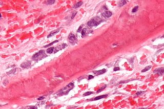
<span class="mw-page-title-main">Osteoblast</span> Cells secreting extracellular matrix

Osteoblasts are cells with a single nucleus that synthesize bone. However, in the process of bone formation, osteoblasts function in groups of connected cells. Individual cells cannot make bone. A group of organized osteoblasts together with the bone made by a unit of cells is usually called the osteon.

<span class="mw-page-title-main">Hyperparathyroidism</span> Increase in parathyroid hormone levels in the blood

Hyperparathyroidism is an increase in parathyroid hormone (PTH) levels in the blood. This occurs from a disorder either within the parathyroid glands or as response to external stimuli. Symptoms of hyperparathyroidism are caused by inappropriately normal or elevated blood calcium excreted from the bones and flowing into the blood stream in response to increased production of parathyroid hormone. In healthy people, when blood calcium levels are high, parathyroid hormone levels should be low. With long-standing hyperparathyroidism, the most common symptom is kidney stones. Other symptoms may include bone pain, weakness, depression, confusion, and increased urination. Both primary and secondary may result in osteoporosis.

<span class="mw-page-title-main">Hypophosphatemia</span> Lack of phosphate in the blood

Hypophosphatemia is an electrolyte disorder in which there is a low level of phosphate in the blood. Symptoms may include weakness, trouble breathing, and loss of appetite. Complications may include seizures, coma, rhabdomyolysis, or softening of the bones.

<span class="mw-page-title-main">Calcitriol</span> Active form of vitamin D

Calcitriol is the active form of vitamin D, normally made in the kidney. It is also known as 1,25-dihydroxycholecalciferol. It is a hormone which binds to and activates the vitamin D receptor in the nucleus of the cell, which then increases the expression of many genes. Calcitriol increases blood calcium (Ca2+) mainly by increasing the uptake of calcium from the intestines.

Renal osteodystrophy is currently defined as an alteration of bone morphology in patients with chronic kidney disease (CKD). It is one measure of the skeletal component of the systemic disorder of chronic kidney disease-mineral and bone disorder (CKD-MBD). The term "renal osteodystrophy" was coined in 1943, 60 years after an association was identified between bone disease and kidney failure.

<span class="mw-page-title-main">Hypophosphatasia</span> Medical condition

Hypophosphatasia (; also called deficiency of alkaline phosphatase, phosphoethanolaminuria, or Rathbun's syndrome; sometimes abbreviated HPP) is a rare, and sometimes fatal, inherited metabolic bone disease. Clinical symptoms are heterogeneous, ranging from the rapidly fatal, perinatal variant, with profound skeletal hypomineralization, respiratory compromise or vitamin B6 dependent seizures to a milder, progressive osteomalacia later in life. Tissue non-specific alkaline phosphatase (TNSALP) deficiency in osteoblasts and chondrocytes impairs bone mineralization, leading to rickets or osteomalacia. The pathognomonic finding is subnormal serum activity of the TNSALP enzyme, which is caused by one of 388 genetic mutations identified to date, in the gene encoding TNSALP. Genetic inheritance is autosomal recessive for the perinatal and infantile forms but either autosomal recessive or autosomal dominant in the milder forms.

<span class="mw-page-title-main">Osteopontin</span> Mammalian protein found in Homo sapiens

Osteopontin (OPN), also known as bone /sialoprotein I, early T-lymphocyte activation (ETA-1), secreted phosphoprotein 1 (SPP1), 2ar and Rickettsia resistance (Ric), is a protein that in humans is encoded by the SPP1 gene. The murine ortholog is Spp1. Osteopontin is a SIBLING (glycoprotein) that was first identified in 1986 in osteoblasts.

<span class="mw-page-title-main">Bone resorption</span> Medical condition

Bone resorption is resorption of bone tissue, that is, the process by which osteoclasts break down the tissue in bones and release the minerals, resulting in a transfer of calcium from bone tissue to the blood.

<span class="mw-page-title-main">Fibroblast growth factor 23</span> Protein-coding gene in the species Homo sapiens

Fibroblast growth factor 23 (FGF23) is a protein and member of the fibroblast growth factor (FGF) family which participates in the regulation of phosphate in plasma and vitamin D metabolism. In humans it is encoded by the FGF23 gene. FGF23 decreases reabsorption of phosphate in the kidney. Mutations in FGF23 can lead to its increased activity, resulting in autosomal dominant hypophosphatemic rickets.

<span class="mw-page-title-main">X-linked hypophosphatemia</span> X-linked dominant disorder that causes rickets

X-linked hypophosphatemia (XLH) is an X-linked dominant form of rickets that differs from most cases of dietary deficiency rickets in that vitamin D supplementation does not cure it. It can cause bone deformity including short stature and genu varum (bow-leggedness). It is associated with a mutation in the PHEX gene sequence (Xp.22) and subsequent inactivity of the PHEX protein. PHEX mutations lead to an elevated circulating (systemic) level of the hormone FGF23 which results in renal phosphate wasting, and local elevations of the mineralization/calcification-inhibiting protein osteopontin in the extracellular matrix of bones and teeth. An inactivating mutation in the PHEX gene results in an increase in systemic circulating FGF23, and a decrease in the enzymatic activity of the PHEX enzyme which normally removes (degrades) mineralization-inhibiting osteopontin protein; in XLH, the decreased PHEX enzyme activity leads to an accumulation of inhibitory osteopontin locally in bones and teeth to block mineralization which, along with renal phosphate wasting, both cause osteomalacia and odontomalacia.

Metabolic bone disease is an abnormality of bones caused by a broad spectrum of disorders. Most commonly these disorders are caused by deficiencies of minerals such as calcium, phosphorus, magnesium or vitamin D leading to dramatic clinical disorders that are commonly reversible once the underlying defect has been treated. These disorders are to be differentiated from a larger group of genetic bone disorders where there is a defect in a specific signaling system or cell type that causes the bone disorder. There may be overlap. For example, genetic or hereditary hypophosphatemia may cause the metabolic bone disorder osteomalacia. Although there is currently no treatment for the genetic condition, replacement of phosphate often corrects or improves the metabolic bone disorder. Metabolic bone disease in captive reptiles is also common, and is typically caused by calcium deficiency in a reptile's diet.

<span class="mw-page-title-main">PHEX</span> Protein-coding gene in the species Homo sapiens

Phosphate-regulating endopeptidase homolog X-linked also known as phosphate-regulating gene with homologies to endopeptidases on the X chromosome or metalloendopeptidase homolog PEX is an enzyme that in humans is encoded by the PHEX gene. This gene contains 18 exons and is located on the X chromosome.

<span class="mw-page-title-main">ALPL</span> Protein-coding gene in the species Homo sapiens

Alkaline phosphatase, tissue-nonspecific isozyme is an enzyme that in humans is encoded by the ALPL gene.

<span class="mw-page-title-main">MEPE</span> Protein-coding gene in the species Homo sapiens

Matrix extracellular phosphoglycoprotein is a protein that in humans is encoded by the MEPE gene. A conserved RGD motif is found in this protein, and this is potentially involved in integrin recognition.

Oncogenic osteomalacia, also known as oncogenic hypophosphatemic osteomalacia, is an uncommon disorder resulting in increased renal phosphate excretion, hypophosphatemia and osteomalacia. It may be caused by a phosphaturic mesenchymal tumor.

<span class="mw-page-title-main">Vitamin D deficiency</span> Human disorder

Vitamin D deficiency or hypovitaminosis D is a vitamin D level that is below normal. It most commonly occurs in people when they have inadequate exposure to sunlight, particularly sunlight with adequate ultraviolet B rays (UVB). Vitamin D deficiency can also be caused by inadequate nutritional intake of vitamin D; disorders that limit vitamin D absorption; and disorders that impair the conversion of vitamin D to active metabolites, including certain liver, kidney, and hereditary disorders. Deficiency impairs bone mineralization, leading to bone-softening diseases, such as rickets in children. It can also worsen osteomalacia and osteoporosis in adults, increasing the risk of bone fractures. Muscle weakness is also a common symptom of vitamin D deficiency, further increasing the risk of fall and bone fractures in adults. Vitamin D deficiency is associated with the development of schizophrenia.

An endocrine bone disease is a bone disease associated with a disorder of the endocrine system. An example is osteitis fibrosa cystica.

Phosphate diabetes is a rare, congenital, hereditary disorder associated with inadequate tubular reabsorption that affects the way the body processes and absorbs phosphate. Also named as X-linked dominant hypophosphatemic rickets (XLH), this disease is caused by a mutation in the X-linked PHEX gene, which encodes for a protein that regulates phosphate levels in the human body. phosphate is an essential mineral which plays a significant role in the formation and maintenance of bones and teeth, energy production and other important cellular processes. phosphate diabetes is a condition that falls under the category of tubulopathies, which refers to the pathologies of the renal tubules. The mutated PHEX gene causes pathological elevations in fibroblast growth factor 23 (FGF23), a hormone that regulates phosphate homeostasis by decreasing the reabsorption of phosphate in the kidneys.

References

  1. McKee, MD; Buss, DJ; Reznikov, N (13 December 2021). "Mineral tessellation in bone and the stenciling principle for extracellular matrix mineralization". Journal of Structural Biology. 214 (1): 107823. doi:10.1016/j.jsb.2021.107823. PMID   34915130. S2CID   245187449.
  2. Buss, DJ; Reznikov, N; McKee, MD (1 November 2020). "Crossfibrillar mineral tessellation in normal and Hyp mouse bone as revealed by 3D FIB-SEM microscopy". Journal of Structural Biology. 212 (2): 107603. doi:10.1016/j.jsb.2020.107603. PMID   32805412. S2CID   221164596.
  3. Salmon, B; Bardet, C; Coyac, BR; Baroukh, B; Naji, J; Rowe, PS; Opsahl Vital, S; Linglart, A; Mckee, MD; Chaussain, C (August 2014). "Abnormal osteopontin and matrix extracellular phosphoglycoprotein localization, and odontoblast differentiation, in X-linked hypophosphatemic teeth". Connective Tissue Research. 55 (Suppl 1): 79–82. doi:10.3109/03008207.2014.923864. PMID   25158186. S2CID   19702315.
  4. Boukpessi, T; Hoac, B; Coyac, BR; Leger, T; Garcia, C; Wicart, P; Whyte, MP; Glorieux, FH; Linglart, A; Chaussain, C; McKee, MD (21 November 2016). "Osteopontin and the dento-osseous pathobiology of X-linked hypophosphatemia". Bone. 95: 151–161. doi:10.1016/j.bone.2016.11.019. PMID   27884786.
  5. Barros, NM; Hoac, B; Neves, RL; Addison, WN; Assis, DM; Murshed, M; Carmona, AK; McKee, MD (March 2013). "Proteolytic processing of osteopontin by PHEX and accumulation of osteopontin fragments in Hyp mouse bone, the murine model of X-linked hypophosphatemia". Journal of Bone and Mineral Research. 28 (3): 688–99. doi: 10.1002/jbmr.1766 . PMID   22991293.
  6. McKee, MD; Hoac, B; Addison, WN; Barros, NM; Millán, JL; Chaussain, C (October 2013). "Extracellular matrix mineralization in periodontal tissues: Noncollagenous matrix proteins, enzymes, and relationship to hypophosphatasia and X-linked hypophosphatemia". Periodontology 2000. 63 (1): 102–22. doi:10.1111/prd.12029. PMC   3766584 . PMID   23931057.
  7. Boukpessi, T; Gaucher, C; Léger, T; Salmon, B; Le Faouder, J; Willig, C; Rowe, PS; Garabédian, M; Meilhac, O; Chaussain, C (August 2010). "Abnormal presence of the matrix extracellular phosphoglycoprotein-derived acidic serine- and aspartate-rich motif peptide in human hypophosphatemic dentin". The American Journal of Pathology. 177 (2): 803–12. doi:10.2353/ajpath.2010.091231. PMC   2913338 . PMID   20581062.
  8. Reznikov, N.; Hoac, B.; Buss, D. J.; Addison, W. N.; Barros NMT; McKee, M. D. (2020). "Biological stenciling of mineralization in the skeleton: Local enzymatic removal of inhibitors in the extracellular matrix". Bone. 138: 115447. doi:10.1016/j.bone.2020.115447. PMID   32454257. S2CID   218909350.
  9. McKee, M. D.; Buss, D. J.; Reznikov, N. (2022). "Mineral tessellation in bone and the Stenciling Principle for extracellular matrix mineralization". Journal of Structural Biology. 214 (1): 107823. doi:10.1016/j.jsb.2021.107823. PMID   34915130. S2CID   245187449.
  10. "Osteomalacia: MedlinePlus Medical Encyclopedia". medlineplus.gov.
  11. 1 2 Longo, Dan L.; et al. (2012). Harrison's principles of internal medicine (18th ed.). New York: McGraw-Hill. ISBN   978-0-07174889-6.
  12. Kennel, KA; Drake, MT; Hurley, DL (August 2010). "Vitamin D deficiency in adults: when to test and how to treat". Mayo Clinic Proceedings. 85 (8): 752–7, quiz 757-8. doi:10.4065/mcp.2010.0138. PMC   2912737 . PMID   20675513.
  13. 1 2 3 4 "Osteomalacia and Rickets". The Lecturio Medical Concept Library. Retrieved 23 August 2021.
  14. Chakravorty, N. K. (1980). "Triradiate deformity of the pelvis in Paget's disease of bone". Postgraduate Medical Journal. 56 (653): 213–5. doi:10.1136/pgmj.56.653.213. PMC   2425842 . PMID   7393817.
  15. "Autoimmunity research foundation, Science behind Vitamin D" . Retrieved 2011-07-19.
  16. Pack, Alison (2008). "Bone health in people with epilepsy: is it impaired and what are the risk factors". Seizure. 17 (2): 181–6. doi: 10.1016/j.seizure.2007.11.020 . PMID   18187347. S2CID   16490292.
  17. "Definition & Facts for Celiac Disease. What are the complications of celiac disease?". NIDDK. June 2016. Retrieved 26 May 2018.
  18. Basu, R.A.; Elmer, K.; Babu, A.; Kelly, C. A. (2000). "Coeliac disease can still present with osteomalacia!". Rheumatology. 39 (3): 335–336. doi: 10.1093/rheumatology/39.3.335 . PMID   10788547.
  19. Holick, Michael F. (19 July 2007). "Vitamin D Deficiency". New England Journal of Medicine. 357 (3): 266–281. doi:10.1056/NEJMra070553. PMID   17634462. S2CID   18566028.
  20. Eisman, John A. (1988). "6 Osteomalacia". Baillière's Clinical Endocrinology and Metabolism. 2 (1): 125–55. doi:10.1016/S0950-351X(88)80011-9. PMID   3044328.