Complex regional pain syndrome

Last updated
Complex regional pain syndrome
Other namesAlgodystrophy; Amplified musculoskeletal pain syndrome; hyponym: reflex sympathetic dystrophy (RSD); hyponym: causalgia; hyponym: reflex neurovascular dystrophy (RND)
Complex regional pain syndrome.jpg
Leg of an individual (left) with complex regional pain syndrome following tibial bone fracture
Specialty Neurology, psychiatry, anesthesiology
Symptoms Pain, allodynia, hypo- or hyperesthesia, skin temperature abnormalities, atrophy, stiffness
TreatmentPhysical therapy (more effective with early diagnosis); medications (e.g., anticonvulsants, opioids, muscle relaxers, etc.); sympathetic nerve blockade; ketamine infusions; lidocaine infusions; implantable pharmaceuticals; amputation
Medication Anticonvulsants (e.g., gabapentin); muscle relaxers (e.g., baclofen), ketamine or lidocaine infusions[ citation needed ]

Complex regional pain syndrome (CRPS Type 1 and Type 2) is a severe form of chronic pain, in which pain from a physical trauma outlasts the expected recovery time. The symptoms of types 1 and 2 are the same except type 2 is associated with nerve injury.

Contents

Usually starting in a limb, CRPS manifests as pain, swelling, limited range of motion, and/or changes to the skin and bones. It may initially affect one limb and then spread throughout the body; 35% of affected individuals report symptoms throughout the body. [1] Two types are thought to exist: CRPS type 1 (previously referred to as reflex sympathetic dystrophy) and CRPS type 2 (previously referred to as causalgia). It is possible to have both types. [2]

Amplified musculoskeletal pain syndrome, a condition that is similar to CRPS, primarily affects pediatric patients, falls under rheumatology and pediatrics, and is generally considered a subset of CRPS type I. [3]

Classification

The classification system in use by the International Association for the Study of Pain (IASP) divides CRPS into two types based on the presence or absence of measurable nerve pathophysiology. [4] [5]

International Association for the Study of Pain Classification
TypeClinical findingsSynonyms
Type ICRPS without evidence of neuropathology in the affected limb. This accounts for about 90% of CRPS.reflex sympathetic dystrophy, RSD, Sudeck's atrophy
Type IICRPS with evidence of neuropathology in the affected limb.causalgia

Signs and symptoms

Severe CRPS of right arm Severe CRPS.jpg
Severe CRPS of right arm
CRPS visible on hands and wrists CRPS 002ms5.jpg
CRPS visible on hands and wrists

Clinical features of CRPS have been found to be inflammation resulting from the release of certain pro-inflammatory chemical signals from surrounding nerve cells; hypersensitization of pain receptors; dysfunction of local vasoconstriction and vasodilation; and maladaptive neuroplasticity. [6]

The signs and symptoms of CRPS usually manifest near the injury site. The most common symptoms are extreme pain, including burning, stabbing, grinding, and throbbing. The pain is out of proportion to the severity of the initial injury. [7] Moving or touching the limb is disproportionately painful (allodynia). Other findings are aspects of disuse including swelling, stiffness (limited range of motion), and disuse related changes to the skin (temperature, color, sweating, texture) and bones (disuse osteoporosis). [8] [9]

A prior concept of CRPS having three stages is no longer in wide use. [10] The trend is now to consider distinct sub-types of CRPS. [10]

Cause

Complex regional pain syndrome is uncommon, and its cause is not clearly understood. CRPS typically develops after an injury, surgery, heart attack, or stroke. [7] [11] Investigators estimate that 2–5% of those with peripheral nerve injury, [12] and 13–70% of those with hemiplegia (paralysis of one side of the body) [13] will develop CRPS. In addition, some studies have indicated that cigarette smoking was strikingly present in patients and is statistically linked to RSD. This may be involved in its pathology by enhancing sympathetic activity, vasoconstriction, or by some other unknown neurotransmitter-related mechanism. This hypothesis was based on a retrospective analysis of 53 patients with RSD, which showed that 68% of patients were smokers, compared to only 37% of the control population. The results are preliminary and are limited by their retrospective nature. [14] 7% of people who have CRPS in one limb later develop it in another limb. [15]

Pathophysiology

Inflammation and alteration of pain perception in the central nervous system are proposed to play important roles. The persistent pain and the perception of nonpainful stimuli as painful are thought to be caused by inflammatory molecules (IL-1, IL-2, TNF-alpha) and neuropeptides (substance P) released from peripheral nerves. This release may be caused by inappropriate cross-talk between sensory and motor fibers at the affected site. [16] CRPS is not a psychological illness, yet pain can cause psychological problems, such as anxiety and depression. Often, impaired social and occupational function occur. [17]

Complex regional pain syndrome is a multifactorial disorder with clinical features of neurogenic inflammation (inflammation mediated by nerve cells), nociceptive sensitisation (which causes extreme sensitivity or allodynia), vasomotor dysfunction (blood flow problems which cause swelling and discolouration) and maladaptive neuroplasticity (where the brain changes and adapts with constant pain signals); CRPS is the result of an "aberrant [inappropriate] response to tissue injury". [6] The "underlying neuronal matrix" of CRPS is seen to involve cognitive and motor as well as nociceptive processing; pinprick stimulation of a CRPS affected limb was painful (mechanical hyperalgesia) and showed a "significantly increased activation" of not just the S1 cortex (contralateral), S2 (bilateral) areas, and insula (bilateral) but also the associative-somatosensory cortices (contralateral), frontal cortices, and parts of the anterior cingulate cortex. [18] In contrast to previous thoughts reflected in the name RSD, it appears that there is reduced sympathetic nervous system outflow, at least in the affected region (although there may be sympatho-afferent coupling). [19] Wind-up (the increased sensation of pain with time) [20] and central nervous system (CNS) sensitization are key neurologic processes that appear to be involved in the induction and maintenance of CRPS. [21]

Compelling evidence shows that the N-methyl-D-aspartate (NMDA) receptor has significant involvement in the CNS sensitization process. [22] It is also hypothesized that elevated CNS glutamate levels promote wind-up and CNS sensitization. [21] In addition, there exists experimental evidence demonstrating the presence of NMDA receptors in peripheral nerves. [23] Because immunological functions can modulate CNS physiology, a variety of immune processes have also been hypothesized to contribute to the initial development and maintenance of peripheral and central sensitization. [24] [25] Furthermore, trauma-related cytokine release, exaggerated neurogenic inflammation, sympathetic afferent coupling, adrenoreceptor pathology, glial cell activation, cortical reorganisation, [26] and oxidative damage (e.g., by free radicals) are all factors which have been implicated in the pathophysiology of CRPS. [27] In addition, autoantibodies are present in a wide number of CRPS patients and IgG has been recognized as one of the causes of hypersensitivity that stimulates A and C nociceptors, attributing to the inflammation. [28]

The mechanisms leading to reduced bone mineral density (up to overt osteoporosis) are still unknown. Potential explanations include a dysbalance of the activities of sympathetic and parasympathetic autonomic nervous system [29] [30] [31] and mild secondary hyperparathyroidism. [32] However, the trigger of secondary hyperparathyroidism has not yet been identified.[ citation needed ]

In summary, the pathophysiology of complex regional pain syndrome has not yet been defined; CRPS, with its variable manifestations, could be the result of multiple pathophysiological processes. [19]

Diagnosis

Diagnosis is primarily based on clinical findings. The original diagnostic criteria for CRPS adopted by the International Association for the Study of Pain (IASP) in 1994 have now been superseded in both clinical practice and research by the "Budapest Criteria" which were created in 2003 and have been found to be more sensitive and specific. [33] They have since been adopted by the IASP. The criteria require there to be pain as well as a history and clinical evidence of sensory, vasomotor, sudomotor, and motor or trophic changes. It is also stated that it is a diagnosis of exclusion. [34]

To make a clinical diagnosis all four of the following criteria must be met: [35]

  1. Continuing pain, which is disproportionate to any inciting event
  2. Must report at least one symptom in three of the four following categories.
    • Sensory: Reports of hyperesthesia
    • Vasomotor: Reports of temperature asymmetry and/or skin color changes and/or skin color asymmetry
    • Sudomotor/Edema: Reports of edema and/or sweating changes and/or sweating asymmetry
    • Motor/Trophic: Reports of decreased range of motion and/or motor dysfunction (weakness, tremor, dystonia) and/or trophic changes (hair, nail, skin)
  3. Must display at least one sign at time of evaluation in two or more of the following categories
    • Sensory: Evidence of hyperalgesia (to pinprick) and/or allodynia (to light touch and/or temperature sensation and/or deep somatic pressure and/or joint movement)
    • Vasomotor: Evidence of temperature asymmetry (>1 °C) and/or skin color changes and/or asymmetry
    • Sudomotor/Edema: Evidence of edema and/or sweating changes and/or sweating asymmetry
    • Motor/Trophic: Evidence of decreased range of motion and/or motor dysfunction (weakness, tremor, dystonia) and/or trophic changes (hair, nail, skin)
  4. There is no other diagnosis that better explains the signs and symptoms

Diagnostic adjuncts

No specific test is available for CRPS, which is diagnosed primarily through observation of the symptoms. However, thermography, sweat testing, X-rays, electrodiagnostics, and sympathetic blocks can be used to build up a picture of the disorder. Diagnosis is complicated by the fact that some patients improve without treatment. A delay in diagnosis and/or treatment for this syndrome can result in severe physical and psychological problems. Early recognition and prompt treatment provide the greatest opportunity for recovery.[ citation needed ]

Thermography

Presently, established empirical evidence suggests against thermography's efficacy as a reliable tool for diagnosing CRPS. Although CRPS may, in some cases, lead to measurably altered blood flow throughout an affected region, many other factors can also contribute to an altered thermographic reading, including the patient's smoking habits, use of certain skin lotions, recent physical activity, and prior history of trauma to the region. Also, not all patients diagnosed with CRPS demonstrate such "vasomotor instability"—particularly those in the later stages of the disease. [36] Thus, thermography alone cannot be used as conclusive evidence for—or against—a diagnosis of CRPS and must be interpreted in light of the patient's larger medical history and prior diagnostic studies. [37]

In order to minimise the confounding influence of external factors, patients undergoing infrared thermographic testing must conform to special restrictions regarding the use of certain vasoconstrictors (namely, nicotine and caffeine), skin lotions, physical therapy, and other diagnostic procedures in the days prior to testing. Patients may also be required to discontinue certain pain medications and sympathetic blockers. After a patient arrives at a thermographic laboratory, he or she is allowed to reach thermal equilibrium in a 16–20 °C, draft-free, steady-state room wearing a loose fitting cotton hospital gown for approximately twenty minutes. A technician then takes infrared images of both the patient's affected and unaffected limbs, as well as reference images of other parts of the patient's body, including his or her face, upper back, and lower back. After capturing a set of baseline images, some labs further require the patient to undergo cold-water autonomic-functional-stress-testing to evaluate the function of their autonomic nervous system's peripheral vasoconstrictor reflex. This is performed by placing a patient's unaffected limb in a cold water bath (approximately 20 °C) for five minutes while collecting images. In a normal, intact, functioning autonomic nervous system, a patient's affected extremity will become colder. Conversely, warming of an affected extremity may indicate a disruption of the body's normal thermoregulatory vasoconstrictor function, which may sometimes indicate underlying CRPS. [38]

Radiography

Scintigraphy, plain radiographs, and magnetic resonance imaging may all be useful diagnostically. Patchy osteoporosis (post-traumatic osteoporosis), which may be due to disuse of the affected extremity, can be detected through X-ray imagery as early as two weeks after the onset of CRPS. A bone scan of the affected limb may detect these changes even sooner and can almost confirm the disease. Bone densitometry can also be used to detect changes in bone mineral density. It can also be used to monitor the results of treatment since bone densitometry parameters improve with treatment.[ citation needed ]

Ultrasound

Ultrasound-based osteodensitometry (ultrasonometry) may be potential future radiation-free technique to identify reduced bone mineral density in CRPS. [32] Additionally, this method promises to quantify the bone architecture in the periphery of affected limbs. [32] This method is still under experimental development.[ citation needed ]

Electrodiagnostic testing

Electromyography (EMG) and nerve conduction studies (NCS) are important ancillary tests in CRPS because they are among the most reliable methods of detecting nerve injury. They can be used as one of the primary methods to distinguish between CRPS types I and II, which differ based on evidence of actual nerve damage. EMG and NCS are also among the best tests for ruling in or out alternative diagnoses. CRPS is a "diagnosis of exclusion", which requires that no other diagnosis can explain the patient's symptoms. This is very important to emphasise because patients otherwise can be given a wrong diagnosis of CRPS when they actually have a treatable condition that better accounts for their symptoms. An example is severe carpal tunnel syndrome (CTS), which can often present in a very similar way to CRPS. Unlike CRPS, CTS can often be corrected with surgery to alleviate the pain and avoid permanent nerve damage and malformation. [39]

Both EMG and NCS involve some measure of discomfort. EMG involves the use of a tiny needle inserted into specific muscles to test the associated muscle and nerve function. Both EMG and NCS involve very mild shocks that in normal patients are comparable to a rubber band snapping on the skin. Although these tests can be very useful in CRPS, thorough informed consent must be obtained prior to the procedure, particularly in patients experiencing severe allodynia. In spite of the utility of the test, these patients may wish to decline the procedure to avoid discomfort.[ citation needed ]

Classification

  • Type I, formerly known as reflex sympathetic dystrophy (RSD), Sudeck's atrophy, or algoneurodystrophy, does not exhibit demonstrable nerve lesions. As the vast majority of patients diagnosed with CRPS have this type, it is most commonly referred to in medical literature as type I.[ citation needed ]
  • Type II, formerly known as causalgia, has evidence of obvious nerve damage. Despite evidence of nerve injury, the cause or the mechanisms of CRPS type II are as unknown, as the mechanisms of type I.[ citation needed ]

Patients are frequently classified into two groups based upon temperature: "warm" or "hot" CRPS in one group and "cold" CRPS in the other group. The majority of patients (about 70%) have the "hot" type, which is said to be an acute form of CRPS. [40] Cold CRPS is said to be indicative of a more chronic CRPS and is associated with poorer McGill Pain Questionnaire scores, increased central nervous system reorganisation, and a higher prevalence of dystonia. [40] Prognosis is not favourable for cold CRPS patients; longitudinal studies suggest these patients have "poorer clinical pain outcomes and show persistent signs of central sensitisation correlating with disease progression". [41]

Prevention

Vitamin C supplementation may be useful in prevention of the syndrome following fracture of the forearm, foot, or ankle. [42]

Treatment

Treatment of CRPS often involves a number of modalities. [43]

Therapy

Physical and occupational therapy have low-quality evidence to support their use. [44] Physical therapy interventions may include transcutaneous electrical nerve stimulation, progressive weight bearing, graded tactile desensitization, massage, and contrast bath therapy. In a retrospective cohort (unblinded, non-randomised and with intention-to-treat) of fifty patients diagnosed with CRPS, the subjective pain and body perception scores of patients decreased after engagement with a two-week multidisciplinary rehabilitation programme. The authors call for randomised controlled trials to probe the true value of multidisciplinary programs for CRPS patients. [45]

Mirror box therapy

Mirror box therapy uses a mirror box, or a stand-alone mirror, to create a reflection of the normal limb such that the patient thinks they are looking at the affected limb. Movement of this reflected normal limb is then performed so that it looks to the patient as though they are performing movement with the affected limb. Mirror box therapy appears to be beneficial at least in early CRPS. [46] However, beneficial effects of mirror therapy in the long term is still unproven. [47]

Graded motor imagery

Graded motor imagery appears to be useful for people with CRPS-1. [48] Graded motor imagery is a sequential process that consists of (a) laterality reconstruction, (b) motor imagery, and (c) mirror therapy. [43] [49]

Transcutaneous Electrical Nerve Stimulation (TENS)

Transcutaneous Electrical Nerve Stimulation (TENS) is a therapy that uses low-voltage electrical signals to provide pain relief through electrodes that are placed on the surface of the skin. Evidence supports its use in treating pain and edema associated with CRPS, but it does not seem to increase functional ability in CRPS patients. [50]

Medications

Tentative evidence supports the use of bisphosphonates, calcitonin, and ketamine. [44] [51] Nerve blocks with guanethidine appear to be harmful. [44] Evidence for sympathetic nerve blocks generally is insufficient to support their use. [52] Intramuscular botulinum injections may benefit people with symptoms localized to one extremity. [53]

Ketamine

Ketamine, a dissociative anesthetic, appears promising as a treatment for CRPS. [54] It may be used in low doses if other treatments have not worked. [55] [56] No benefit on either function or depression, however, has been seen. [56]

Bisphosphonate treatment

As of 2013, high-quality evidence supports the use of bisphosphonates (either orally or via IV infusion) in the treatment of CRPS. [51] Bisphosphonates inhibit osteoclasts: cells involved in the resorption of bone. Bone remodeling (via osteoclast activity in resorption of bone) is thought to sometimes be hyperactive in CRPS. It is hypothesized that bone resorption causes acidification of the intercellular milieu which, in turn, activates nerves involved in nociception that densely innervate bone and causes pain. [51] Therefore, inhibiting bone resorption and remodeling is thought to help with regard to CRPS pain. [51] CRPS involving high levels of bone resorption, as seen on bone scan, is more likely to respond to bisphosphonate therapy. [51]

Opioids

Opioids such as oxycodone, morphine, hydrocodone, and fentanyl have a controversial place in treatment of CRPS. These drugs must be prescribed and monitored under close supervision of a physician as they can quickly lead to physical dependence and addiction. [57] To date so far, no long-term studies of oral opioid use in treating neuropathic pain, including CRPS, have been performed. The consensus among experts is that opioids should not be a first-line therapy and should be considered only after all other modalities (e.g., non-opioid medications, physical therapy, and procedures) have been trialed. [58]

Surgery

Spinal cord stimulators

Spinal cord stimulation appears to be an effective therapy in the management of patients with CRPS type I (level A evidence) and type II (level D evidence). [59] Although they improve patient pain and quality of life, evidence is unclear regarding effects on mental health and general functioning. [60]

Dorsal root ganglion stimulation is a type of neurostimulation that is effective in the management of focal neuropathic pain. The FDA approved its use in February 2016. The ACCURATE Study demonstrated superiority of dorsal root ganglion stimulation over spinal (dorsal column) stimulation in the management of CRPS and causalgia. [61]

Sympathectomy

Surgical, chemical, or radiofrequency sympathectomy—interruption of the affected portion of the sympathetic nervous system—can be used as a last resort in patients with impending tissue loss, edema, recurrent infection, or ischemic necrosis. [62] However, little evidence supports these permanent interventions to alter the pain symptoms of the affected patients, and in addition to the normal risks of surgery, such as bleeding and infection, sympathectomy has several specific risks, such as adverse changes in how nerves function.[ citation needed ]

Amputation

No randomized study in medical literature has studied the response with amputation of patients who have failed the above-mentioned therapies and who continue to be in pain. Nonetheless, on average, about half of the patients will have resolution of their pain, while half will develop phantom limb pain and/or pain at the amputation site. As in any other chronic pain syndrome, the brain likely becomes chronically stimulated with pain, and late amputation may not work as well as it might be expected. In a survey of 15 patients with CRPS type 1, 11 responded that their lives were better after amputation. [63]

Other

Cannabidiol (CBD), despite evidence of very low quality, is proposed to relieve pain. [64]

Prognosis

The prognosis in CRPS is improved with early and aggressive treatment; with the risk of chronic, debilitating pain being reduced with the early treatment. [65] If treatment is delayed, however, the disorder can quickly spread to the entire limb, and changes in bone, nerve, and muscle may become irreversible. The prognosis is worse with the chronic "cold" form of CRPS and with CRPS affecting the upper extremities. [65] Disuse of the limb after an injury or psychological distress related to an injury are also associated with a poorer prognosis in CRPS. [65] Some cases of CRPS may resolve spontaneously (with 74% of patients in a population-based study in Minnesota undergoing complete resolution of symptoms, often spontaneously), but others may develop chronic pain for many years. [65] Once one is diagnosed with CRPS, should it go into remission, the likelihood of it resurfacing after going into remission is significant. Taking precautions and seeking immediate treatment upon any injury is important. [66]

Epidemiology

CRPS can occur at any age, with the average age at diagnosis being 42. [12] It affects both men and women; however, CRPS is three times more frequent in females than males. [12]

CRPS affects both adults and children, and the number of reported CRPS cases among adolescents and young adults has been increasing, [67] with a recent observational study finding an incidence of 1.16/100,000 among children in Scotland. [68]

History

The condition currently known as CRPS was originally described by Ambroise Paré. He successfully treated a severe and persistent pain syndrome that occurred to the French King Charles IX of Valois after a limb phlebotomy . During the American Civil War, Silas Weir Mitchell is sometimes also credited with inventing the name "causalgia". [69] However, this term was actually coined by Mitchell's friend Robley Dunglison from the Greek words for heat and for pain. [70] Contrary to what is commonly accepted, it emerges that these causalgias were certainly major by the importance of the vasomotor and sudomotor symptoms but stemmed from minor neurological lesions. In the 1940s, the term reflex sympathetic dystrophy came into use to describe this condition, based on the theory that sympathetic hyperactivity was involved in the pathophysiology. [71] In 1959, Noordenbos observed in causalgia patients that "the damage of the nerve is always partial." [72] Misuse of the terms, as well as doubts about the underlying pathophysiology, led to calls for better nomenclature. In 1993, a special consensus workshop held in Orlando, Florida, provided the umbrella term "complex regional pain syndrome", with causalgia and RSD as subtypes. [73]

Research

The National Institute of Neurological Disorders and Stroke (NINDS), a part of the National Institutes of Health, supports and conducts research on the brain and central nervous system, including research relevant to RSDS, through grants to major medical institutions across the country. NINDS-supported scientists are working to develop effective treatments for neurological conditions and ultimately, to find ways of preventing them. Investigators are studying new approaches to treat CRPS and intervene more aggressively after traumatic injury to lower the patient's chances of developing the disorder. In addition, NINDS-supported scientists are studying how signals of the sympathetic nervous system cause pain in CRPS patients. Using a technique called microneurography, these investigators are able to record and measure neural activity in single nerve fibers of affected patients. By testing various hypotheses, these researchers hope to discover the unique mechanism that causes the spontaneous pain of CRPS, and that discovery may lead to new ways of blocking pain. Other studies to overcome chronic pain syndromes are discussed in the pamphlet "Chronic Pain: Hope Through Research", published by the NINDS.[ citation needed ]

Research into treating the condition with mirror visual feedback is being undertaken at the Royal National Hospital for Rheumatic Disease in Bath. Patients are taught how to desensitize in the most effective way, then progress to using mirrors to rewrite the faulty signals in the brain that appear responsible for this condition. [74] However, while CRPS can go into remission, the chance of it reoccurring is significant.[ citation needed ]

The Netherlands has the most comprehensive program of research into CRPS, as part of a multimillion-Euro initiative called TREND. [75] German and Australian research teams are also pursuing better understanding and treatments for CRPS.[ citation needed ] [76]

In other animal species

CRPS has also been described in non-human animals, such as cattle. [77]

Notable cases

Related Research Articles

<span class="mw-page-title-main">Bell's palsy</span> Facial paralysis resulting from dysfunction in the cranial nerve VII (facial nerve)

Bell's palsy is a type of facial paralysis that results in a temporary inability to control the facial muscles on the affected side of the face. In most cases, the weakness is temporary and significantly improves over weeks. Symptoms can vary from mild to severe. They may include muscle twitching, weakness, or total loss of the ability to move one or, in rare cases, both sides of the face. Other symptoms include drooping of the eyebrow, a change in taste, and pain around the ear. Typically symptoms come on over 48 hours. Bell's palsy can trigger an increased sensitivity to sound known as hyperacusis.

<span class="mw-page-title-main">Carpal tunnel syndrome</span> Medical condition

Carpal tunnel syndrome (CTS) is the collection of symptoms and signs associated with nerve compression of the median nerve at the carpal tunnel. Most CTS is related to idiopathic compression of the median nerve as it travels through the wrist at the carpal tunnel. Idiopathic means that there is no other disease process contributing to pressure on the nerve. As with most structural issues, it occurs in both hands, and the strongest risk factor is genetics.

<span class="mw-page-title-main">Back pain</span> Area of body discomfort

Back pain is pain felt in the back. It may be classified as neck pain (cervical), middle back pain (thoracic), lower back pain (lumbar) or coccydynia based on the segment affected. The lumbar area is the most common area affected. An episode of back pain may be acute, subacute or chronic depending on the duration. The pain may be characterized as a dull ache, shooting or piercing pain or a burning sensation. Discomfort can radiate to the arms and hands as well as the legs or feet, and may include numbness or weakness in the legs and arms.

<span class="mw-page-title-main">Compartment syndrome</span> Condition in which increased pressure results in insufficient blood supply

Compartment syndrome is a condition in which increased pressure within one of the body's anatomical compartments results in insufficient blood supply to tissue within that space. There are two main types: acute and chronic. Compartments of the leg or arm are most commonly involved.

<span class="mw-page-title-main">Tietze syndrome</span> Inflammation, tenderness, and pain of the chest wall with swelling present

Tietze syndrome is a benign inflammation of one or more of the costal cartilages. It was first described in 1921 by German surgeon Alexander Tietze and was subsequently named after him. The condition is characterized by tenderness and painful swelling of the anterior (front) chest wall at the costochondral, sternocostal, or sternoclavicular junctions. Tietze syndrome affects the true ribs and has a predilection for the 2nd and 3rd ribs, commonly affecting only a single joint.

<span class="mw-page-title-main">Raynaud syndrome</span> Medical condition in which spasm of arteries causes episodes of reduced blood flow

Raynaud syndrome, also known as Raynaud's phenomenon, is a medical condition in which the spasm of small arteries causes episodes of reduced blood flow to end arterioles. Typically the fingers, and, less commonly, the toes, are involved. Rarely, the nose, ears, nipples, or lips are affected. The episodes classically result in the affected part turning white and then blue. Often, numbness or pain occurs. As blood flow returns, the area turns red and burns. The episodes typically last minutes but can last several hours. The condition is named after the physician Auguste Gabriel Maurice Raynaud, who first described it in his doctoral thesis in 1862.

<span class="mw-page-title-main">Erythromelalgia</span> Inflammation due to periodic blood vessel blockage

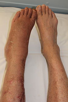
Erythromelalgia or Mitchell's disease is a rare vascular peripheral pain disorder in which blood vessels, usually in the lower extremities or hands, are episodically blocked, then become hyperemic and inflamed. There is severe burning pain and skin redness. The attacks are periodic and are commonly triggered by heat, pressure, mild activity, exertion, insomnia or stress. Erythromelalgia may occur either as a primary or secondary disorder. Secondary erythromelalgia can result from small fiber peripheral neuropathy of any cause, polycythemia vera, essential thrombocythemia, hypercholesterolemia, mushroom or mercury poisoning, and some autoimmune disorders. Primary erythromelalgia is caused by mutation of the voltage-gated sodium channel α-subunit gene SCN9A.

<span class="mw-page-title-main">Piriformis syndrome</span> Medical condition

Piriformis syndrome is a condition which is believed to result from nerve compression at the sciatic nerve by the piriformis muscle. It is a specific case of deep gluteal syndrome. The largest and most bulky nerve in the human body is the sciatic nerve. Starting at its origin it is 2 cm wide and 0.5 cm thick. The sciatic nerve forms the roots of L4-S3 segments of the lumbosacral plexus. The nerve will pass inferiorly to the piriformis muscle, in the direction of the lower limb where it divides into common tibial and fibular nerves. Symptoms may include pain and numbness in the buttocks and down the leg. Often symptoms are worsened with sitting or running.

Endoscopic thoracic sympathectomy (ETS) is a surgical procedure in which a portion of the sympathetic nerve trunk in the thoracic region is destroyed. ETS is used to treat excessive sweating in certain parts of the body, facial blushing, Raynaud's disease and reflex sympathetic dystrophy. By far the most common complaint treated with ETS is sweaty palms. The intervention is controversial and illegal in some jurisdictions. Like any surgical procedure, it has risks; the endoscopic sympathetic block (ESB) procedure and those procedures that affect fewer nerves have lower risks.

<span class="mw-page-title-main">Cauda equina syndrome</span> Nerve damage at the end of the spinal cord

Cauda equina syndrome (CES) is a condition that occurs when the bundle of nerves below the end of the spinal cord known as the cauda equina is damaged. Signs and symptoms include low back pain, pain that radiates down the leg, numbness around the anus, and loss of bowel or bladder control. Onset may be rapid or gradual.

<span class="mw-page-title-main">Neurofibromatosis type I</span> Type of neurofibromatosis disease

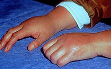
Neurofibromatosis type I (NF-1), or von Recklinghausen syndrome, is a complex multi-system human disorder caused by the mutation of neurofibromin 1 (NF-1), a gene on chromosome 17 that is responsible for production of a protein (neurofibromin) which is needed for normal function in many human cell types. NF-1 causes tumors along the nervous system which can grow anywhere on the body. NF-1 is one of the most common genetic disorders and is not limited to any person's race or sex. NF-1 is an autosomal dominant disorder, which means that mutation or deletion of one copy of the NF-1 gene is sufficient for the development of NF-1, although presentation varies widely and is often different even between relatives affected by NF-1.

<span class="mw-page-title-main">Mirror therapy</span> Treatment for some kinds of pain

Mirror therapy (MT) or mirror visual feedback (MVF) is a therapy for pain or disability that affects one side of the patient more than the other side. It was invented by Vilayanur S. Ramachandran to treat post-amputation patients who had phantom limb pain (PLP). Ramachandran created a visual illusion of two intact limbs by putting the patient's affected limb into a "mirror box," with a mirror down the center.

<span class="mw-page-title-main">Stellate ganglion</span>

The stellate ganglion is a sympathetic ganglion formed by the fusion of the inferior cervical ganglion and the first thoracic ganglion, which is present in 80% of individuals. Sometimes, the second and the third thoracic ganglia are included in this fusion.

<span class="mw-page-title-main">Pelvic pain</span> Medical condition

Pelvic pain is pain in the area of the pelvis. Acute pain is more common than chronic pain. If the pain lasts for more than six months, it is deemed to be chronic pelvic pain. It can affect both the male and female pelvis.

<span class="mw-page-title-main">Radiculopathy</span> Medical condition

Radiculopathy, also commonly referred to as pinched nerve, refers to a set of conditions in which one or more nerves are affected and do not work properly. Radiculopathy can result in pain, weakness, altered sensation (paresthesia) or difficulty controlling specific muscles. Pinched nerves arise when surrounding bone or tissue, such as cartilage, muscles or tendons, put pressure on the nerve and disrupt its function.

<span class="mw-page-title-main">Spinal stenosis</span> Disease of the bony spine that results in narrowing of the spinal canal

Spinal stenosis is an abnormal narrowing of the spinal canal or neural foramen that results in pressure on the spinal cord or nerve roots. Symptoms may include pain, numbness, or weakness in the arms or legs. Symptoms are typically gradual in onset and improve with leaning forward. Severe symptoms may include loss of bladder control, loss of bowel control, or sexual dysfunction.

<span class="mw-page-title-main">Cervicocranial syndrome</span> Medical condition

Cervicocranial syndrome or is a neurological illness. It is a combination of symptoms that are caused by an abnormality in the neck. The bones of the neck that are affected are cervical vertebrae (C1-C7). This syndrome can be identified by confirming cervical bone shifts, collapsed cervical bones or misalignment of the cervical bone leading to improper functioning of cervical spinal nerves. Cervicocranial syndrome is either congenital or acquired. Some examples of diseases that could result in cervicocranial syndrome are Chiari disease, Klippel-Feil malformation osteoarthritis, and trauma. Treatment options include neck braces, pain medication and surgery. The quality of life for individuals suffering from CCJ syndrome can improve through surgery.

Barré–Liéou syndrome is a traditional medical diagnosis that is not utilized frequently in modern medicine. It is a complex combination of symptoms, amounting to a headache syndrome, that was originally hypothesized to be due to cervical spondylosis. Damage to the posterior cervical sympathetic chain due to the degeneration of the cervical vertebra was theorized to play a role in this syndrome by the prolapsing of disc in the mid-cervical spine. However, the medical theory as was originally postulated was found to contain inconsistencies.

Peripheral mononeuropathy is a nerve related disease where a single nerve, that is used to transport messages from the brain to the peripheral body, is diseased or damaged. Peripheral neuropathy is a general term that indicates any disorder of the peripheral nervous system. The name of the disorder itself can be broken down in order to understand this better; peripheral: in regard to peripheral neuropathy, refers to outside of the brain and spinal cord; neuro: means nerve related; -pathy; means disease. Peripheral mononeuropathy is a disorder that links to Peripheral Neuropathy, as it only effects a single peripheral nerve rather than several damaged or diseased nerves throughout the body. Healthy peripheral nerves are able to “carry messages from the brain and spinal cord to muscles, organs, and other body tissues”.

<span class="mw-page-title-main">Amplified musculoskeletal pain syndrome</span> Condition characterized by amplified and chronic pain

Amplified musculoskeletal pain syndrome (AMPS) is an illness characterized by notable pain intensity without an identifiable physical cause.

References

  1. Schwartzman, R.J.; Erwin, K.L.; Alexander, G.M. (May 2009). "The natural history of complex regional pain syndrome". The Clinical Journal of Pain. 25 (4): 273–280. doi:10.1097/AJP.0b013e31818ecea5. PMID   19590474. S2CID   10909080.
  2. Harden, R.N.; Bruehl, S.; Stanton-Hicks, M.; Wilson, P.R. (May 2007). "Proposed new diagnostic criteria for complex regional pain syndrome". Pain Medicine. 8 (4): 326–331. doi: 10.1111/j.1526-4637.2006.00169.x . PMID   17610454.
  3. "Amplified Musculoskeletal Pain Syndrome (AMPS) in Children". Cleveland Clinic. Retrieved 2024-02-10.
  4. Young-Do, K (1970). "Diagnosis of complex regional pain syndrome". Annals of Clinical Neurophysiology. 24 (2): 35–45. doi: 10.14253/acn.2022.24.2.35 . S2CID   253273786.
  5. Goebel, A; Birklein, F; Brunner, F; Clark, JD; Gierthmühlen, J; Harden, N; Huygen, F; Knudsen, L; McCabe, C; Lewis, J; Maihöfner, C; Magerl, W; Moseley, GL; Terkelsen, A; Thomassen, I; Bruehl, S (2021). "The Valencia consensus-based adaptation of the IASP complex regional pain syndrome diagnostic criteria". Pain. 162 (9): 2346–2348. doi:10.1097/j.pain.0000000000002245. PMC   8374712 . PMID   33729210. S2CID   232263568.
  6. 1 2 Marinus J, Moseley GL, Birklein F, Baron R, Maihöfner C, Kingery WS, van Hilten JJ (July 2011). "Clinical features and pathophysiology of complex regional pain syndrome". The Lancet. Neurology. 10 (7): 637–648. doi:10.1016/S1474-4422(11)70106-5. PMC   5511749 . PMID   21683929.
  7. 1 2 "Complex regional pain syndrome - Symptoms and causes". Mayo Clinic.
  8. Taylor SS, Noor N, Urits I, Paladini A, Sadhu MS, Gibb C, et al. (December 2021). "Complex Regional Pain Syndrome: A Comprehensive Review". Pain and Therapy. 10 (2) (published 24 June 2021): 875–892. doi:10.1007/s40122-021-00279-4. PMC   8586273 . PMID   34165690.
  9. Rolvien, T; Amling, M (2022). "Disuse Osteoporosis: Clinical and Mechanistic Insights". Calcif Tissue Int. 110 (5): 592–604. doi: 10.1007/s00223-021-00836-1 . PMC   9013332 . PMID   33738515. S2CID   232304611.
  10. 1 2 Bruehl S, Harden RN, Galer BS, Saltz S, Backonja M, Stanton-Hicks M (January 2002). "Complex regional pain syndrome: are there distinct subtypes and sequential stages of the syndrome?". Pain. 95 (1–2): 119–124. doi:10.1016/s0304-3959(01)00387-6. PMID   11790474. S2CID   20773804.
  11. "Complex regional pain syndrome". nhs.uk. 2017-10-19.
  12. 1 2 3 Veldman PH, Reynen HM, Arntz IE, Goris RJ (October 1993). "Signs and symptoms of reflex sympathetic dystrophy: prospective study of 829 patients". Lancet. 342 (8878): 1012–1016. doi:10.1016/0140-6736(93)92877-V. PMID   8105263. S2CID   39843988.
  13. Yu D (August 2004). "Shoulder pain in hemiplegia". Physical Medicine and Rehabilitation Clinics of North America. 15 (3): vi–vii, 683–97. doi:10.1016/S1047-9651(03)00130-X. PMID   15219895.
  14. Pawelka S, Fialka V, Ernst E (January 1993). "Reflex sympathetic dystrophy and cigarette smoking". The Journal of Hand Surgery. 18 (1): 168–169. doi:10.1016/0363-5023(93)90273-6. PMID   8423309.
  15. Goebel A, Turner-Stokes LF (May 2012). Complex regional pain syndrome in adults: UK guidelines for diagnosis, referral and management in primary and secondary care. Royal College of Physicians London.
  16. Bussa M, Guttilla D, Lucia M, Mascaro A, Rinaldi S (July 2015). "Complex regional pain syndrome type I: a comprehensive review". Acta Anaesthesiologica Scandinavica. 59 (6): 685–697. doi:10.1111/aas.12489. PMID   25903457. S2CID   20134091.
  17. Lohnberg JA, Altmaier EM (June 2013). "A review of psychosocial factors in complex regional pain syndrome". Journal of Clinical Psychology in Medical Settings. 20 (2): 247–254. doi:10.1007/s10880-012-9322-3. PMID   22961122. S2CID   14756892.
  18. Maihöfner C, Forster C, Birklein F, Neundörfer B, Handwerker HO (March 2005). "Brain processing during mechanical hyperalgesia in complex regional pain syndrome: a functional MRI study". Pain. 114 (1–2): 93–103. doi:10.1016/j.pain.2004.12.001. PMID   15733635. S2CID   19187294.
  19. 1 2 Howard W (2012). "Complex regional pain syndrome (CRPS), a brief review" (PDF). Australasian anaesthesia 2011: invited papers and selected continuing education lectures. Melbourne, Vic.: Australian and New Zealand College of Anaesthetists. pp. 1–6. ISBN   978-0-9775174-7-3. Archived from the original (PDF) on 3 February 2015.
  20. "Pain". Courses.washington.edu. University of Washington. Archived from the original on 14 February 2005. Retrieved 2013-12-23.
  21. 1 2 Correll GE, Maleki J, Gracely EJ, Muir JJ, Harbut RE (September 2004). "Subanesthetic ketamine infusion therapy: a retrospective analysis of a novel therapeutic approach to complex regional pain syndrome". Pain Medicine. 5 (3): 263–275. doi: 10.1111/j.1526-4637.2004.04043.x . PMID   15367304.
  22. Kiefer RT, Rohr P, Ploppa A, Nohé B, Dieterich HJ, Grothusen J, et al. (2008). "A pilot open-label study of the efficacy of subanesthetic isomeric S(+)-ketamine in refractory CRPS patients". Pain Medicine. 9 (1): 44–54. doi: 10.1111/j.1526-4637.2006.00223.x . PMID   18254766.
  23. Pöyhiä R, Vainio A (January 2006). "Topically administered ketamine reduces capsaicin-evoked mechanical hyperalgesia". The Clinical Journal of Pain. 22 (1): 32–36. doi:10.1097/01.ajp.0000149800.39240.95. PMID   16340591. S2CID   14035667.
  24. Watkins LR, Maier SF (February 2005). "Immune regulation of central nervous system functions: from sickness responses to pathological pain". Journal of Internal Medicine. 257 (2): 139–155. doi: 10.1111/j.1365-2796.2004.01443.x . PMID   15656873. S2CID   24853745.
  25. Koffler SP, Hampstead BM, Irani F, Tinker J, Kiefer RT, Rohr P, Schwartzman RJ (August 2007). "The neurocognitive effects of 5 day anesthetic ketamine for the treatment of refractory complex regional pain syndrome". Archives of Clinical Neuropsychology. 22 (6): 719–729. doi: 10.1016/j.acn.2007.05.005 . PMID   17611073.
  26. Birklein F (February 2005). "Complex regional pain syndrome". Journal of Neurology. 252 (2): 131–138. CiteSeerX   10.1.1.483.1324 . doi:10.1007/s00415-005-0737-8. PMID   15729516. S2CID   2965351.
  27. Zollinger PE, Tuinebreijer WE, Breederveld RS, Kreis RW (July 2007). "Can vitamin C prevent complex regional pain syndrome in patients with wrist fractures? A randomized, controlled, multicenter dose-response study". The Journal of Bone and Joint Surgery. American Volume. 89 (7): 1424–1431. doi:10.2106/JBJS.F.01147. PMID   17606778. S2CID   7200059.
  28. Cuhadar U, Gentry C, Vastani N, Sensi S, Bevan S, Goebel A, Andersson DA (December 2019). "Autoantibodies produce pain in complex regional pain syndrome by sensitizing nociceptors". Pain. 160 (12): 2855–2865. doi: 10.1097/j.pain.0000000000001662 . PMID   31343542.
  29. Bajayo A, Bar A, Denes A, Bachar M, Kram V, Attar-Namdar M, et al. (September 2012). "Skeletal parasympathetic innervation communicates central IL-1 signals regulating bone mass accrual". Proceedings of the National Academy of Sciences of the United States of America. 109 (38): 15455–15460. Bibcode:2012PNAS..10915455B. doi: 10.1073/pnas.1206061109 . PMC   3458367 . PMID   22949675.
  30. Kondo H, Takeuchi S, Togari A (March 2013). "β-Adrenergic signaling stimulates osteoclastogenesis via reactive oxygen species". American Journal of Physiology. Endocrinology and Metabolism. 304 (5): E507–E515. doi:10.1152/ajpendo.00191.2012. PMID   23169789.
  31. Nencini S, Ivanusic JJ (2016). "The Physiology of Bone Pain. How Much Do We Really Know?". Frontiers in Physiology. 7: 157. doi: 10.3389/fphys.2016.00157 . PMC   4844598 . PMID   27199772.
  32. 1 2 3 Bazika-Gerasch B, Maier C, Kumowski N, Fiege C, Kaisler M, Vollert J, Dietrich JW (June 2019). "Compared to limb pain of other origin, ultrasonographic osteodensitometry reveals loss of bone density in complex regional pain syndrome". Pain. 160 (6): 1261–1269. doi:10.1097/j.pain.0000000000001520. PMID   30747906. S2CID   73428940.
  33. Harden NR, Bruehl S, Perez RS, Birklein F, Marinus J, Maihofner C, et al. (August 2010). "Validation of proposed diagnostic criteria (the "Budapest Criteria") for Complex Regional Pain Syndrome". Pain. 150 (2): 268–274. doi:10.1016/j.pain.2010.04.030. PMC   2914601 . PMID   20493633.
  34. Frontera WR, Silver JK, Rizzo TD (2014-09-05). Essentials of Physical Medicine and Rehabilitation E-Book. Elsevier Health Sciences. ISBN   978-0-323-22272-3.
  35. Harden NR, Bruehl S, Perez RS, Birklein F, Marinus J, Maihofner C, et al. (August 2010). "Validation of proposed diagnostic criteria (the "Budapest Criteria") for Complex Regional Pain Syndrome". Pain. 150 (2): 268–274. doi:10.1016/j.pain.2010.04.030. PMC   2914601 . PMID   20493633. S2CID   30417585.
  36. Birklein F, Künzel W, Sieweke N (August 2001). "Despite clinical similarities there are significant differences between acute limb trauma and complex regional pain syndrome I (CRPS I)". Pain. 93 (2): 165–171. doi:10.1016/s0304-3959(01)00309-8. PMID   11427328. S2CID   20172363.
  37. Wasner G, Schattschneider J, Baron R (July 2002). "Skin temperature side differences--a diagnostic tool for CRPS?". Pain. 98 (1–2): 19–26. doi:10.1016/s0304-3959(01)00470-5. PMID   12098613. S2CID   24474769.
  38. Gulevich SJ, Conwell TD, Lane J, Lockwood B, Schwettmann RS, Rosenberg N, Goldman LB (March 1997). "Stress infrared telethermography is useful in the diagnosis of complex regional pain syndrome, type I (formerly reflex sympathetic dystrophy)". The Clinical Journal of Pain. 13 (1): 50–59. doi:10.1097/00002508-199703000-00008. PMID   9084952. S2CID   21310682.
  39. "Reflex Sympathetic Dystrophy Clinical Practice Guidelines". Rsdfoundation.org. 2003-01-01. Archived from the original on 2013-11-02. Retrieved 2013-12-23.
  40. 1 2 Eberle T, Doganci B, Krämer HH (February 2009). "Warm and cold complex regional pain syndromes: differences beyond skin temperature?". Neurology. 72 (6): 505–12. doi:10.1212/01.wnl.0000341930.35494.66. PMID   19204260. S2CID   6670243.
  41. Vaneker M, Wilder-Smith OH, Schrombges P, de Man-Hermsen I, Oerlemans HM (May 2005). "Patients initially diagnosed as "warm" or "cold" CRPS 1 show differences in central sensory processing some eight years after diagnosis: a quantitative sensory testing study". Pain. 115 (1–2): 204–11. doi:10.1016/j.pain.2005.02.031. PMID   15836983. S2CID   11529390.
  42. Meena S, Sharma P, Gangary SK, Chowdhury B (May 2015). "Role of vitamin C in prevention of complex regional pain syndrome after distal radius fractures: a meta-analysis". European Journal of Orthopaedic Surgery & Traumatology. 25 (4): 637–641. doi:10.1007/s00590-014-1573-2. PMID   25488053. S2CID   22016034.
  43. 1 2 Shah A, Kirchner JS (June 2011). "Complex regional pain syndrome". Foot and Ankle Clinics. 16 (2): 351–66. CiteSeerX   10.1.1.483.1324 . doi:10.1016/j.fcl.2011.03.001. PMID   21600455.
  44. 1 2 3 Ferraro, Michael C.; Cashin, Aidan G.; Wand, Benedict M.; Smart, Keith M.; Berryman, Carolyn; Marston, Louise; Moseley, G. Lorimer; McAuley, James H.; O'Connell, Neil E. (2023-06-12). "Interventions for treating pain and disability in adults with complex regional pain syndrome- an overview of systematic reviews". The Cochrane Database of Systematic Reviews. 2023 (6): CD009416. doi:10.1002/14651858.CD009416.pub3. ISSN   1469-493X. PMC  10259367. PMID   37306570.
  45. Lewis JS, Kellett S, McCullough R, Tapper A, Tyler C, Viner M, Palmer S (November 2019). "Body Perception Disturbance and Pain Reduction in Longstanding Complex Regional Pain Syndrome Following a Multidisciplinary Rehabilitation Program". Pain Medicine. 20 (11): 2213–2219. doi:10.1093/pm/pnz176. PMID   31373373.
  46. O'Connell NE, Wand BM, McAuley J, Marston L, Moseley GL (April 2013). "Interventions for treating pain and disability in adults with complex regional pain syndrome". The Cochrane Database of Systematic Reviews. 2013 (4): CD009416. doi:10.1002/14651858.CD009416.pub2. PMC   6469537 . PMID   23633371.
  47. Moseley LG, Zalucki NM, Wiech K (July 2008). "Tactile discrimination, but not tactile stimulation alone, reduces chronic limb pain". Pain. 137 (3): 600–608. doi:10.1016/j.pain.2007.10.021. PMID   18054437. S2CID   2757963.
  48. Daly AE, Bialocerkowski AE (April 2009). "Does evidence support physiotherapy management of adult Complex Regional Pain Syndrome Type One? A systematic review". European Journal of Pain. 13 (4): 339–53. doi:10.1016/j.ejpain.2008.05.003. PMID   18619873. S2CID   207607466.
  49. "Graded Motor Imagery". Graded Motor Imagery. Archived from the original on 2014-01-02. Retrieved 2013-12-23.
  50. Duong S, Bravo D, Todd KJ, Finlayson RJ, Tran Q (June 2018). "Treatment of complex regional pain syndrome: an updated systematic review and narrative synthesis". Canadian Journal of Anaesthesia. 65 (6): 658–684. doi: 10.1007/s12630-018-1091-5 . PMID   29492826.
  51. 1 2 3 4 5 Harden RN, Oaklander AL, Burton AW, Perez RS, Richardson K, Swan M, et al. (February 2013). "Complex regional pain syndrome: practical diagnostic and treatment guidelines, 4th edition". Pain Medicine. 14 (2): 180–229. doi: 10.1111/pme.12033 . PMID   23331950.
  52. O'Connell NE, Wand BM, Gibson W, Carr DB, Birklein F, Stanton TR (July 2016). "Local anaesthetic sympathetic blockade for complex regional pain syndrome". The Cochrane Database of Systematic Reviews (Submitted manuscript). 7 (7): CD004598. doi:10.1002/14651858.CD004598.pub4. PMC   7202132 . PMID   27467116.
  53. Kharkar S, Ambady P, Venkatesh Y, Schwartzman RJ (2011). "Intramuscular botulinum toxin in complex regional pain syndrome: case series and literature review". Pain Physician. 14 (5): 419–424. PMID   21927045. Archived from the original on 2015-02-03.
  54. Azari P, Lindsay DR, Briones D, Clarke C, Buchheit T, Pyati S (March 2012). "Efficacy and safety of ketamine in patients with complex regional pain syndrome: a systematic review". CNS Drugs. 26 (3): 215–228. doi:10.2165/11595200-000000000-00000. PMID   22136149. S2CID   26780542.
  55. Schwartzman RJ, Alexander GM, Grothusen JR (May 2011). "The use of ketamine in complex regional pain syndrome: possible mechanisms". Expert Review of Neurotherapeutics. 11 (5): 719–734. doi:10.1586/ern.11.31. PMID   21539489. S2CID   18063794.
  56. 1 2 Niesters M, Martini C, Dahan A (February 2014). "Ketamine for chronic pain: risks and benefits". British Journal of Clinical Pharmacology. 77 (2): 357–367. doi:10.1111/bcp.12094. PMC   4014022 . PMID   23432384.
  57. "Complex Regional Pain Syndrome Fact Sheet". National Institute of Neurological Disorders and Stroke.
  58. Stengel M, Binder A, Baron R (2007). "Update on the diagnosis and management of complex regional pain syndrome". Adv Pain Manage. 3 (1): 96–104.
  59. Taylor RS, Van Buyten JP, Buchser E (February 2006). "Spinal cord stimulation for complex regional pain syndrome: a systematic review of the clinical and cost-effectiveness literature and assessment of prognostic factors". European Journal of Pain. 10 (2): 91–101. doi:10.1016/j.ejpain.2005.02.004. PMID   16310712. S2CID   27988384.
  60. Visnjevac O, Costandi S, Patel BA, Azer G, Agarwal P, Bolash R, Mekhail NA (April 2017). "A Comprehensive Outcome-Specific Review of the Use of Spinal Cord Stimulation for Complex Regional Pain Syndrome". Pain Practice. 17 (4): 533–545. doi:10.1111/papr.12513. PMID   27739179. S2CID   20443171.
  61. Brucăr M (1969). "The cleavable hydatiform polycystic pseudotumoural disease". Romanian Medical Review. 13 (3): 52–61. PMID   5359787.
  62. Stanton-Hicks M, Baron R, Boas R, Gordh T, Harden N, Hendler N, et al. (June 1998). "Complex Regional Pain Syndromes: guidelines for therapy". The Clinical Journal of Pain. 14 (2): 155–166. doi:10.1097/00002508-199806000-00012. PMID   9647459.
  63. Bodde MI, Dijkstra PU, den Dunnen WF, Geertzen JH (October 2011). "Therapy-resistant complex regional pain syndrome type I: to amputate or not?". The Journal of Bone and Joint Surgery. American Volume. 93 (19): 1799–1805. doi:10.2106/JBJS.J.01329. hdl:11370/64f97da0-c435-4382-bd23-e0d91dad0c7e. PMID   22005865. S2CID   12589466.
  64. "WorkSafeBC". www.worksafebc.com. Retrieved 2023-07-20.
  65. 1 2 3 4 Lloyd EC, Dempsey B, Romero L (July 2021). "Complex Regional Pain Syndrome". American Family Physician. 104 (1): 49–55. PMID   34264598.
  66. "Complex Regional Pain Syndrome (CRPS): Management and Treatment". Cleveland Clinic. Archived from the original on 7 August 2020. Retrieved 10 August 2020.
  67. "RSDSA :: Reflex Sympathetic Dystrophy Syndrome Association". Rsds.org. 2010-01-21. Archived from the original on 2010-03-24. Retrieved 2010-04-10.
  68. Abu-Arafeh H, Abu-Arafeh I (August 2016). "Complex regional pain syndrome in children: incidence and clinical characteristics". Archives of Disease in Childhood. 101 (8): 719–723. doi:10.1136/archdischild-2015-310233. PMID   27005945. S2CID   35072465.
  69. Mitchell SE (1872). Injuries of Nerves and their Consequences. Philadelphia: JB Lippincott. 377 pages.
  70. Richards RL (January 1967). "The term 'causalgia'". Medical History. 11 (1): 97–99. doi:10.1017/s0025727300011789. PMC   1033672 . PMID   5341040.
  71. Evans JA (March 1947). "Reflex sympathetic dystrophy; report on 57 cases". Annals of Internal Medicine. 26 (3): 417–426. doi:10.7326/0003-4819-26-3-417. PMID   20288177.
  72. Noordenbos W (1959). PAIN Problems pertaining to the transmission of nerve impulses which give rise to pain. Amsterdam: Elsevier.[ page needed ]
  73. Stanton-Hicks M, Jänig W, Hassenbusch S, Haddox JD, Boas R, Wilson P (October 1995). "Reflex sympathetic dystrophy: changing concepts and taxonomy". Pain. 63 (1): 127–133. doi:10.1016/0304-3959(95)00110-E. PMID   8577483. S2CID   1085473.
  74. McCabe CS, Haigh RC, Ring EF, Halligan PW, Wall PD, Blake DR (January 2003). "A controlled pilot study of the utility of mirror visual feedback in the treatment of complex regional pain syndrome (type 1)". Rheumatology. 42 (1). Oxford University Press (OUP): 97–101. doi: 10.1093/rheumatology/keg041 . PMID   12509620.
  75. "TREND homepage". Archived from the original on December 24, 2009..
  76. Raja SN, Grabow TS (2003). "Complex regional pain syndrome". Anesthesiology. 98 (3): 800. doi: 10.1097/00000542-200303000-00048 .
  77. Bergadano A, Moens Y, Schatzmann U (May 2006). "Continuous extradural analgesia in a cow with complex regional pain syndrome". Veterinary Anaesthesia and Analgesia. 33 (3): 189–192. doi:10.1111/j.1467-2995.2005.00245.x. PMID   16634945.
  78. Paula Abdul "Paula Abdul: Putting the Spotlight on Rare Disease". Archived from the original on 2015-06-02. Retrieved 2015-06-02.
  79. Crowe J (22 May 2011). "Jill Kinmont Boothe is still going strong". LA Times.
  80. "Paralympic ban devastating - Brown". BBC Sport. 20 April 2014.
  81. "Gemma Collis-McCann". British Paralympic Association.
  82. "Radene Marie Cook". U.S. Pain Foundation. 2017-12-02.
  83. "Actor Shin Dong Wook suffering from rare disease".
  84. Tennant F (July–August 2007). "Howard Hughes & Pseudoaddiction" (PDF). Practical Pain Management. 6 (7): 12–29. Archived from the original (PDF) on September 25, 2007. Retrieved January 7, 2011.
  85. Etienne, Vanessa; Roedel, Abby (7 June 2023). "A Shocking Accusation of Munchausen by Proxy Leads to a Mom's Death by Suicide: 'I Want Justice' (Exclusive)". People. Retrieved 28 September 2023.
  86. "Rachel Morris". British Paralympic Association. Archived from the original on 2012-09-01.
  87. Burtt K (13 March 2015). Geehr EC (ed.). "Cynthia Toussaint". Lifescript. Archived from the original on 2015-06-27.
  88. "Rolstoelatlete Marieke Vervoort is overleden". De Standaard. 22 October 2019.