Renal osteodystrophy

Last updated
Renal osteodystrophy
Other namesRenal rickets
Specialty Nephrology, orthopedic surgery   OOjs UI icon edit-ltr-progressive.svg

Renal osteodystrophy is currently defined as an alteration of bone morphology in patients with chronic kidney disease (CKD). [1] It is one measure of the skeletal component of the systemic disorder of chronic kidney disease-mineral and bone disorder (CKD-MBD). [1] [2] [3] The term "renal osteodystrophy" was coined in 1943, [4] 60 years after an association was identified between bone disease and kidney failure. [5]

Contents

The traditional types of renal osteodystrophy [6] have been defined on the basis of turnover and mineralization as follows: 1) mild, slight increase in turnover and normal mineralization; 2) osteitis fibrosa, increased turnover and normal mineralization; 3) osteomalacia, decreased turnover and abnormal mineralization; 4) adynamic, decreased turnover and acellularity; and, 5) mixed, increased turnover with abnormal mineralization. [1] A Kidney Disease: Improving Global Outcomes report has suggested that bone biopsies in patients with CKD should be characterized by determining bone turnover, mineralization, and volume (TMV system). [2]

On the other hand, CKD-MBD is defined as a systemic disorder of mineral and bone metabolism due to CKD manifested by either one or a combination of the following: 1) abnormalities of calcium, phosphorus, PTH, or vitamin D metabolism; 2) abnormalities in bone turnover, mineralization, volume, linear growth, or strength (renal osteodystrophy); and 3) vascular or other soft-tissue calcification. [1] [7]

Signs and symptoms

Renal osteodystrophy may exhibit no symptoms; if it does show symptoms, they can include:

Pathogenesis

Renal osteodystrophy has been classically described to be the result of hyperparathyroidism secondary to hyperphosphatemia combined with hypocalcemia, both of which are due to decreased excretion of phosphate by the damaged kidney.[ citation needed ]

Low activated vitamin D3 levels are a result of the damaged kidneys' inability to convert vitamin D3 into its active form, calcitriol, and result in further hypocalcemia. High levels of fibroblast growth factor 23 seem now to be the most important cause of decreased calcitriol levels in CKD patients.[ citation needed ]

In CKD, the excessive production of parathyroid hormone increases the bone resorption rate and leads to histologic bone signs of secondary hyperparathyroidism. [16] However, in other situations, the initial increase in parathyroid hormone and bone remodeling may be slowed excessively by a multitude of factors, including age, ethnic origin, sex, and treatments such as vitamin D, calcium salts, calcimimetics, steroids, and so forth, leading to low bone turnover or adynamic bone disease. [16] [17]

Both high and low bone turnover diseases are currently observed equally in CKD patients treated by dialysis, and all types of renal osteodystrophy are associated with an increased risk of skeletal fractures, reduced quality of life, and poor clinical outcomes. [16]

Diagnosis

Renal osteodystrophy is usually diagnosed after treatment for end-stage kidney disease begins; however the CKD-MBD starts early in the course of CKD. [1] [6] In advanced stages, blood tests will indicate decreased calcium and calcitriol (vitamin D) and increased phosphate, and parathyroid hormone levels. In earlier stages, serum calcium, phosphate levels are normal at the expense of high parathyroid hormone and fibroblast growth factor-23 levels. X-rays will also show bone features of renal osteodystrophy (subperiostic bone resorption, chondrocalcinosis at the knees and pubic symphysis, osteopenia and bone fractures) but may be difficult to differentiate from other conditions. Since the diagnosis of these bone abnormalities cannot be obtained correctly by current clinical, biochemical, and imaging methods (including measurement of bone-mineral density), bone biopsy has been, and still remains, the gold standard analysis for assessing the exact type of renal osteodystrophy. [6] [16]

Differential diagnosis

To confirm the diagnosis, renal osteodystrophy must be characterized by determining bone turnover, mineralization, and volume (TMV system) [2] (bone biopsy). [16] All forms of renal osteodystrophy should also be distinguished from other bone diseases which may equally result in decreased bone density (related or unrelated to CKD):

Treatment

Treatment for renal osteodystrophy includes the following:[ citation needed ]

Prognosis

Recovery from renal osteodystrophy has been observed following kidney transplantation. Renal osteodystrophy is a chronic condition with a conventional hemodialysis schedule. [24] Nevertheless, it is important to consider that the broader concept of CKD-MBD, [1] which includes renal osteodystrophy, is not only associated with bone disease and increased risk of fractures but also with cardiovascular calcification, poor quality of life and increased morbidity and mortality in CKD patients (the so-called bone-vascular axis). [13] Actually, bone may now be considered a new endocrine organ at the heart of CKD-MBD. [26]

Related Research Articles

<span class="mw-page-title-main">Parathyroid hormone</span> Mammalian protein found in Homo sapiens

Parathyroid hormone (PTH), also called parathormone or parathyrin, is a peptide hormone secreted by the parathyroid glands that regulates the serum calcium concentration through its effects on bone, kidney, and intestine.

<span class="mw-page-title-main">Calcium metabolism</span> Movement and regulation of calcium ions in and out of the body

Calcium metabolism is the movement and regulation of calcium ions (Ca2+) in (via the gut) and out (via the gut and kidneys) of the body, and between body compartments: the blood plasma, the extracellular and intracellular fluids, and bone. Bone acts as a calcium storage center for deposits and withdrawals as needed by the blood via continual bone remodeling.

Hypercalcemia, also spelled hypercalcaemia, is a high calcium (Ca2+) level in the blood serum. The normal range is 2.1–2.6 mmol/L (8.8–10.7 mg/dL, 4.3–5.2 mEq/L), with levels greater than 2.6 mmol/L defined as hypercalcemia. Those with a mild increase that has developed slowly typically have no symptoms. In those with greater levels or rapid onset, symptoms may include abdominal pain, bone pain, confusion, depression, weakness, kidney stones or an abnormal heart rhythm including cardiac arrest.

<span class="mw-page-title-main">Hyperparathyroidism</span> Increase in parathyroid hormone levels in the blood

Hyperparathyroidism is an increase in parathyroid hormone (PTH) levels in the blood. This occurs from a disorder either within the parathyroid glands or as response to external stimuli.

<span class="mw-page-title-main">Chronic kidney disease</span> Medical condition

Chronic kidney disease (CKD) is a type of kidney disease in which a gradual loss of kidney function occurs over a period of months to years. Initially generally no symptoms are seen, but later symptoms may include leg swelling, feeling tired, vomiting, loss of appetite, and confusion. Complications can relate to hormonal dysfunction of the kidneys and include high blood pressure, bone disease, and anemia. Additionally CKD patients have markedly increased cardiovascular complications with increased risks of death and hospitalization.

<span class="mw-page-title-main">Hyperphosphatemia</span> Medical condition

Hyperphosphatemia is an electrolyte disorder in which there is an elevated level of phosphate in the blood. Most people have no symptoms while others develop calcium deposits in the soft tissue. Often there is also low calcium levels which can result in muscle spasms.

<span class="mw-page-title-main">Parathyroidectomy</span> Surgical removal of one or more of the parathyroid glands

Parathyroidectomy is the surgical removal of one or more of the (usually) four parathyroid glands. This procedure is used to remove an adenoma or hyperplasia of these glands when they are producing excessive parathyroid hormone (PTH): hyperparathyroidism. The glands are usually four in number and located adjacent to the posterior surface of the thyroid gland, but their exact location is variable. When an elevated PTH level is found, a sestamibi scan or an ultrasound may be performed in order to confirm the presence and location of abnormal parathyroid tissue.

<span class="mw-page-title-main">Metabolic acidosis</span> Medical condition

Metabolic acidosis is a serious electrolyte disorder characterized by an imbalance in the body's acid-base balance. Metabolic acidosis has three main root causes: increased acid production, loss of bicarbonate, and a reduced ability of the kidneys to excrete excess acids. Metabolic acidosis can lead to acidemia, which is defined as arterial blood pH that is lower than 7.35. Acidemia and acidosis are not mutually exclusive – pH and hydrogen ion concentrations also depend on the coexistence of other acid-base disorders; therefore, pH levels in people with metabolic acidosis can range from low to high.

<span class="mw-page-title-main">Calcitriol</span> Active form of vitamin D

Calcitriol is the active form of vitamin D, normally made in the kidney. It is also known as 1,25-dihydroxycholecalciferol. It is a hormone which binds to and activates the vitamin D receptor in the nucleus of the cell, which then increases the expression of many genes. Calcitriol increases blood calcium (Ca2+) mainly by increasing the uptake of calcium from the intestines.

<span class="mw-page-title-main">Osteitis fibrosa cystica</span> Medical condition

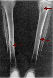
Osteitis fibrosa cystica is a skeletal disorder resulting in a loss of bone mass, a weakening of the bones as their calcified supporting structures are replaced with fibrous tissue, and the formation of cyst-like brown tumors in and around the bone. Osteitis fibrosis cystica (OFC), also known as osteitis fibrosa, osteodystrophia fibrosa, and von Recklinghausen's disease of bone, is caused by hyperparathyroidism, which is a surplus of parathyroid hormone from over-active parathyroid glands. This surplus stimulates the activity of osteoclasts, cells that break down bone, in a process known as osteoclastic bone resorption. The hyperparathyroidism can be triggered by a parathyroid adenoma, hereditary factors, parathyroid carcinoma, or renal osteodystrophy. Osteoclastic bone resorption releases minerals, including calcium, from the bone into the bloodstream, causing both elevated blood calcium levels, and the structural changes which weaken the bone. The symptoms of the disease are the consequences of both the general softening of the bones and the excess calcium in the blood, and include bone fractures, kidney stones, nausea, moth-eaten appearance in the bones, appetite loss, and weight loss.

<span class="mw-page-title-main">Secondary hyperparathyroidism</span> Medical condition

Secondary hyperparathyroidism is the medical condition of excessive secretion of parathyroid hormone (PTH) by the parathyroid glands in response to hypocalcemia, with resultant hyperplasia of these glands. This disorder is primarily seen in patients with chronic kidney failure. It is sometimes abbreviated "SHPT" in medical literature.

<span class="mw-page-title-main">Tertiary hyperparathyroidism</span> Medical condition

Tertiary hyperparathyroidism is a condition involving the overproduction of the hormone, parathyroid hormone, produced by the parathyroid glands. The parathyroid glands are involved in monitoring and regulating blood calcium levels and respond by either producing or ceasing to produce parathyroid hormone.

<span class="mw-page-title-main">Calciphylaxis</span> Medical condition

Calciphylaxis, also known as calcific uremic arteriolopathy (CUA) or “Grey Scale”, is a rare syndrome characterized by painful skin lesions. The pathogenesis of calciphylaxis is unclear but believed to involve calcification of the small blood vessels located within the fatty tissue and deeper layers of the skin, blood clots, and eventual death of skin cells due to lack of blood flow. It is seen mostly in people with end-stage kidney disease but can occur in the earlier stages of chronic kidney disease and rarely in people with normally functioning kidneys. Calciphylaxis is a rare but serious disease, believed to affect 1-4% of all dialysis patients. It results in chronic non-healing wounds and indicates poor prognosis, with typical life expectancy of less than one year.

Bone disease refers to the medical conditions which affect the bone.

<span class="mw-page-title-main">Alfacalcidol</span> Chemical compound

Alfacalcidol is an analogue of vitamin D used for supplementation in humans and as a poultry feed additive.

Calcium acetate/magnesium carbonate is a fixed-dose combination drug that contains 110 mg calcium and 60 mg magnesium ions and is indicated as a phosphate binder for dialysis patients with hyperphosphataemia. It is registered by Fresenius Medical Care under the trade names Renepho (Belgium) and OsvaRen.

Sucroferric oxyhydroxide, sold under the brand name Velphoro, is a non-calcium, iron-based phosphate binder used for the control of serum phosphorus levels in adults with chronic kidney disease (CKD) on haemodialysis (HD) or peritoneal dialysis (PD). It is used in form of chewable tablets.

Chronic kidney disease–mineral and bone disorder (CKD–MBD) is one of the many complications associated with chronic kidney disease. It represents a systemic disorder of mineral and bone metabolism due to CKD manifested by either one or a combination of the following:

A renal diet is a diet aimed at keeping levels of fluids, electrolytes, and minerals balanced in the body in individuals with chronic kidney disease or who are on dialysis. Dietary changes may include the restriction of fluid intake, protein, and electrolytes including sodium, phosphorus, and potassium. Calories may also be supplemented if the individual is losing weight undesirably.

<span class="mw-page-title-main">Idiopathic hypercalcinuria</span>

Idiopathic hypercalcinuria (IH) is a condition including an excessive urinary calcium level with a normal blood calcium level resulting from no underlying cause. IH has become the most common cause of hypercalciuria and is the most serious metabolic risk factor for developing nephrolithiasis. IH can predispose individuals to osteopenia or osteoporosis, and affects the entire body. IH arises due to faulty calcium homeostasis, a closely monitored process, where slight deviations in calcium transport in the intestines, blood, and bone can lead to excessive calcium excretion, bone mineral density loss, or kidney stone formation. 50%-60% of nephrolithiasis patients suffer from IH and have 5%-15% lower bone density than those who do not.

References

  1. 1 2 3 4 5 6 7 Eckardt, Kai-Uwe; Kasiske, Bertram L. (August 2009). "KDIGO clinical practice guideline for the diagnosis, evaluation, prevention, and treatment of Chronic Kidney Disease-Mineral and Bone Disorder (CKD-MBD)" (PDF). Kidney International Supplements. 76 (113). Kidney Disease: Improving Global Outcomes (KDIGO) CKD-MBD Work Group: S1–130. doi: 10.1038/ki.2009.188 . PMID   19644521.
  2. 1 2 3 Moe S, Drüeke T, Cunningham J, Goodman W, Martin K, Olgaard K, Ott S, Sprague S, Lameire N, Eknoyan G (June 2006). "Definition, evaluation, and classification of renal osteodystrophy: a position statement from Kidney Disease: Improving Global Outcomes (KDIGO)". Kidney International. 69 (11): 1945–53. doi: 10.1038/sj.ki.5000414 . PMID   16641930.
  3. "ERA-EDTA Working Group on Chronic Kidney Disease and Mineral Bone Disorder (CKD-MBD)" . Retrieved 4 February 2016.
  4. Lui S, Chu H (1943). "Studies in calcium and phosphorus metabolism with special reference to pathogenesis and effects of dihydrotachysterol (A.T.10) and iron". Medicine. 22 (2): 103–107. doi: 10.1097/00005792-194305000-00002 .
  5. Lucas RC (1833). "Form of late rickets associated with albuminuria, rickets of adolescents". The Lancet. 1 (3119): 993–994. doi:10.1016/S0140-6736(02)37965-0.
  6. 1 2 3 Llach F, Bover J (2000). "Renal Osteodystrophies" . In Brenner BM (ed.). The Kidney. Philadelphia: W.B. Saunders Company. pp.  2103–2186. ISBN   978-0-7216-7998-3.
  7. Cozzolino M, Ureña-Torres P, Vervloet MG, Brandenburg V, Bover J, Goldsmith D, Larsson TE, Massy ZA, Mazzaferro S (October 2014). "Is chronic kidney disease-mineral bone disorder (CKD-MBD) really a syndrome?" (PDF). Nephrology, Dialysis, Transplantation. 29 (10): 1815–20. doi: 10.1093/ndt/gft514 . PMID   24516228.
  8. Luger, N. Mach, D. Sevcik, M. Mantyh, P. (2005). Bone cancer pain: From mechanism to model to therapy. Journal of Pain and Symptom Management. 29(5): 32-46.
  9. "Arthralgia Definition". MedicineNet.com. Archived from the original on 11 October 2007. Retrieved 2007-09-20.
  10. Joe G. Hardin. "Arthralgia". Clinical Methods - The History, Physical, and Laboratory Examinations. Retrieved 2007-09-20.
  11. 1 2 Levy, Adrian R.; Xing, Shan; Brunelli, Steven M.; Cooper, Kerry; Finkelstein, Fredric O.; Germain, Michael J.; Kimel, Miriam; Platt, Robert W.; Belozeroff, Vasily (March 2020). "Symptoms of Secondary Hyperparathyroidism in Patients Receiving Maintenance Hemodialysis: A Prospective Cohort Study". American Journal of Kidney Diseases. 75 (3): 373–383. doi:10.1053/j.ajkd.2019.07.013.
  12. "Overview of Bone Fractures". The Lecturio Medical Concept Library. Retrieved 27 August 2021.
  13. 1 2 London GM (February 2009). "Bone-vascular axis in chronic kidney disease: a reality?". Clinical Journal of the American Society of Nephrology. 4 (2): 254–7. doi: 10.2215/CJN.06661208 . PMID   19176792.
  14. "EraEdta Scientific Working Groups". era-edtaworkinggroups.org.
  15. "Kidney Disease: Improving Global Outcomes (KDIGO)". kdigo.org.
  16. 1 2 3 4 5 Torres PU, Bover J, Mazzaferro S, de Vernejoul MC, Cohen-Solal M (November 2014). "When, how, and why a bone biopsy should be performed in patients with chronic kidney disease". Seminars in Nephrology. 34 (6): 612–25. doi:10.1016/j.semnephrol.2014.09.004. PMID   25498380.
  17. Bover J, Ureña P, Brandenburg V, Goldsmith D, Ruiz C, DaSilva I, Bosch RJ (November 2014). "Adynamic bone disease: from bone to vessels in chronic kidney disease". Seminars in Nephrology. 34 (6): 626–40. doi:10.1016/j.semnephrol.2014.09.008. PMID   25498381.
  18. 1 2 3 4 5 6 7 Patel, Anish A.; Ramanathan, Rohit; Kuban, Joshua; Willis, Marc H. (2015). "Imaging Findings and Evaluation of Metabolic Bone Disease". Advances in Radiology. 2015: 1–21. doi: 10.1155/2015/812794 . ISSN   2356-6876.
  19. "Osteoporosis". The Lecturio Medical Concept Library. Retrieved 27 August 2021.
  20. staff, familydoctor org editorial. "What Is Osteopenia?". familydoctor.org. Retrieved 2021-03-12.
  21. "Osteomalacia and Rickets". The Lecturio Medical Concept Library. Retrieved 27 August 2021.
  22. Nassar, George M.; Ayus, Juan Carlos (1999-11-25). "Brown Tumor in End-Stage Renal Disease". The New England Journal of Medicine. 341 (22): 1652. doi:10.1056/NEJM199911253412204. ISSN   0028-4793. PMID   10572153.
  23. Barsić, Neven; Cala, Kresimir; Pavlović, Drasko (September 2010). "Brown tumor--a rare manifestation of renal osteodystrophy and severe secondary hyperparathyroidism: case report". Acta Clinica Croatica. 49 (3): 299–304. ISSN   0353-9466. PMID   21462819.
  24. 1 2 Bonomini V, Mioli V, Albertazzi A, Scolari P (November 1998). "Daily-dialysis programme: indications and results". Nephrology, Dialysis, Transplantation. 13 (11): 2774–7, discussion 2777–8. doi: 10.1093/ndt/13.11.2774 . PMID   9829478.
  25. "Parathyroidectomy in end stage renal disease". UpToDate.
  26. Vervloet MG, Massy ZA, Brandenburg VM, Mazzaferro S, Cozzolino M, Ureña-Torres P, Bover J, Goldsmith D (May 2014). "Bone: a new endocrine organ at the heart of chronic kidney disease and mineral and bone disorders". The Lancet Diabetes & Endocrinology. 2 (5): 427–36. doi:10.1016/S2213-8587(14)70059-2. PMID   24795256.