Shoulder problem

Last updated
Shoulder problem
Shoulder joint.svg
Diagram of the human shoulder joint
Specialty Orthopedic surgery   OOjs UI icon edit-ltr-progressive.svg

Shoulder problems including pain, are one of the more common reasons for physician visits for musculoskeletal symptoms. The shoulder is the most movable joint in the body. However, it is an unstable joint because of the range of motion allowed. This instability increases the likelihood of joint injury, often leading to a degenerative process in which tissues break down and no longer function well.

Contents

Shoulder pain may be localized or may be referred to areas around the shoulder or down the arm. Other regions within the body (such as gallbladder, liver, or heart disease, or disease of the cervical spine of the neck) also may generate pain that the brain may interpret as arising from the shoulder. [1]

Shoulder structures and functions

The shoulder joint is composed of three bones: the clavicle (collarbone), the scapula (shoulder blade), and the humerus (upper arm bone) (see diagram). Two joints facilitate shoulder movement. The acromioclavicular (AC) joint is located between the acromion (part of the scapula that forms the highest point of the shoulder) and the clavicle. The glenohumeral joint, to which the term "shoulder joint" commonly refers, is a ball-and-socket joint that allows the arm to rotate in a circular fashion or to hinge out and up away from the body. The "ball" is the top, rounded portion of the upper arm bone or humerus; the "socket," or glenoid, is a dish-shaped part of the outer edge of the scapula into which the ball fits. Arm movement is further facilitated by the ability of the scapula itself to slide along the rib cage. The capsule is a soft tissue envelope that encircles the glenohumeral joint. It is lined by a thin, smooth synovial membrane.[ citation needed ]

The bones of the shoulder are held in place by muscles, tendons, and ligaments. Tendons are tough cords of tissue that attach the shoulder muscles to bone and assist the muscles in moving the shoulder. Ligaments attach shoulder bones to each other, providing stability. For example, the front of the joint capsule is anchored by three glenohumeral ligaments.[ citation needed ]

The rotator cuff is a structure composed of tendons that, with associated muscles, holds the ball at the top of the humerus in the glenoid socket and provides mobility and strength to the shoulder joint. [2]

Four filmy sac-like structures called bursa permit smooth gliding between bone, muscle, and tendon. They cushion and protect the rotator cuff from the bony arch of the acromion.[ citation needed ]

Diagnosis

Following are some of the ways doctors diagnose shoulder problems:

Medical history and physical exam

Diagnostic tests

Dislocation

Description

The shoulder joint is the most frequently dislocated major joint of the body. In a typical case of a dislocated shoulder, a strong force that pulls the shoulder outward (abduction) or extreme rotation of the joint pops the ball of the humerus out of the shoulder socket. Dislocation commonly occurs when there is a backward pull on the arm that either catches the muscles unprepared to resist or overwhelms the muscles. When a shoulder dislocates frequently, the condition is referred to as shoulder instability. A partial dislocation where the upper arm bone is partially in and partially out of the socket is called a subluxation. In the medical community, dislocation is commonly referred to as luxation.[ citation needed ]

Signs and diagnosis

Almost all shoulder dislocations are downwards (inferior) and of these, 95 percent are in a forward direction. Clinically this is referred to as an anterior dislocation of the glenohumeral joint. Not only does the arm appear out of position when the shoulder dislocates, but the dislocation also produces pain. Muscle spasms may increase the intensity of pain. Swelling and bruising normally develop, and in some cases there may be numbness and muscle weakness. Problems seen with a dislocated shoulder are tearing of the ligaments or tendons reinforcing the joint capsule and, less commonly, nerve damage. Doctors usually diagnose a dislocation by a physical examination, but X-rays are taken to confirm the diagnosis and to rule out a related fracture and other complications. X-rays are also taken after relocation to ensure it is in the correct place.[ citation needed ]

Treatment

Doctors treat a dislocation by putting the head of the humerus back into the joint socket (glenoid fossa) of the scapula—a procedure called manipulation and reduction. This is usually followed up with an X-ray to make sure the reduction did not fracture the surrounding bones. The arm is then immobilized in a sling or a device called a shoulder immobilizer for several days. Usually the doctor recommends resting the shoulder and applying ice three or four times a day. After pain and swelling have been controlled, the patient enters a rehabilitation program that includes exercises to restore the range of motion of the shoulder and strengthen the muscles to prevent future dislocations. These exercises may progress from simple motion to the use of weights.[ citation needed ]

After treatment and recovery, a previously dislocated shoulder may remain more susceptible to reinjury, especially in young, active individuals. Ligaments are stretched and may tear due to dislocation. Torn ligaments and other problems resulting from dislocation can increase the chance of repeated dislocation. A shoulder that dislocates severely or often, injuring surrounding tissues or nerves, usually requires surgery to repair the damaged parts of the shoulder.[ citation needed ]

Sometimes the doctor performs surgery through a tiny incision into which a small scope (arthroscope) is inserted to observe the inside of the joint. After this procedure, called arthroscopic surgery, the shoulder is generally restrained by a sling for three to six weeks, while full recovery, including physical therapy, takes several months. Arthroscopic techniques involving the shoulder are relatively new and many surgeons prefer to repair a recurrent dislocating shoulder by the time-tested open surgery under direct vision. There are usually fewer repeat dislocations and improved movement following open surgery, but it may take a little longer to regain motion.[ citation needed ]

Separation

Description

A shoulder separation occurs where the collarbone (clavicle) meets the shoulder blade (scapula). When ligaments that hold the AC (acromioclavicular) joint together are partially or completely torn, the outer end of the clavicle may slip out of place, preventing it from properly meeting the scapula. Most often the injury is caused by a blow to the shoulder or by falling on an outstretched hand. After injury it is hard to do a 180 degrees rotation.[ citation needed ]

Signs and diagnosis

Shoulder pain or tenderness and, occasionally, a bump in the middle of the top of the shoulder (over the AC joint) are signs that a separation may have occurred. Sometimes the severity of a separation can be detected by taking X-rays while the patient holds a light weight that pulls on the muscles, making a separation more pronounced.[ citation needed ]

Treatment

A shoulder separation is usually treated conservatively by rest and wearing a sling. Soon after injury, an ice bag may be applied to relieve pain and swelling. After a period of rest, a therapist helps the patient perform exercises that put the shoulder through its range of motion. Most shoulder separations heal within two or three months without further intervention. However, if ligaments are severely torn, surgical repair may be required to hold the clavicle in place. A doctor may wait to see if conservative treatment works before deciding whether surgery is required.[ citation needed ]

Sternoclavicular separation

Description

While not directly a shoulder problem, this may affect shoulder functionality due to problems with sternoclavicular rotation. A sternoclavicular separation occurs when the sternum separates from the clavicle at the sternoclavicular joint. Sternoclavicular separations (dislocation and subluxation) are rare [6] and generally caused by accident. If the clavicle is separated posteriorly (i.e. the clavicle separates and goes behind the sternum) the situation can be dangerous and the clavicle can cause damage to interior arteries, veins or organs.[ citation needed ]

Signs and diagnosis

An X-ray or CT Scan may be necessary to accurately diagnose a sternoclavicular separation.[ citation needed ]

Treatment

Treatment consists of the standard use of plenty of rest, icing, NSAIDs and a sling. The joint may need to be reduced (i.e. put back in place), especially after posterior separations. In severe cases, surgery may be advised.[ citation needed ]

Rotator cuff tendinopathy (tendinitis, bursitis, impingement syndrome, and rotator cuff tears)

Anatomy

The "rotator cuff" is a group of four tendons that blend together as they attach to the upper end of the arm bone (humerus). These tendons transmit the force of muscles originating on the shoulder blade (scapula) to the arm providing rotational motion and centering or stability of the joint.[ citation needed ]

Pathology

The rotator cuff tendons degenerate with age. [7] [8] [9] [10] A group of respected scientists wrote in a review of existing evidence that, the theory that this degeneration is related to pinching (or impingement) between the head of the humerus and the acromion is now considered inaccurate. [11] Rotator cuff pathology is similar in non-dominant compared to dominant and symptomatic compared to asymptomatic shoulder. [8] [9] [12] [13] [14] [15] About two-thirds of all humans develop rotator cuff tendinopathy if they live to 70 years of age. [7] The pathology is mucoid degeneration, not inflammation. [11] The process can involve the intra-articular part of the long head of biceps in addition to the supraspinatus, infraspinatus, and subscapularis tendons. [16]

Tendinitis is inflammation (redness, soreness, and swelling) of a tendon. In tendinitis of the shoulder, the rotator cuff and/or biceps tendon become inflamed, usually as a result of being pinched by surrounding structures. The injury may vary from mild inflammation to involvement of most of the rotator cuff. When the rotator cuff tendon becomes inflamed and thickened, it may get trapped under the acromion. Squeezing of the rotator cuff is called impingement syndrome.[ citation needed ]

An inflamed bursa is called bursitis. Tendinitis and impingement syndrome are often accompanied by inflammation of the bursa sacs that protect the shoulder. Inflammation caused by a disease such as rheumatoid arthritis may cause rotator cuff tendinitis and bursitis. Sports involving overuse of the shoulder and occupations requiring frequent overhead reaching are other potential causes of irritation to the rotator cuff or bursa and may lead to inflammation and impingement.[ citation needed ]

Rotator cuff tear

The most commonly affected tendon is that of the supraspinatus muscle. Defects in the rotator cuff can come from an injury (cuff tear) or from degeneration (cuff wear). The degree to which a tendon is reparable depends on its quantity and quality. Degenerated tendons are often frail and retracted and may not be amenable to repair. Individuals that are elderly, smokers, or those having had cortisone injections often have weaker tendon tissue that fails without a significant injury. By contrast those whose tendon was torn by a substantial fall often have good quality tendon that can be repaired if surgery is performed promptly after the injury. The symptoms of rotator cuff disease include difficulty lifting the arm. Repair of a rotator cuff requires that the tendon be securely anchored to the bone at surgery and that the repair be protected for several months during healing.[ citation needed ]

Signs

Signs of these conditions include the slow onset of discomfort and pain in the upper shoulder or upper third of the arm and/or difficulty sleeping on the shoulder, similar condition can have sharp pain or discomfort when the upper shoulder is positioned at certain angles. Tendinitis and bursitis also cause pain when the arm is lifted away from the body or overhead. If tendinitis involves the biceps tendon (the tendon located in front of the shoulder that helps bend the elbow and turn the forearm), pain will occur in the front or side of the shoulder and may travel down to the elbow and forearm. Pain may also occur when the arm is forcefully pushed upward overhead.[ citation needed ]

Diagnosis

Diagnosis of tendinitis and bursitis begins with a medical history and physical examination. X-rays do not show tendons or the bursae but may be helpful in ruling out bony abnormalities or arthritis. The doctor may remove and test fluid from the inflamed area to rule out infection. Ultrasound scans are frequently used to confirm a suspected tendinitis or bursitis as well as rule out a tear in the rotator cuff muscles. Impingement syndrome may be confirmed when injection of a small amount of anesthetic (lidocaine hydrochloride) into the space under the acromion relieves pain.[ citation needed ]

Treatment

Anti-inflammatory medicines such as aspirin, naproxen or ibuprofen among others can be taken to help with pain. In some cases the physical therapist will use ultrasound and electrical stimulation, as well as manipulation. Gentle stretching and strengthening exercises are added gradually. If there is no improvement, the doctor may inject a corticosteroid medicine into the space under the acromion. However, recent level one evidence showed limited efficacy of corticosteroid injections for pain relief. [17] While steroid injections are a common treatment, they must be used with caution because they may lead to tendon rupture. If there is still no improvement after six to 12 months, the doctor may perform either arthroscopic or open surgery to repair damage and relieve pressure on the tendons and bursae.[ citation needed ]

In those with calcific tendinitis of the shoulder high energy extracorporeal shock-wave therapy can be useful. [18] It is not useful in other types of tendonitis. [18] For a rotator cuff tear, tentative evidence suggests exercise may reduce pain in the short-term. [19] Combination of exercise and joint mobilization can result in long term benefits. [19] Other evidence demonstrates the use of corticosteroids injections to be more effective. [19]

SLAP tear (lesion)

A SLAP (superior labrum anterior to posterior) tear occurs when the cartilage of the shoulder (labrum) delaminates from glenoid. This causes an instability of the shoulder, typically in overhead movements.[ citation needed ]

Symptoms

Symptoms include a dull ache deep in the shoulder joint, trouble sleeping due to the instability and discomfort, and extreme weakness in overhead activities.[ citation needed ]

Diagnosis

The best diagnosis for a SLAP tear is a clinical exam followed by an MRI combined with a contrast agent.[ citation needed ]

Treatment

Very few cases recover to complete mobility without surgical intervention. Some patients are able to strengthen their shoulders to limit the day to day dull ache, but with limited mobility and function. Surgery reattaches the labrum to the glenoid through the use of surgical anchors.[ citation needed ]

Recovery

Recovery is often a lengthy process. The first four or so weeks the patient is required to wear a sling. Following this, there is a month of physical therapy to regain range of motion. At two months limited strength training occurs. At six months the patient is typically released to full active use, although many patients find that full recovery takes longer than this.[ citation needed ]

Frozen shoulder (adhesive capsulitis)

As the name implies, movement of the shoulder is severely restricted in people with a "frozen shoulder". This condition, which doctors call adhesive capsulitis, is frequently caused by injury that leads to lack of use due to pain. Rheumatic disease progression and recent shoulder surgery can also cause frozen shoulder. Intermittent periods of use may cause inflammation. Adhesions (abnormal bands of tissue) grow between the joint surfaces, restricting motion. There is also a lack of synovial fluid, which normally lubricates the gap between the arm bone and socket to help the shoulder joint move. It is this restricted space between the capsule and ball of the humerus that distinguishes adhesive capsulitis from a less complicated painful, stiff shoulder. People with diabetes, stroke, lung disease, rheumatoid arthritis, and heart disease, or who have been in an accident, are at a higher risk for frozen shoulder. The condition rarely appears in people under the age of 40.[ citation needed ]

Fracture

Description

A bone fracture of the shoulder involves a partial or total crack through one of the three bones in the shoulder, the clavicle, the scapula, and the humerus. The break in a bone usually occurs as a result of an impact injury, such as a fall or blow to the shoulder. Fractures usually involve the clavicle or the neck (area below the ball) of the humerus. Fractures of the scapula sometimes occur through the coracoid process.[ citation needed ]

Signs and diagnosis

A shoulder fracture that occurs after a major injury is usually accompanied by severe pain. Within a short time, there may be redness and bruising around the area. Sometimes a fracture is obvious because the bones appear out of position. (However this occur in non-involved dislocations and separations.) Both diagnosis and severity can be confirmed by X-rays.[ citation needed ]

Treatment

When a fracture occurs, the doctor tries to bring the bones into a position that will promote healing and restore arm movement. If the clavicle is fractured, the patient must at first wear a strap and sling around the chest to keep the clavicle in place. After removing the strap and sling, the doctor will prescribe exercises to strengthen the shoulder and restore movement. Surgery is occasionally needed for certain clavicle fractures, especially for disunions.[ citation needed ]

Fracture of the neck of the humerus is usually treated with a sling or shoulder immobilizer. If the bones are out of position, surgery may be necessary to reset them. Exercises are also part of restoring shoulder strength and motion.[ citation needed ]

Arthritis of the shoulder (glenohumeral joint)

Description

In arthritis of the shoulder, the cartilage of the ball and socket (glenohumeral joint) is lost so that bone rubs on bone. It may be caused by wear and tear (degenerative joint disease), injury (traumatic arthritis), surgery (secondary degenerative joint disease), inflammation (rheumatoid arthritis) or infection (septic arthritis).[ citation needed ]

Signs and diagnosis

X-ray of shoulder osteoarthritis. X-ray of osteoarthritis of the shoulder.jpg
X-ray of shoulder osteoarthritis.

Arthritis of the shoulder causes pain and loss of motion and use of the shoulder. X-rays of the shoulder show loss of the normal space between the ball and socket. X-ray can provide radiographic staging of shoulder osteoarthritis.[ citation needed ]

Treatment

Early on arthritis of the shoulder can be managed with mild analgesics and gentle exercises. [20] Known gentle exercises include warm water therapy pool exercises that are provided by a trained and licensed physical therapist; approved land exercises to assure free movement of the arthritic area; cortisone injections (administered at the minimum of every six months according to orthopedic physicians) to reduce inflammation; ice and hot moist pact application are very effective. Moist heat is preferred over ice whereas ice is preferred if inflammation occurs during the daytime hours. Local analgesics along with ice or moist heat are adequate treatments for acute pain.[ citation needed ]

In the case of rheumatoid arthritis, specific medications selected by a rheumatologist may offer substantial relief.[ citation needed ]

When exercise and medication are no longer effective, shoulder replacement surgery for arthritis may be considered. In this operation, a surgeon replaces the shoulder joint with an artificial ball for the top of the humerus and a cap (glenoid) for the scapula. Passive shoulder exercises (where someone else moves the arm to rotate the shoulder joint) are started soon after surgery. Patients begin exercising on their own about three to six weeks after surgery. Eventually, stretching and strengthening exercises become a major part of the rehabilitation programme. The success of the operation often depends on the condition of rotator cuff muscles prior to surgery and the degree to which the patient follows the exercise programme.[ citation needed ]

In young and active patients a partial shoulder replacement with a non-prosthetic glenoid arthroplasty may also be a consideration

Arthritis or osteolysis of the AC (acromioclavicular) joint

Description

The acromioclavicular articulation consists of the acromioclavicular ligament and a small disk of cartilage located in between the acromion and the clavicle. This disk can wear down through injury, extreme joint stress (via bodybuilding) or normal wear.[ citation needed ]

Signs and diagnosis

Pain is perceived on shoulder motion, especially on certain movements. Often a crossover arm test is utilized in diagnosis because this compresses the AC joint, exacerbating the symptoms. X-rays of the shoulder joint may show either arthritic changes of the ac joint or osteolysis.

Treatment

Conservative treatment for this joint is similar to treatments for other types of arthritis, including restricting activity, anti-inflammatory medications (or supplements), physical therapy, and occasionally cortisone shots. If the pain is severe, surgery may be an option. The most common surgical treatment, known as resection arthroplasty, involves cutting a very small portion off the clavicle end and letting scar tissue fill in its place. Some portions of the acromioclavicular ligament may still remain attached.[ citation needed ]

Treatment

A mnemonic for the basic treatment principles of any musculoskeletal problems is PRICE : Protection, Rest, Ice, Compression, and Elevation:[ citation needed ]

If pain and stiffness persist, see a doctor.

According to the American Academy of Orthopaedic Surgeons (AAOS) visits to orthopedic specialists for shoulder pain has been rising since 1998 and in 2005 over 13 million patients sought medical care for shoulder pain, of which only 34 percent were related to injury. [21]

Related Research Articles

<span class="mw-page-title-main">Clavicle</span> Long bone that serves as a strut between the scapula and the sternum

The clavicle, or collarbone, is a slender, S-shaped long bone approximately 6 inches (15 cm) long that serves as a strut between the shoulder blade and the sternum (breastbone). There are two clavicles, one on the left and one on the right. The clavicle is the only long bone in the body that lies horizontally. Together with the shoulder blade, it makes up the shoulder girdle. It is a palpable bone and, in people who have less fat in this region, the location of the bone is clearly visible. It receives its name from Latin clavicula 'little key' because the bone rotates along its axis like a key when the shoulder is abducted. The clavicle is the most commonly fractured bone. It can easily be fractured by impacts to the shoulder from the force of falling on outstretched arms or by a direct hit.

<span class="mw-page-title-main">Rotator cuff</span> Group of muscles

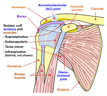
The rotator cuff is a group of muscles and their tendons that act to stabilize the human shoulder and allow for its extensive range of motion. Of the seven scapulohumeral muscles, four make up the rotator cuff. The four muscles are:

<span class="mw-page-title-main">Shoulder</span> Part of the body

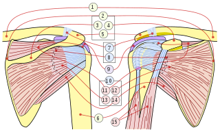
The human shoulder is made up of three bones: the clavicle (collarbone), the scapula, and the humerus as well as associated muscles, ligaments and tendons.

<span class="mw-page-title-main">Deltoid muscle</span> Shoulder muscle

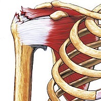
The deltoid muscle is the muscle forming the rounded contour of the human shoulder. It is also known as the 'common shoulder muscle', particularly in other animals such as the domestic cat. Anatomically, the deltoid muscle appears to be made up of three distinct sets of muscle fibers, namely the

  1. anterior or clavicular part
  2. posterior or scapular part
  3. intermediate or acromial part
<span class="mw-page-title-main">Rotator cuff tear</span> Shoulder injury

Rotator cuff tendinopathy is a process of senescence. The pathophysiology is mucoid degeneration. Most people develop rotator cuff tendinopathy within their lifetime.

<span class="mw-page-title-main">Clavicle fracture</span> Medical condition

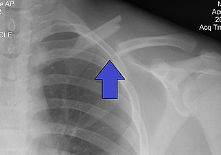
A clavicle fracture, also known as a broken collarbone, is a bone fracture of the clavicle. Symptoms typically include pain at the site of the break and a decreased ability to move the affected arm. Complications can include a collection of air in the pleural space surrounding the lung (pneumothorax), injury to the nerves or blood vessels in the area, and an unpleasant appearance.

<span class="mw-page-title-main">Acromioclavicular joint</span> Shoulder junction between the scapula and the clavicle

The acromioclavicular joint, or AC joint, is a joint at the top of the shoulder. It is the junction between the acromion and the clavicle. It is a plane synovial joint.

<span class="mw-page-title-main">Supraspinatus muscle</span> Muscle of the upper back

The supraspinatus is a relatively small muscle of the upper back that runs from the supraspinous fossa superior portion of the scapula to the greater tubercle of the humerus. It is one of the four rotator cuff muscles and also abducts the arm at the shoulder. The spine of the scapula separates the supraspinatus muscle from the infraspinatus muscle, which originates below the spine.

<span class="mw-page-title-main">Shoulder joint</span> Synovial ball and socket joint in the shoulder

The shoulder joint is structurally classified as a synovial ball-and-socket joint and functionally as a diarthrosis and multiaxial joint. It involves an articulation between the glenoid fossa of the scapula and the head of the humerus. Due to the very loose joint capsule that gives a limited interface of the humerus and scapula, it is the most mobile joint of the human body.

<span class="mw-page-title-main">Shoulder girdle</span> Set of bones which connects the arm to the axial skeleton on each side

The shoulder girdle or pectoral girdle is the set of bones in the appendicular skeleton which connects to the arm on each side. In humans it consists of the clavicle and scapula; in those species with three bones in the shoulder, it consists of the clavicle, scapula, and coracoid. Some mammalian species have only the scapula.

<span class="mw-page-title-main">Sternoclavicular joint</span> Joint between the manubrium of the sternum and the clavicle bone.

The sternoclavicular joint or sternoclavicular articulation is a synovial saddle joint between the manubrium of the sternum, and the clavicle, and the first costal cartilage. The joint possesses a joint capsule, and an articular disc, and is reinforced by multiple ligaments.

<span class="mw-page-title-main">Glenoid fossa</span> Part of the shoulder

The glenoid fossa of the scapula or the glenoid cavity is a bone part of the shoulder. The word glenoid is pronounced or and is from Greek: gléne, "socket", reflecting the shoulder joint's ball-and-socket form. It is a shallow, pyriform articular surface, which is located on the lateral angle of the scapula. It is directed laterally and forward and articulates with the head of the humerus; it is broader below than above and its vertical diameter is the longest.

<span class="mw-page-title-main">Coracoacromial ligament</span> Ligament between the coracoid process and the acromion of the scapula

The coracoacromial ligament is a strong triangular ligament between the coracoid process and the acromion. It protects the head of the humerus. Its acromial attachment may be repositioned to the clavicle during reconstructive surgery of the acromioclavicular joint.

<span class="mw-page-title-main">Separated shoulder</span> Medical condition

A separated shoulder, also known as acromioclavicular joint injury, is a common injury to the acromioclavicular joint. The AC joint is located at the outer end of the clavicle where it attaches to the acromion of the scapula. Symptoms include non-radiating pain which may make it difficult to move the shoulder. The presence of swelling or bruising and a deformity in the shoulder is also common depending on how severe the dislocation is.

<span class="mw-page-title-main">Subacromial bursitis</span> Medical condition

Subacromial bursitis is a condition caused by inflammation of the bursa that separates the superior surface of the supraspinatus tendon from the overlying coraco-acromial ligament, acromion, and coracoid and from the deep surface of the deltoid muscle. The subacromial bursa helps the motion of the supraspinatus tendon of the rotator cuff in activities such as overhead work.

<span class="mw-page-title-main">Dislocated shoulder</span> Injury

A dislocated shoulder is a condition in which the head of the humerus is detached from the shoulder joint. Symptoms include shoulder pain and instability. Complications may include a Bankart lesion, Hill-Sachs lesion, rotator cuff tear, or injury to the axillary nerve.

Shoulder surgery is a means of treating injured shoulders. Many surgeries have been developed to repair the muscles, connective tissue, or damaged joints that can arise from traumatic or overuse injuries to the shoulder.

<span class="mw-page-title-main">Shoulder replacement</span>

Shoulder replacement is a surgical procedure in which all or part of the glenohumeral joint is replaced by a prosthetic implant. Such joint replacement surgery generally is conducted to relieve arthritis pain or fix severe physical joint damage.

<span class="mw-page-title-main">Elbow</span> Joint between the upper and lower parts of the arm

The elbow is the region between the upper arm and the forearm that surrounds the elbow joint. The elbow includes prominent landmarks such as the olecranon, the cubital fossa, and the lateral and the medial epicondyles of the humerus. The elbow joint is a hinge joint between the arm and the forearm; more specifically between the humerus in the upper arm and the radius and ulna in the forearm which allows the forearm and hand to be moved towards and away from the body. The term elbow is specifically used for humans and other primates, and in other vertebrates forelimb plus joint is used.

<span class="mw-page-title-main">Shoulder impingement syndrome</span> Medical condition

Shoulder impingement syndrome is a syndrome involving tendonitis of the rotator cuff muscles as they pass through the subacromial space, the passage beneath the acromion. It is particularly associated with tendonitis of the supraspinatus muscle. This can result in pain, weakness, and loss of movement at the shoulder.

References

  1. "Referred Shoulder Pain - Topic Overview". WebMD, LLC. 14 November 2014. Retrieved 28 July 2016.
  2. Zadro, Joshua; Rischin, Adam; Johnston, Renea V; Buchbinder, Rachelle (2021-08-26). Cochrane Musculoskeletal Group (ed.). "Image-guided glucocorticoid injection versus injection without image guidance for shoulder pain". Cochrane Database of Systematic Reviews. 2021 (9). doi:10.1002/14651858.CD009147.pub3. PMC   8407470 . PMID   34435661.
  3. 1 2 Woodward, T. W.; Best, T. M. (15 May 2000). "The painful shoulder: part I. Clinical evaluation". American Family Physician. 61 (10): 3079–88. PMID   10839557.
  4. "Penn Shoulder Score description and calculator – orthotoolkit" . Retrieved January 10, 2018.
  5. E. J. Hegedus; A. Goode; S. Campbell; A. Morin; M. Tamaddoni; C. T. Moorman III; C. Cook (2007). "Physical Examination Tests of the Shoulder: A Systematic Review with Meta-analysis of Individual Tests". Br J Sports Med. 42 (2): 80–92. doi: 10.1136/bjsm.2007.038406 . PMID   17720798.
  6. Sternoclavicular Joint Dislocations http://lifeinthefastlane.com/2010/02/sternoclavicular-joint-dislocation/
  7. 1 2 Teunis, Teun; Lubberts, Bart; Reilly, Brian T.; Ring, David (December 2014). "A systematic review and pooled analysis of the prevalence of rotator cuff disease with increasing age". Journal of Shoulder and Elbow Surgery. 23 (12): 1913–1921. doi:10.1016/j.jse.2014.08.001. ISSN   1532-6500. PMID   25441568.
  8. 1 2 Yamaguchi, Ken; Ditsios, Konstantinos; Middleton, William D.; Hildebolt, Charles F.; Galatz, Leesa M.; Teefey, Sharlene A. (August 2006). "The demographic and morphological features of rotator cuff disease. A comparison of asymptomatic and symptomatic shoulders". The Journal of Bone and Joint Surgery. American Volume. 88 (8): 1699–1704. doi:10.2106/JBJS.E.00835. ISSN   0021-9355. PMID   16882890.
  9. 1 2 Liu, Tiffany C.; Leung, Nina; Edwards, Leonard; Ring, David; Bernacki, Edward; Tonn, Melissa D. (October 2017). "Patients Older Than 40 Years With Unilateral Occupational Claims for New Shoulder and Knee Symptoms Have Bilateral MRI Changes". Clinical Orthopaedics and Related Research. 475 (10): 2360–2365. doi:10.1007/s11999-017-5401-y. ISSN   1528-1132. PMC   5599397 . PMID   28600690.
  10. Vincent, Karl; Leboeuf-Yde, Charlotte; Gagey, Olivier (May 2017). "Are degenerative rotator cuff disorders a cause of shoulder pain? Comparison of prevalence of degenerative rotator cuff disease to prevalence of nontraumatic shoulder pain through three systematic and critical reviews". Journal of Shoulder and Elbow Surgery. 26 (5): 766–773. doi:10.1016/j.jse.2016.09.060. ISSN   1532-6500. PMID   28089260.
  11. 1 2 McFarland, Edward G.; Maffulli, Nicola; Del Buono, Angelo; Murrell, George A. C.; Garzon-Muvdi, Juan; Petersen, Steve A. (July 2013). "Impingement is not impingement: the case for calling it "Rotator Cuff Disease"". Muscles, Ligaments and Tendons Journal. 3 (3): 196–200. doi:10.32098/mltj.03.2013.11. ISSN   2240-4554. PMC   3838328 . PMID   24367779.
  12. Barreto, Rodrigo Py Gonçalves; Braman, Jonathan P.; Ludewig, Paula M.; Ribeiro, Larissa Pechincha; Camargo, Paula Rezende (September 2019). "Bilateral magnetic resonance imaging findings in individuals with unilateral shoulder pain". Journal of Shoulder and Elbow Surgery. 28 (9): 1699–1706. doi:10.1016/j.jse.2019.04.001. ISSN   1532-6500. PMID   31279721. S2CID   195830915.
  13. Eliason, Anna; Harringe, Marita; Engström, Björn; Sunding, Kerstin; Werner, Suzanne (2021-08-17). "Bilateral ultrasound findings in patients with unilateral subacromial pain syndrome". Physiotherapy Theory and Practice. 38 (13): 2568–2579. doi: 10.1080/09593985.2021.1962462 . ISSN   1532-5040. PMID   34402715. S2CID   237149226.
  14. Yamaguchi, K.; Tetro, A. M.; Blam, O.; Evanoff, B. A.; Teefey, S. A.; Middleton, W. D. (May 2001). "Natural history of asymptomatic rotator cuff tears: a longitudinal analysis of asymptomatic tears detected sonographically". Journal of Shoulder and Elbow Surgery. 10 (3): 199–203. doi:10.1067/mse.2001.113086. ISSN   1058-2746. PMID   11408898.
  15. Ranebo, Mats C.; Björnsson Hallgren, Hanna C.; Adolfsson, Lars E. (March 2018). "Patients with a long-standing cuff tear in one shoulder have high rates of contralateral cuff tears: a study of patients with arthroscopically verified cuff tears 22 years ago". Journal of Shoulder and Elbow Surgery. 27 (3): e68–e74. doi:10.1016/j.jse.2017.10.007. ISSN   1532-6500. PMID   29249548.
  16. Ahrens, P. M.; Boileau, P. (August 2007). "The long head of biceps and associated tendinopathy". The Journal of Bone and Joint Surgery. British Volume. 89 (8): 1001–1009. doi: 10.1302/0301-620X.89B8.19278 . ISSN   0301-620X. PMID   17785735.
  17. Mohamadi, Amin; Chan, Jimmy J.; Claessen, Femke M. A. P.; Ring, David; Chen, Neal C. (January 2017). "Corticosteroid Injections Give Small and Transient Pain Relief in Rotator Cuff Tendinosis: A Meta-analysis". Clinical Orthopaedics and Related Research. 475 (1): 232–243. doi:10.1007/s11999-016-5002-1. ISSN   1528-1132. PMC   5174041 . PMID   27469590.
  18. 1 2 Bannuru, R. R.; Flavin, N. E.; Vaysbrot, E.; Harvey, W.; McAlindon, T. (15 April 2014). "High-energy extracorporeal shock-wave therapy for treating chronic calcific tendinitis of the shoulder: a systematic review". Annals of Internal Medicine. 160 (8): 542–9. doi:10.7326/m13-1982. PMID   24733195. S2CID   6164436.
  19. 1 2 3 Green, Sally; Buchbinder, Rachelle; Hetrick, Sarah E (2003-04-22). "Physiotherapy interventions for shoulder pain". Cochrane Database of Systematic Reviews. 2013 (2): CD004258. doi:10.1002/14651858.cd004258. ISSN   1465-1858. PMC   8769566 . PMID   12804509.
  20. "Introduction - Home Exercises for the Rough Shoulder". Orthop.washington.edu. 2006-10-09. Retrieved 2010-01-10.
  21. American Academy of Orthopaedic Surgeons Physician Visits for Musculoskeletal Symptoms http://www.aaos.org/Research/stats/Common%20Orthopaedic%20Symptoms%20Seen%20by%20a%20Physician.pdf

This article contains and extends text from the public domain document "Questions and Answers about Shoulder Problems", NIH Publication No. 01-4865, available from URL http://www.niams.nih.gov/hi/topics/shoulderprobs/shoulderqa.htm