Animal heme-dependent peroxidases

Last updated
Animal heme-dependent peroxidase
Idnu.png
Crystal structure of the human myeloperoxidase-thiocyanate complex. [1]
Identifiers
SymbolAn_peroxidase
Pfam PF03098
InterPro IPR019791
PROSITE PDOC00394
SCOP2 1mhl / SCOPe / SUPFAM
OPM superfamily 36
OPM protein 1q4g
CDD cd05396
Available protein structures:
Pfam   structures / ECOD  
PDB RCSB PDB; PDBe; PDBj
PDBsum structure summary

Animal heme-dependent peroxidases is a family of peroxidases. Peroxidases are found in bacteria, fungi, plants and animals. On the basis of sequence similarity, a number of animal heme peroxidases can be categorized as members of a superfamily: myeloperoxidase (MPO); eosinophil peroxidase (EPO); lactoperoxidase (LPO); thyroid peroxidase (TPO); prostaglandin H synthase (PGHS); and peroxidasin. [2] [3] [4]

Contents

Function

Myeloperoxidase (MPO) plays a major role in the oxygen-dependent microbicidal system of neutrophils. EPO from eosinophilic granulocytes participates in immunological reactions, and potentiates tumor necrosis factor (TNF) production and hydrogen peroxide release by human monocyte-derived macrophages. [5] [6] MPO (and possibly EPO) primarily use Clions and H2O2 to form hypochlorous acid (HOCl), which can effectively kill bacteria or parasites. In secreted fluids, LPO catalyses the oxidation of thiocyanate ions (SCN) by H2O2, producing the weak oxidizing agent hypothiocyanite (OSCN), which has bacteriostatic activity. [7] TPO uses I ions and H2O2 to generate iodine, and plays a central role in the biosynthesis of thyroid hormones T3 and T4. Myeloperoxidase ( PDB: 1dnu ), for example, resides in the human nucleus and lysosome and acts as a defense response to oxidative stress, preventing apoptosis of the cell. [1]

Structure

3D structures of MPO and PGHS have been reported. MPO is a homodimer: each monomer consists of a light (A or B) and a heavy (C or D) chain resulting from post-translational excision of 6 residues from the common precursor. Monomers are linked by a single inter-chain disulfide. Each monomer includes a bound calcium ion. [8] PGHS exists as a symmetric homodimer, each monomer of which consists of 3 domains: an N-terminal epidermal growth factor (EGF) like module; a membrane-binding domain; and a large C-terminal catalytic domain containing the cyclooxygenase and the peroxidase active sites. The catalytic domain shows striking structural similarity to MPO. The image at the top of this page is an example of Myeloperoxidase 1dnu derived from X-ray diffraction with resolution 1.85 angstrom. [1]

Active site

The cyclooxygenase active site, which catalyzes the formation of prostaglandin G2 (PGG2) from arachidonic acid, resides at the apex of a long hydrophobic channel, extending from the membrane-binding domain to the center of the molecule. The peroxidase active site, which catalyzes the reduction of PGG2 to PGH2, is located on the other side of the molecule, at the heme binding site. [9] Both MPO and the catalytic domain of PGHS are mainly alpha-helical, 19 helices being identified as topologically and spatially equivalent; PGHS contains 5 additional N-terminal helices that have no equivalent in MPO. In both proteins, three Asn residues in each monomer are glycosylated.

Human proteins containing this domain

The following is a list of human proteins containing this domain: [10]

DUOX1; DUOX2; EPX; LPO; MPO; PTGS1; PTGS2; PXDNL; TPO

Related Research Articles

<span class="mw-page-title-main">Peroxidase</span> Peroxide-decomposing enzyme

Peroxidases or peroxide reductases are a large group of enzymes which play a role in various biological processes. They are named after the fact that they commonly break up peroxides.

<span class="mw-page-title-main">Heme</span> Chemical coordination complex of an iron ion chelated to a porphyrin

Heme, or haem, is a precursor to hemoglobin, which is necessary to bind oxygen in the bloodstream. Heme is biosynthesized in both the bone marrow and the liver.

<span class="mw-page-title-main">Cyclooxygenase</span> Class of enzymes

Cyclooxygenase (COX), officially known as prostaglandin-endoperoxide synthase (PTGS), is an enzyme that is responsible for formation of prostanoids, including thromboxane and prostaglandins such as prostacyclin, from arachidonic acid. A member of the animal-type heme peroxidase family, it is also known as prostaglandin G/H synthase. The specific reaction catalyzed is the conversion from arachidonic acid to prostaglandin H2 via a short-living prostaglandin G2 intermediate.

<span class="mw-page-title-main">Cytochrome c peroxidase</span>

Cytochrome c peroxidase, or CCP, is a water-soluble heme-containing enzyme of the peroxidase family that takes reducing equivalents from cytochrome c and reduces hydrogen peroxide to water:

<span class="mw-page-title-main">Thiocyanate</span> Ion (S=C=N, charge –1)

Thiocyanate is the anion [SCN], a salt or an ester of thiocyanic acid. It is the conjugate base of thiocyanic acid. Common derivatives include the colourless salts potassium thiocyanate and sodium thiocyanate. Mercury(II) thiocyanate was formerly used in pyrotechnics.

<span class="mw-page-title-main">Myeloperoxidase</span> Mammalian protein found in Homo sapiens

Myeloperoxidase (MPO) is a peroxidase enzyme that in humans is encoded by the MPO gene on chromosome 17. MPO is most abundantly expressed in neutrophil granulocytes, and produces hypohalous acids to carry out their antimicrobial activity, including hypochlorous acid, the sodium salt of which is the chemical in bleach. It is a lysosomal protein stored in azurophilic granules of the neutrophil and released into the extracellular space during degranulation. Neutrophil myeloperoxidase has a heme pigment, which causes its green color in secretions rich in neutrophils, such as mucus and sputum. The green color contributed to its outdated name verdoperoxidase.

<span class="mw-page-title-main">Thyroid peroxidase</span> Enzyme expressed mainly in the thyroid gland

Thyroid peroxidase, also called thyroperoxidase (TPO) or iodide peroxidase, is an enzyme expressed mainly in the thyroid where it is secreted into colloid. Thyroid peroxidase oxidizes iodide ions to form iodine atoms for addition onto tyrosine residues on thyroglobulin for the production of thyroxine (T4) or triiodothyronine (T3), the thyroid hormones. In humans, thyroperoxidase is encoded by the TPO gene.

<span class="mw-page-title-main">Thromboxane-A synthase</span> Mammalian protein found in Homo sapiens

Thromboxane A synthase 1 , also known as TBXAS1, is a cytochrome P450 enzyme that, in humans, is encoded by the TBXAS1 gene.

<span class="mw-page-title-main">Horseradish peroxidase</span> Chemical compound and enzyme

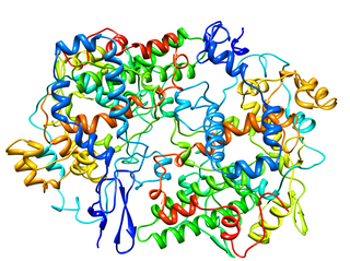
The enzyme horseradish peroxidase (HRP), found in the roots of horseradish, is used extensively in biochemistry applications. It is a metalloenzyme with many isoforms, of which the most studied type is C. It catalyzes the oxidation of various organic substrates by hydrogen peroxide.

Prostaglandin H<sub>2</sub> Chemical compound

Prostaglandin H2 is a type of prostaglandin and a precursor for many other biologically significant molecules. It is synthesized from arachidonic acid in a reaction catalyzed by a cyclooxygenase enzyme. The conversion from Arachidonic acid to Prostaglandin H2 is a two step process. First, COX-1 catalyzes the addition of two free oxygens to form the 1,2-Dioxane bridge and a peroxide functional group to form Prostaglandin G2. Second, COX-2 reduces the peroxide functional group to a Secondary alcohol, forming Prostaglandin H2. Other peroxidases like Hydroquinone have been observed to reduce PGG2 to PGH2. PGH2 is unstable at room temperature, with a half life of 90-100 seconds, so it is often converted into a different prostaglandin.

<span class="mw-page-title-main">Prostaglandin-endoperoxide synthase 2</span> Human enzyme involved in inflammation

Prostaglandin-endoperoxide synthase 2, also known as cyclooxygenase-2 or COX-2, is an enzyme that in humans is encoded by the PTGS2 gene. In humans it is one of two cyclooxygenases. It is involved in the conversion of arachidonic acid to prostaglandin H2, an important precursor of prostacyclin, which is expressed in inflammation.

In enzymology, a manganese peroxidase (EC 1.11.1.13) is an enzyme that catalyzes the chemical reaction

<span class="mw-page-title-main">PTGS1</span>

Cyclooxygenase 1 (COX-1), also known as prostaglandin G/H synthase 1, prostaglandin-endoperoxide synthase 1 or prostaglandin H2 synthase 1, is an enzyme that in humans is encoded by the PTGS1 gene. In humans it is one of two cyclooxygenases.

<span class="mw-page-title-main">Dual oxidase 1</span> Protein-coding gene in the species Homo sapiens

Dual oxidase 1, also known as DUOX1 or ThOX1, is an enzyme which in humans is encoded by the DUOX1 gene. DUOX1 was first identified in the mammalian thyroid gland. In humans, two isoforms are found; hDUOX1 and hDUOX2. Human DUOX protein localization is not exclusive to thyroid tissue; hDUOX1 is prominent in airway epithelial cells and hDUOX2 in the salivary glands and gastrointestinal tract.

Haloperoxidases are peroxidases that are able to mediate the oxidation of halides by hydrogen peroxide. Both halides and hydrogen peroxide are widely available in the environment.

Hypothiocyanite is the anion [OSCN] and the conjugate base of hypothiocyanous acid (HOSCN). It is an organic compound part of the thiocyanates as it contains the functional group SCN. It is formed when an oxygen is singly bonded to the thiocyanate group. Hypothiocyanous acid is a fairly weak acid; its acid dissociation constant (pKa) is 5.3.

The respiratory tract antimicrobial defense system is a layered defense mechanism which relies on components of both the innate and adaptive immune systems to protect the lungs and the rest of the respiratory tract against inhaled microorganisms.

<span class="mw-page-title-main">Lactoperoxidase</span> Mammalian protein found in Homo sapiens

Lactoperoxidase is a peroxidase enzyme secreted from mammary, salivary and other mucosal glands including the lungs, bronchii and nose that functions as a natural and the first line of defense against bacteria and viruses. Lactoperoxidase is a member of the heme peroxidase family of enzymes. In humans, lactoperoxidase is encoded by the LPO gene.

<span class="mw-page-title-main">Eosinophil peroxidase</span> Protein-coding gene in the species Homo sapiens

Eosinophil peroxidase is an enzyme found within the eosinophil granulocytes, innate immune cells of humans and mammals. This oxidoreductase protein is encoded by the gene EPX, expressed within these myeloid cells. EPO shares many similarities with its orthologous peroxidases, myeloperoxidase (MPO), lactoperoxidase (LPO), and thyroid peroxidase (TPO). The protein is concentrated in secretory granules within eosinophils. Eosinophil peroxidase is a heme peroxidase, its activities including the oxidation of halide ions to bacteriocidal reactive oxygen species, the cationic disruption of bacterial cell walls, and the post-translational modification of protein amino acid residues.

In enzymology, a prostaglandin-F synthase (PGFS; EC 1.1.1.188) is an enzyme that catalyzes the chemical reaction:

References

  1. 1 2 3 PDB: 1dnu ; Blair-Johnson M, Fiedler T, Fenna R (November 2001). "Human myeloperoxidase: structure of a cyanide complex and its interaction with bromide and thiocyanate substrates at 1.9 A resolution". Biochemistry. 40 (46): 13990–7. doi:10.1021/bi0111808. PMID   11705390.
  2. Nelson RE, Fessler LI, Takagi Y, Blumberg B, Keene DR, Olson PF, Parker CG, Fessler JH (1994). "Peroxidasin: a novel enzyme-matrix protein of Drosophila development". EMBO J. 13 (15): 3438–3447. doi:10.1002/j.1460-2075.1994.tb06649.x. PMC   395246 . PMID   8062820.
  3. Poulos TL, Li H (1994). "Structural variation in heme enzymes: a comparative analysis of peroxidase and P450 crystal structures". Structure. 2 (6): 461–464. doi: 10.1016/S0969-2126(00)00046-0 . PMID   7922023.
  4. Kimura S, Ikeda-Saito M (1988). "Human myeloperoxidase and thyroid peroxidase, two enzymes with separate and distinct physiological functions, are evolutionarily related members of the same gene family". Proteins. 3 (2): 113–120. doi:10.1002/prot.340030206. PMID   2840655. S2CID   29575992.
  5. Kimura S, Hong YS, Kotani T, Ohtaki S, Kikkawa F (1989). "Structure of the human thyroid peroxidase gene: comparison and relationship to the human myeloperoxidase gene". Biochemistry. 28 (10): 4481–4489. doi:10.1021/bi00436a054. PMID   2548579.
  6. Spessotto P, Dri P, Bulla R, Zabucchi G, Patriarca P (1995). "Human eosinophil peroxidase enhances tumor necrosis factor and hydrogen peroxide release by human monocyte-derived macrophages". Eur. J. Immunol. 25 (5): 1366–1373. doi:10.1002/eji.1830250535. PMID   7774640. S2CID   33362887.
  7. Wever R, Kast WM, Kasinoedin JH, Boelens R (1982). "The peroxidation of thiocyanate catalysed by myeloperoxidase and lactoperoxidase". Biochim. Biophys. Acta. 709 (2): 212–219. doi:10.1016/0167-4838(82)90463-0. PMID   6295491.
  8. Fenna RE, Zeng J (1992). "X-ray crystal structure of canine myeloperoxidase at 3 A resolution". J. Mol. Biol. 226 (1): 185–207. doi: 10.1016/0022-2836(92)90133-5 . PMID   1320128.
  9. Picot D, Loll PJ, Garavito RM (1994). "The X-ray crystal structure of the membrane protein prostaglandin H2 synthase-1". Nature. 367 (6460): 243–249. Bibcode:1994Natur.367..243P. doi:10.1038/367243a0. PMID   8121489. S2CID   4340064.
  10. Zamocky M, Jakopitsch C, Furtmüller PG, Dunand C, Obinger C (August 2008). "The peroxidase-cyclooxygenase superfamily: Reconstructed evolution of critical enzymes of the innate immune system". Proteins. 72 (2): 589–605. doi:10.1002/prot.21950. PMID   18247411. S2CID   19832239.