Panniculitis is a group of diseases whose hallmark is inflammation of subcutaneous adipose tissue. Symptoms include tender skin nodules, and systemic signs such as weight loss and fatigue.
Malignant histiocytosis is a rare hereditary disease found in the Bernese Mountain Dog and humans, characterized by histiocytic infiltration of the lungs and lymph nodes. The liver, spleen, and central nervous system can also be affected. Histiocytes are a component of the immune system that proliferate abnormally in this disease. In addition to its importance in veterinary medicine, the condition is also important in human pathology.

Eosinophilic granuloma is a form of Langerhans cell histiocytosis. It is a condition of both human and veterinary pathology.
In medicine, histiocytosis is an excessive number of histiocytes, and the term is also often used to refer to a group of rare diseases which share this sign as a characteristic. Occasionally and confusingly, the term "histiocytosis" is sometimes used to refer to individual diseases.

Neonatal acne is an acneiform eruption that occurs in newborns or infants, and is often seen on the nose and adjacent portions of the cheeks.

Nasal glioma is a rare benign congenital lesion, usually a firm, incompressible, reddish-blue to purple lesion occurring on the nasal bridge or midline near the root.
Non-Langerhans cell histiocytosis refers to a family of histiocytosis characterized by the absence of Langerhans cells.
Generalized eruptive histiocytoma is a rare cutaneous condition characterized by widespread, erythematous, essentially symmetrical papules, particularly involving the trunk and proximal extremities.
Xanthoma disseminatum is a rare cutaneous condition that preferentially affects males in childhood, characterized by the insidious onset of small, yellow-red to brown papules and nodules that are discrete and disseminated.
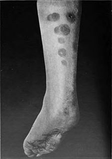
Progressive nodular histiocytosis is a cutaneous condition clinically characterized by the development of two types of skin lesions: superficial papules and deeper larger subcutaneous nodules.
Papular xanthoma is a cutaneous condition that is a rare form of non-X histiocytosis.

Hereditary progressive mucinous histiocytosis is a very rare, benign, non-Langerhans' cell histiocytosis. An autosomal dominant or X-linked hereditary disease described on the skin, it has been found almost exclusively in women. One case of the disease in a male patient has been reported.
Reticulohistiocytosis is a cutaneous condition of which there are two distinct forms:
Reticulohistiocytoma is a cutaneous condition characterized by a solitary, firm, dermal skin lesion of less than 1 cm in diameter. It usually occurs in young adults or middle aged people, most commonly in females. Affected regions include the head and neck region and the upper part of the trunk. It may coexist with certain neoplasms or vasculitis, and in 30 percent of patients with xanthelasma.
Indeterminate cell histiocytosis is a cutaneous condition felt to be caused by dermal precursors of Langerhans cells.
Sea-blue histiocytosis is a cutaneous condition that may occur as a familial inherited syndrome or as an acquired secondary or systemic infiltrative process.
X-type histiocytoses are a clinically well-defined group of cutaneous syndromes characterized by infiltrates of Langerhans cells, as opposed to Non-X histiocytosis in which the infiltrates contain monocytes/macrophages. Conditions included in this group are:
Congenital self-healing reticulohistiocytosis is a condition that is a self-limited form of Langerhans cell histiocytosis.
Giant-cell reticulohistiocytoma is a cutaneous condition characterized by a solitary skin lesion.