Eosinophilic granuloma

Last updated

Humans

Lesion on a nose. Facial eosinophilic granuloma.jpg
Lesion on a nose.

Human eosinophilic granuloma is characterized by abnormal proliferation of Langerhans cells (LCs). LCs are antigen-presenting cells derived from dendritic cells. In humans, eosinophilic granulomas are considered as a benign tumors that occurs mainly in children and adolescents. EG is a quite rare condition, and its incidence is higher in white than in black population, also slightly more affecting males than females. EG develops in 4-5 children (aged under 15) per million/year and in 1 or 2 adults per million/year. [1]

Contents

The etiology of EG is not fully understood yet. However, the onset of abnormal LC proliferation may be triggered by viral stimuli (EBV, Human Herpes virus 6), bacterial toxins or defective regulation of IL-1 and IL-10 production. [2] Another possible explanation may be a defect in Ras/MAPK signaling pathway due to mutation of signaling proteins. Particularly, it was published that about 50% of the EG cases had mutated BRAF V600 E gene [3] and about 21% displayed a mutation in MAP2K1. [4]

Signs and symptoms

Clinically, single or multiple lesions are present mostly on the axial skeleton - on the skull, spine, ribs, pelvis and long bones. Specifically, thoracic spine and frontal bone within the skull are the most affected in children. On the other hand, cervical spine and jaw bone are the major spots of EG lesions in adults. Symptoms of EG include stiffness, local pain, edema of surrounding tissues, posture change, and many others, depending on the affected bone. [1]

Lung EG is even less common. Its prevalence has not been published, however, lung EG was already diagnosed in approximately 5% of all lung biotic samples. Such condition may be asymptomatic, but also a non-productive cough, dyspnea, chest pain, fatigue, and spontaneous pneumothorax can occur. [1] Higher risk of lung EG is being associated with smoking. [5]

Diagnosis

Diagnostic options of EG comprise X-ray, CT and MR. Histopathological examination of the bioptic sample is focused on the presence of mononuclear LCs with prominent nuclear grooves (coffee bean shaped nuclei) with addition of eosinophils. Such examination also utilizes CD1 and CD207 (Langerin) staining. [6] Electron microscopy examination of the sample is based on detection of Birbeck granules, specific "tennis racquet" shaped inclusion within cytoplasm of LCs. [1]

Treatment

Single lesions spontaneously regress often, especially in children, thus are followed up in regular intervals. Patients with spinal lesions are immobilized to minimize the pain. More complicated cases with multiple lesions should be treated with corticosteroids, particularly with an intralesional injection of methylprednisolon. Corticosteroids also represent an option for pulmonary EG treatment. [1] Operative treatment includes bone grafting and surgical fixation. In advanced stages of the disease, lung transplantation may be required. High-risk patients with multiple lesions (CNS-risk bones, lungs) may undergo chemotherapy. [7]

Cats and Dogs

Lesion on the upper lip of a cat. Eosinophiles Granulom 2.jpg
Lesion on the upper lip of a cat.

Feline EG

Feline eosinophilic granuloma complex (EGC) is relatively common condition, characterized by number of patterns affecting oral cavity, skin and mucocutaneous junctions of cats. EGC can be triggered by various stimuli, however, the feline allergic disease is the most relevant one. Among the others, insect bites (fleas), fungi infections and viruses (Feline leukemia virus, feline immunodeficiency virus) may be responsible. [8]

Three primary clinical lesions of EGC include indolent (also referred to as eosinophilic or rodent) ulcer, eosinophilic plaque and eosinophilic granuloma. [9]

Signs and symptoms

The lesions mostly appear on the upper lip as ulcers, but they also develop on the gums and palate. Other locations like foot pads are not exceptional and are typical for developing plaques instead of ulcers. [9] Such plaques may be itchy and swelling. In the case of oral ulceration, bleeding, pain and loss of appetite may be present. [10]

Treatment

The treatment of the EGC generally consists of hypoallergenic diet combined with insect bite prevention. Corticosteroids are administered either orally or by long-acting intralesional injections. Alternatively, cyclosporine provides similar effect. [10]

Canine EG

Canine eosinophilic granuloma (CEG) is extremely rare autoinflammatory state affecting primarily oral cavity and surrounding areas of transition between mucosa and hairy skin. However, there has also been described forms affecting lungs or digits. [11] It may appear independently on the breed or age, although, particular breeds are more prone to the disease, such German shepherds, Labrador retrievers, Huskies and Cavalier King Charles spaniels. The disease also tends to appear mainly in dogs aged under 3 years. [12]

Eosinophilic granuloma lesions in dogs primarily consists of eosinophils with addition of various cell subtypes such macrophages, neutrophils, plasmocytes, lymphocytes, mast cells and many others. [12]

Signs and symptoms

CEG usually manifests as a plaque on the palate, gums, lips or tongue. These are proliferative white or reddish lesions that may be painful and can be the cause of halitosis and hemorrhage that are often present. Loss of appetite is the most obvious symptom indicating the illness. The lesions may spontaneously appear and wane. [13] Biopsy is usually required for conformation of the diagnosis.

Treatment

Although the etiology of the disease is not understood completely, it is believed that the most relevant triggers of the autoimmune reaction are various environmental stimuli, especially food allergens and insect bites. Thus, the treatment usually includes transition to hypoallergenic diet and protection from ectoparasites. Medication includes immunosuppressives (prednisone, azathioprine) either alone, or in combination with antibiotics or other drugs (cytostatics - chlorambucil). Remission of the lesions may occur in some cases, but in the others low doses of life-long immunosuppression may be necessary. [12] Recent publications mention electrochemotherapy as a novel therapeutical method of CEG providing selective disappearance of the granuloma mass. [13]

Related Research Articles

<span class="mw-page-title-main">Eosinophilia</span> Blood condition

Eosinophilia is a condition in which the eosinophil count in the peripheral blood exceeds 5×10⁸/L (500/μL). Hypereosinophilia is an elevation in an individual's circulating blood eosinophil count above 1.5 × 109/L (i.e. 1,500/μL). The hypereosinophilic syndrome is a sustained elevation in this count above 1.5 × 109/L (i.e. 1,500/μL) that is also associated with evidence of eosinophil-based tissue injury.

<span class="mw-page-title-main">Granuloma</span> Aggregation of macrophages in response to chronic inflammation

A granuloma is an aggregation of macrophages that forms in response to chronic inflammation. This occurs when the immune system attempts to isolate foreign substances that it is otherwise unable to eliminate. Such substances include infectious organisms including bacteria and fungi, as well as other materials such as foreign objects, keratin, and suture fragments.

<span class="mw-page-title-main">Granulomatosis with polyangiitis</span> Medical condition

Granulomatosis with polyangiitis (GPA), also known as Wegener's granulomatosis (WG), after the Nazi German physician Friedrich Wegener, is a rare long-term systemic disorder that involves the formation of granulomas and inflammation of blood vessels (vasculitis). It is an autoimmune disease and a form of vasculitis that affects small- and medium-size vessels in many organs but most commonly affects the upper respiratory tract, lungs and kidneys. The signs and symptoms of GPA are highly varied and reflect which organs are supplied by the affected blood vessels. Typical signs and symptoms include nosebleeds, stuffy nose and crustiness of nasal secretions, and inflammation of the uveal layer of the eye. Damage to the heart, lungs and kidneys can be fatal.

<span class="mw-page-title-main">Interstitial lung disease</span> Group of diseases

Interstitial lung disease (ILD), or diffuse parenchymal lung disease (DPLD), is a group of respiratory diseases affecting the interstitium and space around the alveoli of the lungs. It concerns alveolar epithelium, pulmonary capillary endothelium, basement membrane, and perivascular and perilymphatic tissues. It may occur when an injury to the lungs triggers an abnormal healing response. Ordinarily, the body generates just the right amount of tissue to repair damage, but in interstitial lung disease, the repair process is disrupted, and the tissue around the air sacs (alveoli) becomes scarred and thickened. This makes it more difficult for oxygen to pass into the bloodstream. The disease presents itself with the following symptoms: shortness of breath, nonproductive coughing, fatigue, and weight loss, which tend to develop slowly, over several months. The average rate of survival for someone with this disease is between three and five years. The term ILD is used to distinguish these diseases from obstructive airways diseases.

<span class="mw-page-title-main">Chest radiograph</span> Projection X-ray of the chest

A chest radiograph, called a chest X-ray (CXR), or chest film, is a projection radiograph of the chest used to diagnose conditions affecting the chest, its contents, and nearby structures. Chest radiographs are the most common film taken in medicine.

<span class="mw-page-title-main">Langerhans cell</span> Cell type

A Langerhans cell (LC) is a tissue-resident macrophage of the skin once thought to be a resident dendritic cell. These cells contain organelles called Birbeck granules. They are present in all layers of the epidermis and are most prominent in the stratum spinosum. They also occur in the papillary dermis, particularly around blood vessels, as well as in the mucosa of the mouth, foreskin, and vaginal epithelium. They can be found in other tissues, such as lymph nodes, particularly in association with the condition Langerhans cell histiocytosis (LCH).

<span class="mw-page-title-main">Langerhans cell histiocytosis</span> Medical condition

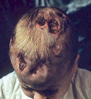
Langerhans cell histiocytosis (LCH) is an abnormal clonal proliferation of Langerhans cells, abnormal cells deriving from bone marrow and capable of migrating from skin to lymph nodes.

Eosinophilic pneumonia is a disease in which an eosinophil, a type of white blood cell, accumulates in the lungs. These cells cause disruption of the normal air spaces (alveoli) where oxygen is extracted from the atmosphere. Several different kinds of eosinophilic pneumonia exist and can occur in any age group. The most common symptoms include cough, fever, difficulty breathing, and sweating at night. Eosinophilic pneumonia is diagnosed by a combination of characteristic symptoms, findings on a physical examination by a health provider, and the results of blood tests and X-rays. Prognosis is excellent once most eosinophilic pneumonia is recognized and treatment with corticosteroids is begun.

Malignant histiocytosis is a rare hereditary disease found in the Bernese Mountain Dog and humans, characterized by histiocytic infiltration of the lungs and lymph nodes. The liver, spleen, and central nervous system can also be affected. Histiocytes are a component of the immune system that proliferate abnormally in this disease. In addition to its importance in veterinary medicine, the condition is also important in human pathology.

<span class="mw-page-title-main">Letterer–Siwe disease</span> Medical condition

Letterer–Siwe disease, (LSD) or Abt-Letterer-Siwe disease, is one of the four recognized clinical syndromes of Langerhans cell histiocytosis (LCH) and is the most severe form, involving multiple organ systems such as the skin, bone marrow, spleen, liver, and lung. Oral cavity and gastrointestinal involvement may also be seen. LCH and all its subtypes are characterized by monoclonal migration and proliferation of specific dendritic cells.

Loeffler endocarditis is a form of heart disease characterized by a stiffened, poorly-functioning heart caused by infiltration of the heart by white blood cells known as eosinophils. Restrictive cardiomyopathy is a disease of the heart muscle which results in impaired diastolic filling of the heart ventricles, i.e. the large heart chambers which pump blood into the pulmonary or systemic circulation. Diastole is the part of the cardiac contraction-relaxation cycle in which the heart fills with venous blood after the emptying done during its previous systole.

<span class="mw-page-title-main">Erdheim–Chester disease</span> Medical condition

Erdheim–Chester disease (ECD) is an extremely rare disease characterized by the abnormal multiplication of a specific type of white blood cells called histiocytes, or tissue macrophages. It was declared a histiocytic neoplasm by the World Health Organization in 2016. Onset typically is in middle age, although younger patients have been documented. The disease involves an infiltration of lipid-laden macrophages, multinucleated giant cells, an inflammatory infiltrate of lymphocytes and histiocytes in the bone marrow, and a generalized sclerosis of the long bones.

<span class="mw-page-title-main">Chronic multifocal Langerhans cell histiocytosis</span> Medical condition

Chronic multifocal Langerhans cell histiocytosis, previously known as Hand–Schüller–Christian disease, is a type of Langerhans cell histiocytosis (LCH), which can affect multiple organs. The condition is traditionally associated with a combination of three features; bulging eyes, breakdown of bone, and diabetes insipidus, although around 75% of cases do not have all three features. Other features may include a fever and weight loss, and depending on the organs involved there may be rashes, asymmetry of the face, ear infections, signs in the mouth and the appearance of advanced gum disease. Features relating to lung and liver disease may occur.

<span class="mw-page-title-main">Kimura's disease</span> Medical condition

Kimura's disease is a benign rare chronic inflammatory disorder. Its primary symptoms are subdermal lesions in the head or neck or painless unilateral inflammation of cervical lymph nodes.

<span class="mw-page-title-main">Langerhans cell sarcoma</span> Medical condition

Langerhans cell sarcoma (LCS) is a rare form of malignant histiocytosis. It should not be confused with Langerhans cell histiocytosis, which is cytologically benign. It can present most commonly in the skin and lymphatic tissue, but may also present in the lung, liver, and bone marrow. Treatment is most commonly with surgery or chemotherapy.

Histiocytic diseases in dogs are a group of diseases in dogs which may involve the skin, and which can be difficult to differentiate from granulomatous, reactive inflammatory or lymphoproliferative diseases. The clinical presentation and behaviour as well as response to therapy vary greatly among the syndromes.

V600E is a mutation of the BRAF gene in which valine (V) is substituted by glutamic acid (E) at amino acid 600. It is a driver mutation in a proportion of certain diagnoses, including melanoma, hairy cell leukemia, papillary thyroid carcinoma, colorectal cancer, non-small-cell lung cancer, Langerhans cell histiocytosis, Erdheim–Chester disease and ameloblastoma.

Eosinophilic myocarditis is inflammation in the heart muscle that is caused by the infiltration and destructive activity of a type of white blood cell, the eosinophil. Typically, the disorder is associated with hypereosinophilia, i.e. an eosinophil blood cell count greater than 1,500 per microliter. It is distinguished from non-eosinophilic myocarditis, which is heart inflammation caused by other types of white blood cells, i.e. lymphocytes and monocytes, as well as the respective descendants of these cells, NK cells and macrophages. This distinction is important because the eosinophil-based disorder is due to a particular set of underlying diseases and its preferred treatments differ from those for non-eosinophilic myocarditis.

Oral manifestations of systematic disease are signs and symptoms of disease occurring elsewhere in the body detected in the oral cavity and oral secretions. High blood sugar can be detected by sampling saliva. Saliva sampling may be a non-invasive way to detect changes in the gut microbiome and changes in systemic disease. Another example is tertiary syphilis, where changes to teeth can occur. Syphilis infection can be associated with longitudinal furrows of the tongue.

Crystal-storing histiocytosis is a form of histiocytosis which mostly occurs in people with monoclonal gammopathies. Histiocytosis is an excessive number of histiocytes. In the vast majority of crystal-storing histiocytosis cases, immunoglobulins accumulate within the cytoplasm of histiocytes; in rare cases clofazimine, cystine, silica, or Charcot–Leyden crystals may be found in the histiocytes instead. Non-immunoglobulin crystal-storing histiocytosis is mostly associated with non-malignant disorders, such as chronic inflammation or autoimmune abnormality conditions such as rheumatoid arthritis, Crohn's disease, or Helicobacter pylori gastritis. It may be a localised or generalised disease. Examples of locations where histiocytosis may occur include the lungs, pleura, stomach, kidney, bone marrow, thyroid, thymus, and parotid gland. The disease is described as generalised if two or more unrelated sites are involved.

References

  1. 1 2 3 4 5 Jha, Suman K.; De Jesus, Orlando (2022), "Eosinophilic Granuloma", StatPearls, Treasure Island (FL): StatPearls Publishing, PMID   32644464 , retrieved 2022-01-31
  2. Chadha, Manish; Agarwal, Anil; Agarwal, Nidhi; Singh, Manoj Kumar (June 2007). "Solitary eosinophilic granuloma of the radius. An unusual differential diagnosis". Acta Orthopaedica Belgica. 73 (3): 413–417. ISSN   0001-6462. PMID   17715738.
  3. Badalian-Very, Gayane; Vergilio, Jo-Anne; Degar, Barbara A.; MacConaill, Laura E.; Brandner, Barbara; Calicchio, Monica L.; Kuo, Frank C.; Ligon, Azra H.; Stevenson, Kristen E.; Kehoe, Sarah M.; Garraway, Levi A. (2010-09-16). "Recurrent BRAF mutations in Langerhans cell histiocytosis". Blood. 116 (11): 1919–1923. doi:10.1182/blood-2010-04-279083. ISSN   1528-0020. PMC   3173987 . PMID   20519626.
  4. Tran, Gary; Huynh, Thy N.; Paller, Amy S. (March 2018). "Langerhans cell histiocytosis: A neoplastic disorder driven by Ras-ERK pathway mutations". Journal of the American Academy of Dermatology. 78 (3): 579–590.e4. doi:10.1016/j.jaad.2017.09.022. ISSN   1097-6787. PMID   29107340. S2CID   46767736.
  5. Ma, Jennifer; Laird, James H.; Chau, Karen W.; Chelius, Monica R.; Lok, Benjamin H.; Yahalom, Joachim (January 2019). "Langerhans cell histiocytosis in adults is associated with a high prevalence of hematologic and solid malignancies". Cancer Medicine. 8 (1): 58–66. doi:10.1002/cam4.1844. ISSN   2045-7634. PMC   6346231 . PMID   30597769.
  6. Kumar, Neeta; Sayed, Shahin; Vinayak, Sudhir (2011-01-20). "Diagnosis of Langerhans cell histiocytosis on fine needle aspiration cytology: a case report and review of the cytology literature". Pathology Research International. 2011: 439518. doi: 10.4061/2011/439518 . ISSN   2042-003X. PMC   3034992 . PMID   21331166.
  7. Kobayashi, Masayuki; Tojo, Arinobu (December 2018). "Langerhans cell histiocytosis in adults: Advances in pathophysiology and treatment". Cancer Science. 109 (12): 3707–3713. doi:10.1111/cas.13817. ISSN   1349-7006. PMC   6272080 . PMID   30281871.
  8. Muse, Russell; Williamson, Nicola (2008-01-01), Morgan, Rhea V. (ed.), "CHAPTER 91 - Erosive and Ulcerative Diseases of the Skin", Handbook of Small Animal Practice (Fifth Edition), Saint Louis: W.B. Saunders, pp. 885–900, ISBN   978-1-4160-3949-5 , retrieved 2022-01-31
  9. 1 2 Hopke, Kaitlin P; Sargent, Sandra J (2019-12-16). "Novel presentation of eosinophilic granuloma complex in a cat". JFMS Open Reports. 5 (2): 2055116919891548. doi:10.1177/2055116919891548. ISSN   2055-1169. PMC   6918502 . PMID   31897305.
  10. 1 2 "Feline Eosinophilic Granuloma Complex In Cats | VCA Animal Hospitals". vcahospitals.com. Retrieved 2022-01-31.
  11. Abbott, Dawn E. E.; Allen, Andrew L. (March 2020). "Canine eosinophilic pulmonary granulomatosis: case report and literature review". Journal of Veterinary Diagnostic Investigation. 32 (2): 329–335. doi:10.1177/1040638720907659. ISSN   1040-6387. PMC   7081515 . PMID   32180541.
  12. 1 2 3 Mendelsohn, Danielle; Lewis, John R.; Scott, Kristin Iglesias; Brown, Dorothy C.; Reiter, Alexander M. (March 2019). "Clinicopathological Features, Risk Factors and Predispositions, and Response to Treatment of Eosinophilic Oral Disease in 24 Dogs (2000-2016)". Journal of Veterinary Dentistry. 36 (1): 25–31. doi: 10.1177/0898756419834785 . ISSN   0898-7564. PMID   31138045. S2CID   169032822.
  13. 1 2 Tellado, Matías Nicolás; Michinski, Sebastián Diego; Olaiz, Nahuel; Maglietti, Felipe; Marshall, Guillermo (2014-10-16). "Canine Oral Eosinophilic Granuloma Treated with Electrochemotherapy". Case Reports in Veterinary Medicine. 2014: e519197. doi: 10.1155/2014/519197 . hdl: 11336/19287 . ISSN   2090-7001.